INTRODUCTION
In tropical and subtropical waters, some colonial ascidians of the family Didemnidae obligately harbour symbiotic cyanophytes such as Prochloron (reviewed by Hirose et al., Reference Hirose, Neilan, Schmidt, Murakami, Gault and Marler2009a). These photosymbiotic ascidians are exclusively present at low latitudes, and species richness gradually decreases toward higher latitudes (Hirose, Reference Hirose2013 and references therein). Prochloron is affected by low temperature; the cyanobacterial cells exhibited no photosynthetic activity at ≤20°C in vitro (Dionisio-Sese et al., Reference Dionisio-Sese, Maruyama and Miyachi2001). Therefore, water temperature is a critical factor in limiting the distribution of host ascidians, and the range of the species may shift to higher latitudes because of global warming (e.g. Walther et al., Reference Walther, Post, Convey, Menzel, Parmesank, Beebee, Fromentin, Hoegh-Guldberg and Bairlein2002).
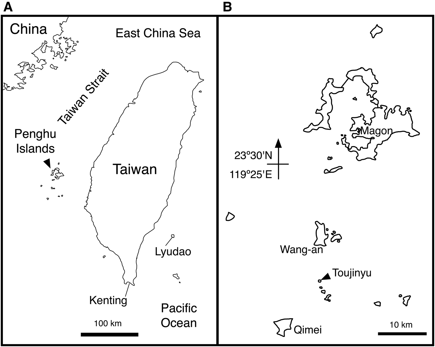
We recorded a total of 11 species of photosymbiotic ascidians from Kenting and Lyudao (Green Island) in south-eastern Taiwan (Figure 1A), but no photosymbiotic species were found in Keelung in northern Taiwan (Hirose & Nozawa, Reference Hirose and Nozawa2010; Hirose & Su, Reference Hirose and Su2011; Hirose et al., Reference Hirose, Nozawa and Hirose2014b). This is likely because the surface water temperature on the northern coasts of Taiwan is too low in the winter (minimum 16°C) due to the influx of the cold China Coastal Current. This photosymbiotic ascidian distribution pattern is consistent with the composition of the coral community: non-reef corals are present on the northern coast of Taiwan, whereas south-eastern Taiwan contains tropical reef corals (Chen, Reference Chen1999; Chen & Shashank, Reference Chen and Shashank2009).
(A) Map of Taiwan. (B) Map of the Penghu Islands showing the Diplosoma gumavirens collection site (Toujinyu).

The Penghu Islands are a major group of islands in the Taiwan Strait, where the average surface water temperature in February is 20.4°C (Figure 1A). However, the temperature can fall because of intrusion of the China Coastal Current, recorded to drop to as low as 12.6°C in 2008 (Chang et al., Reference Chang, Lee, Lee and Lan2009). The coral community in this area is characterized by non-reef species (Chen, Reference Chen1999; Chen & Shashank, Reference Chen and Shashank2009), indicating that the presence of photosymbiotic ascidians is unlikely in Penghu. Surprisingly, however, we found an aggregate colony of photosymbiotic ascidians in Penghu during the International Joint Field Course held by National Taiwan University, Tunghai University, University of the Ryukyus, Bogor Agricultural University and Prince of Songkla University. This is the first record of photosymbiotic ascidians in the Taiwan Strait.
MATERIALS AND METHODS
Animals
Colonies of Diplosoma gumavirens Hirose & Oka, 2009 were collected by the authors in the tide pools of Toujinyu (23°17′27.1″N 119°30′05.6″E, Wang-an, Penghu, Taiwan) on 28 August 2014 (Figure 1B). The colonies were photographed using a digital camera, and subsequently preserved in 99% (v/v) ethanol at −30°C for DNA analysis. Some colonies originally preserved in ethanol were transferred to 70% (v/v) ethanol for morphological observation. Some zooids dissected from the colonies were examined under a light microscope equipped with differential interference contrast optics. In some photomicrographs, several images were combined to increase the depth of field using the post-processing image software Helicon Focus Pro 4.2.8 (Helicon Soft, Ltd., Kharkov, Ukraine). Some colonies were deposited in the National Museum of Nature and Science, Taichung, Taiwan (NMNS-7417-001).
DNA extraction, amplification and sequencing
One colony aggregate was regarded as a clone, since each colony was too small to extract genomic DNA for this study. Three ethanol-preserved colonies from one colony aggregate were dissected under a stereomicroscope to pick out zooids from the colonies. Genomic DNA was extracted from a total of ~40 zooids using a DNeasy Tissue Kit (Qiagen K.K., Tokyo, Japan) following the manufacturer's protocol. A segment of the mitochondrial cytochrome oxidase subunit I (COI) gene was amplified using the primers UroCox1-244F (5′-CATTTWTTTTGATTWTTTRGWCATCCNGA-3′) and UroCox1-387R (5′-GCWCYTATWSWWAAWACATAATGAAARTG-3′), following the method described by Hirose et al. (Reference Hirose, Yokobori and Hirose2009c) for amplification and sequencing of the partial COI fragments. Five clones were randomly selected and sequenced. PCR products were treated with ExoSAP-IT (Affymetrix, California, USA) prior to sequencing reactions. Sequencing reactions were performed using Operon Biotechnology (Tokyo, Japan). The sequences determined in this study have been deposited in the following DNA databases: GenBank, the European Molecular Biology Laboratory and the DNA Data Bank of Japan, under the accession number LC018353.
Phylogenetic analyses
Partial COI gene sequences of 11 didemnid ascidian specimens (eight from the genus Diplosoma) were obtained from GenBank and aligned with the present sequence for molecular analysis. Initial alignments were performed using MUSCLE (Edgar, Reference Edgar2004); these were visually inspected and manually edited. Maximum likelihood (ML) analysis using raxmlGUI version 1.3 (Silvestro & Michalak, Reference Silvestro and Michalak2012) and phylogenetic analysis based on Bayesian inference (BI) (using MrBayes 3.12; Ronquist & Huelsenbeck, Reference Ronquist and Huelsenbeck2003) were performed on the aligned DNA sequences of the partial COI gene. In the BI analysis, a Markov Chain Monte Carlo analysis was run for 1,000,000 generations, and trees were built at 100-generation intervals (burn-in = 2500 runs). Statistical support for the ML tree was evaluated using a nonparametric bootstrap test with 1000 resampling events.
RESULTS
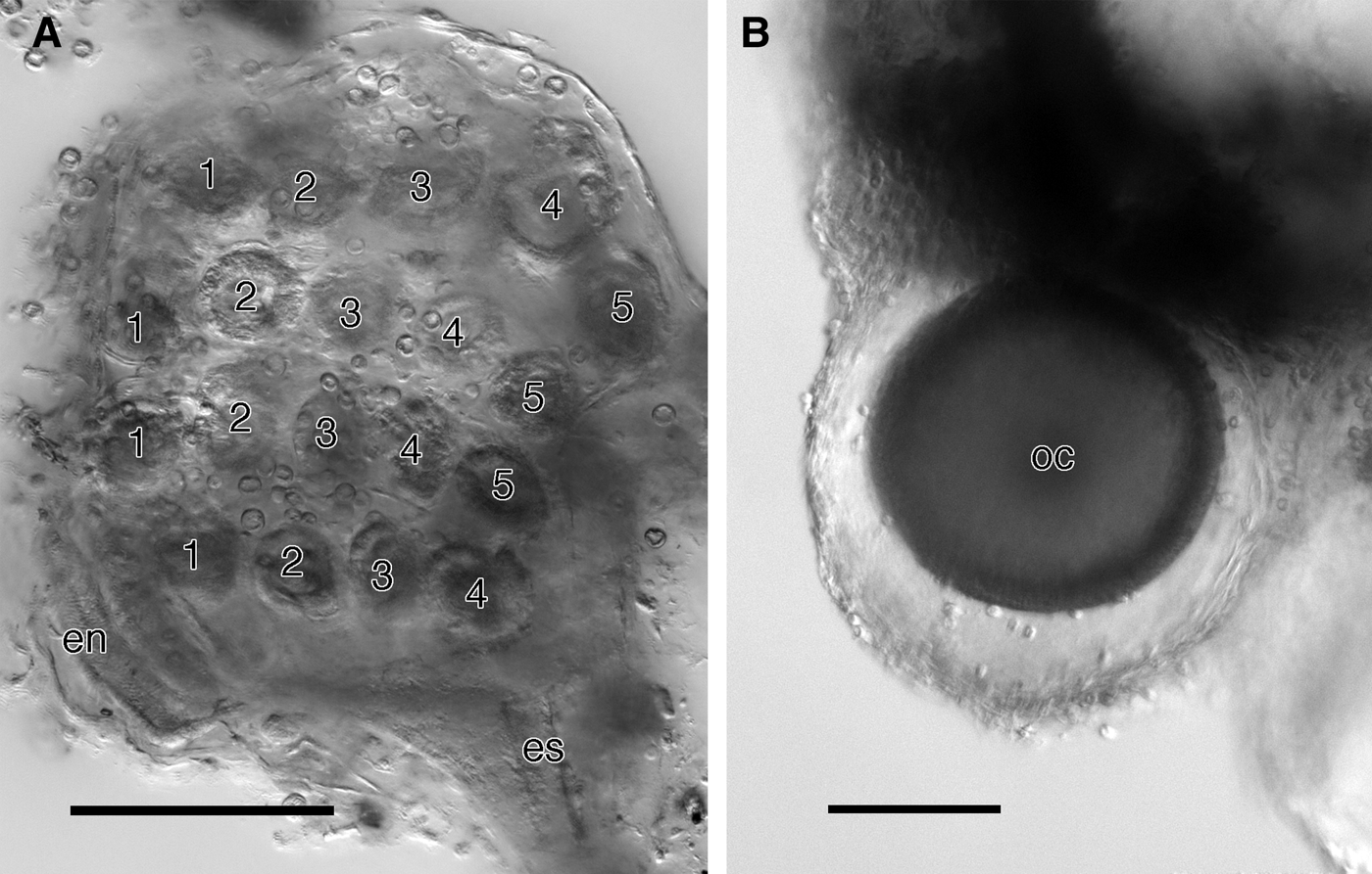
The colonies were found in tide pools approximately 0.5 m deep (Figure 2A). Green colonies were crowded onto rock surfaces and other sessile organisms such as algae encrusting the rocks (Figure 2B). Each colony consisted of an elliptical or irregularly shaped sheet approximately 1–2 cm in diameter and 1–1.5 mm thick (Figure 2C). When the colonies were crowded they formed a large green sheet covering the substratum. Although the tunic of each colony is transparent, the colony always appears green because of the presence of symbiotic Prochloron in the common cloacal cavity. The lack of tunic spicules, as well as zooid morphology, indicates that these specimens belong to the genus Diplosoma. Zooids are sparsely embedded in the colony, and the oral aperture of each zooid is bordered with a light blue ring. The common cloacal aperture is round or elongated and opens at the approximate centre of each colony. We were unable to prepare anesthetized formalin-fixed specimens for microscopic observation, and thus, most zooids severely contracted and poorly preserved in ethanol-fixed specimens. The branchial sac contains four stigmatal rows, with five stigmata on each side in the first (top) row, the second row and the third row, and four stigmata per side in the fourth (bottom) row (i.e. a 5-5-5-4 stigmatal pattern) (Figure 3A). The point of emergence of the retractor muscle could not be confirmed in our specimens. Some zooids have a large oocyte at the posterior end of the abdomen (Figure 3B).
Diplosoma gumavirens. (A) Tide pool containing colonies. (B) Green colonies crowded onto rocks and other sessile organisms in the tide pool. (C) Enlargement of ovoid colonies. Oral apertures are bordered with a light blue ring. A common cloacal opening is present at the approximate centre of each colony.

Diplosoma gumavirens. (A) Branchial sac with four stigmatal rows (left-side view). There are five stigmata per side in the first (top), second and third rows, and four in the fourth (bottom) row. (B) Oocyte (oc) associated with the posterior end of the abdomen. en, endostyle; es, oesophagus. Scale bars = 0.1 mm.
Molecular identification
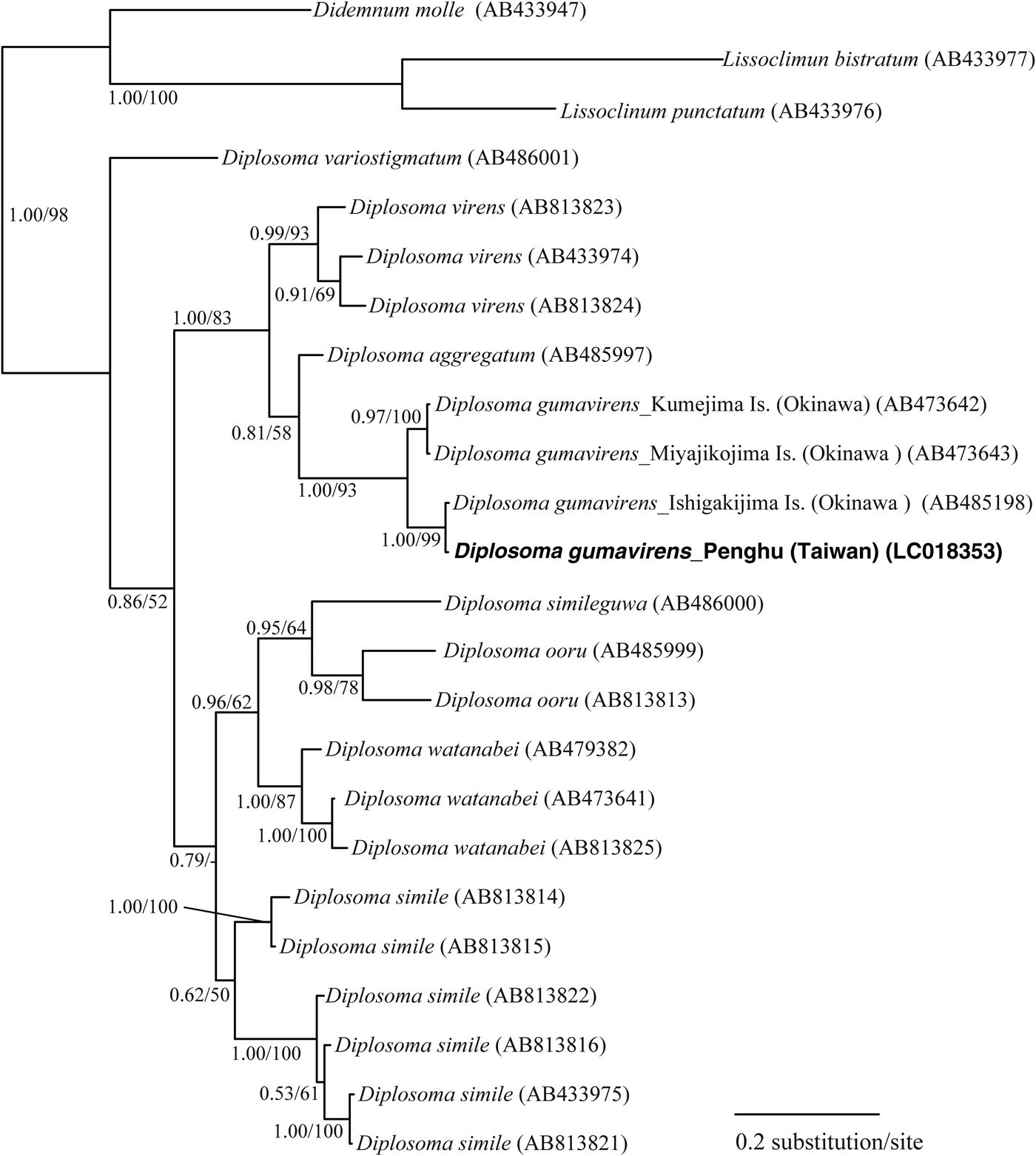
The genetic differences of the partial mtCOI DNA sequences of Diplosoma gumavirens specimens from three sites in the Ryukyu Archipelago (Kumejima Island, Miyakojima Island and Ishigakijima Island, Japan) and Toujinyu (Penghu, Taiwan) was 0–22/401 base pair differences (0–5.5%) and the genetic distance with the uncorrected pairwise distance was 0–0.0549. Figure 4 shows the BI tree generated using a general time-reversible model with an invariable sites and gamma distribution substitution (GTR + I + G). The topology of the ML phylogenetic tree was nearly identical. The phylogenetic trees inferred from the partial COI sequences supported the monophyly of D. gumavirens with a high Bayes posterior probability and bootstrap probability.
Bayesian inference consensus tree of selected didemnid species based on partial COI sequences. The GTR + I + G model was used for analysis. Bayes posterior probability and bootstrap probabilities greater than 50% upon maximum likelihood analyses are noted. New sequences from this study are shown in bold.
DISCUSSION
Diplosoma gumavirens was originally described from Miyakojima Island (Okinawa, Japan) and is widely distributed in the Ryukyu Archipelago (Hirose et al., Reference Hirose, Oka and Hirose2009b). This species was also reported from Kenting and Lyudao (Green Island) in Taiwan, and Pari Island in the Thousand Islands, Indonesia (Hirose & Nozawa, Reference Hirose and Nozawa2010; Hirose et al., Reference Hirose, Iskandar and Wardiatno2014a). Previously the colonies were generally found on rocks or on macro-algae in shallow coral reefs (<1 m deep). The habitat and morphological characteristics of the present specimens were consistent with D. gumavirens. The stigmatal pattern of our specimens was 5-5-5-4, and D. gumavirens is the only species known to have this pattern among the eight photosymbiotic Diplosoma recorded from the Ryukyus and Taiwan (Hirose & Su, Reference Hirose and Su2011). The retractor muscle emerges midway along the oesophageal neck in D. gumavirens, but we could not determine this state in our specimens because they were poorly preserved in ethanol. Some colonies contained oocytes, indicating the occurrence of sexual reproduction in summer. This is also consistent with the behaviour of D. gumavirens at the type locality (Miyakojima Island), where some embryos were found in colonies collected in August (Hirose et al., Reference Hirose, Oka and Hirose2009b).
We developed a dataset of the partial sequences of the COI gene (Yokobori's region) of photosymbiotic ascidians recorded in the Ryukyus–Taiwan region, to allow for DNA barcode species identification. The intraspecific divergence of D. gumavirens among four collection sites (0–5.5%) was less than the interspecific divergence among eight photosymbiotic Diplosoma species (10.2–22.4%) (Hirose & Hirose, Reference Hirose and Hirose2013). Molecular analyses based on partial sequences of the COI gene were consistent with identifications based on morphological characters. This is the first record of photosymbiotic ascidians in the Taiwan Strait.
Photosymbiotic ascidians have never been found on the northern coast of Taiwan, most likely because the water is too cold in winter (lows of 16°C). In contrast, such ascidians have been recorded in south and south-east regions (i.e. Kenting and Lyudao) where the Kuroshio Current warms the water. In the Taiwan Strait, the China Coastal Current cools the water in the winter to at least 16°C, and the coral community in Penghu is characterized by non-reef species (Chen, Reference Chen1999; Chen & Shashank, Reference Chen and Shashank2009). Moreover, although the average temperature of the surface water is 20.4°C in February around Penghu, an exceptional intrusion of cold water caused an extremely low temperature (a low of 12.6°C) to be recorded in 2008 (Chang et al., Reference Chang, Lee, Lee and Lan2009). Given the sensitivity of the cyanobacterial symbiont Prochloron to low temperatures (Dionisio-Sese et al., Reference Dionisio-Sese, Maruyama and Miyachi2001), the winter around Penghu is probably too cold for any photosymbiotic ascidians. This may indicate that D. gumavirens occurs seasonally in Penghu, where larvae drifting from the south settle in the area and the colonies grow and produce gametes during the warm season, but disappear in winter. However, their larval period is probably too short for long-distance dispersal, since photosymbiotic didemnids are always viviparous and the most larvae settle within a few hours after the release from the mother colony (van Duyl et al., Reference van Duyl, Bak and Sybesma1981; Olson, Reference Olson1983). Therefore, it is likely that the colonies in Penghu originated from larvae released from other colonies located on drifting materials from the south. Alternatively, it is possible that the colonies regress and hibernate during low temperature periods, and then regrow when the temperature increases above a critical threshold. Increases in water temperatures caused by global warming will facilitate stable colonization by this species and the recruitment of other tropical species in the future. Some photosymbiotic ascidians are reported to overgrow live corals and occasionally damage coral reefs (Littler & Littler, Reference Littler and Littler1995; Vargas-Ángel et al., Reference Vargas-Ángel, Asher, Godwin and Brainard2009; Sommer et al., Reference Sommer, Harrison and Scheffers2010; Rodriguez-Martinez et al., Reference Rodriguez-Martinez, Jordan-Garza, Baker and Jordan-Dahlgren2012). Therefore, we need to anticipate the potential risks of changes in community structure caused by colonization by photosymbiotic ascidians from the south, and the field monitoring of biota is, therefore, important to establish current ecological conditions.
ACKNOWLEDGEMENTS
The present study was supported by the International Research Hub Project for Climate Change and Coral Reef/Island Dynamics from The University of the Ryukyus, and by the Japan Society for the Promotion of Science (grant no. 23370013). We thank the organizers and members of the 2014 International Joint Field Course.