Introduction
Digeneans parasitizing the eyes of fish, known as eyeflukes, are metacercaria larval stages of species belonging to several genera of the Diplostomoidea Poirier, 1886, including Tylodelphys Diesing, 1850. However, Tylodelphys species can also infect the brain (or cranial cavity), the pericardial sac or the body cavity of their second intermediate hosts, which are typically fish, but sometimes amphibians. Like other diplostomids, Tylodeplphys spp. have a complex three-host life cycle, with a variety of fish- and amphibian-eating birds such as ciconiiforms, suliforms, falconiforms and podicipediforms acting as definitive hosts, and lymnaeid or planorbid gastropods as first intermediate hosts (e.g. Dubois, Reference Dubois1970; Niewiadomska & Laskowski, Reference Niewiadomska and Laskowski2002). Tylodelphys has a cosmopolitan distribution, with four nominal species in Africa (T. aegyptius El-Naffar, Khalifa & Sakla, 1980, T. grandis Zhokov, Morozova & Tessema 2010, T. mashonense Beverley-Burton, 1963 and T. xenopi (Nigrelli & Maraventano, 1944)); seven in Asia (T. cerebralis (Chakrabarti, 1968), T. chandrapali Jain & Gupta, 1970, T. darteri Mehra, 1962, T. duboisilla Mehra, 1962, T. excavata spinnata (Gupta, 1962), T. ophthalmi (Pandey, 1970), T. rauschi (Singh, 1956)); six in Europe (T. clavata (von Nordmann 1832), T. conifera (Mehlis, 1846), T. craniaria (Diesing, 1858), T. excavata excavata (Rudolphi, 1803), T. glossoides (Dubois, 1928) and T. podicipina podicipina Kozicka & Niewiadomska, 1960); four in North America (T. aztecae Garcia-Varela et al., 2015, T. immer Dubois, 1961, T. podicipina robrauschi Dubois, 1969, T. scheuringi (Hughes 1929)) and ten in South America (T. adulta Lunaschi & Drago, 2004, T. americana (Dubois, 1936), T. argentinus Quaggiotto & Valverde, 1992, T. barilochensis Quaggiotto & Valverde, 1992, T. brevis Drago & Lunaschi 2008, T. cardiophilus Szidat 1969, T. crubensis Quaggiotto & Valverde, 1992, T. destructor Szidat & Nani, 1951, T. elongata (Lutz, 1928), T. jenynsiae Szidat, 1969). Unexpectedly, only two records of this genus exist in the whole Australasian region, Tylodelphys sp. and T. podicipina (Dubois & Angel, Reference Dubois and Angel1972; Kennedy, Reference Kennedy1995), which is most likely due to a more limited research effort rather than a distribution gap.
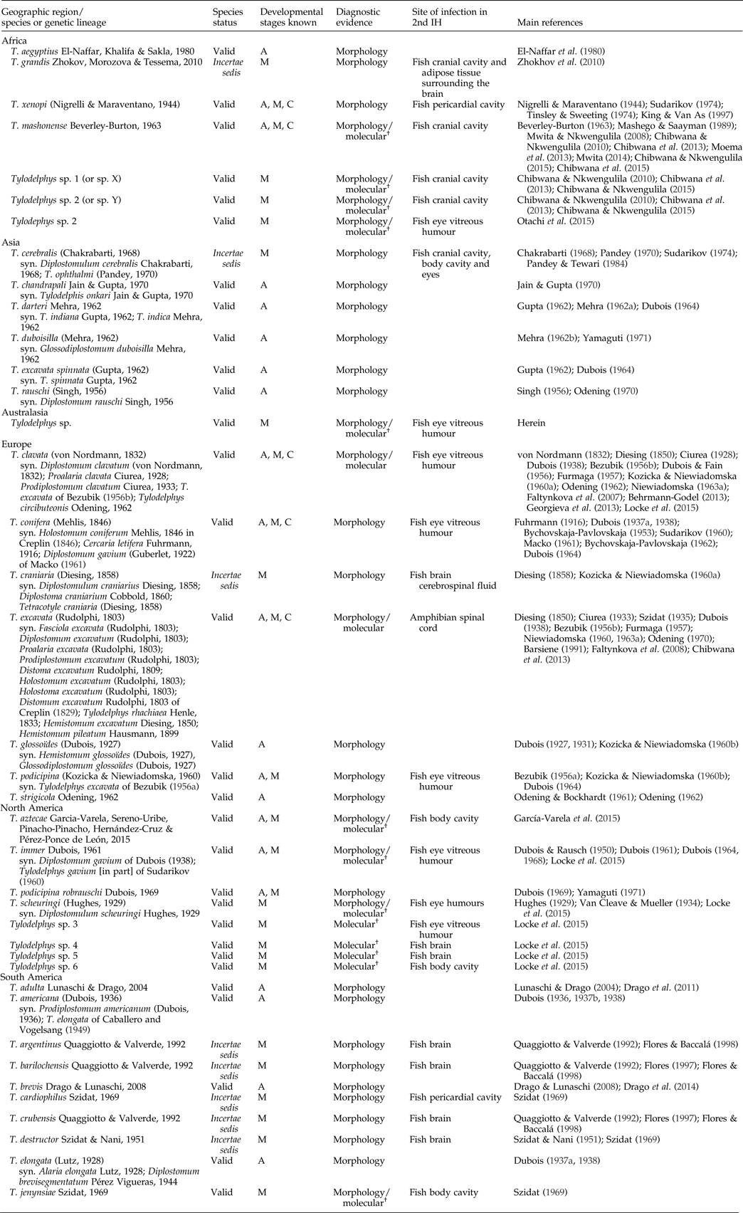
The genus Tylodelphys was erected by Diesing (Reference Diesing1850) for the metacercarial stages of two species, T. clavata (as original combination Diplostomum clavatum von Nordmann, 1832) and T. excavata (as original combination D. rhachiaeum Henle, 1833), the adults of which were later discovered by Ciurea (Reference Ciurea1928) who erected another genus (Prodiplostomum Ciurea, 1933) to accommodate these adults with distinctive metacercarial stages. Dubois (Reference Dubois1937a) reinstated the name Tylodelphys according to the ICZN Principle of Priority. Since then a total of 17 species has been described morphologically from adult specimens, and 11 solely on the basis of the metacercarial stage (table 1). Clearly, the limitations of obtaining adults from birds and the difficulty of completing the life cycles in the laboratory have hampered taxonomic progress in this group. However, recent barcoding surveys have successfully characterized molecularly the cercariae, metacercariae and/or adults of T. aztecae, T. clavata, T. excavata, T. immer, T. jenynsiae, T. mashonense and T. scheuringi (Locke et al., Reference Locke, McLaughlin and Marcogliese2010, Reference Locke, Al-Nasiri, Caffara, Drago, Kalbe, Lapierre, McLaughlin, Nie, Overstreet, Souza, Takemoto and Marcogliese2015; Behrmann-Godel, Reference Behrmann-Godel2013; Georgieva et al., Reference Georgieva, Soldánová, Pérez-del-Olmo, Dangel, Sitko, Sures and Kostadinova2013; Chibwana et al., Reference Chibwana, Nkwengulila, Locke, McLaughlin and Marcogliese2015; García-Varela et al., Reference García-Varela, Sereno-Uribe, Pinacho-Pinacho, Hernández-Cruz and Pérez-Ponce de León2015). Moreover, they have discovered three distinct lineages of unidentified Tylodelphys metacercariae from Africa (Chibwana et al., Reference Chibwana, Blasco-Costa, Georgieva, Hosea, Nkvvengulila, Scholz and Kostadinova2013; Otachi et al., Reference Otachi, Locke, Jirsa, Fellner-Frank and Marcogliese2015) and four from North America (Locke et al., Reference Locke, Al-Nasiri, Caffara, Drago, Kalbe, Lapierre, McLaughlin, Nie, Overstreet, Souza, Takemoto and Marcogliese2015). Only in a couple of studies was molecular characterization of metacercariae carried out in association with evaluation of morphological variation (Chibwana & Nkwengulila (Reference Chibwana and Nkwengulila2010) in conjunction with Chibwana et al. (Reference Chibwana, Blasco-Costa, Georgieva, Hosea, Nkvvengulila, Scholz and Kostadinova2013); and Otachi et al. (Reference Otachi, Locke, Jirsa, Fellner-Frank and Marcogliese2015)). In both cases, the authors found that morphological variation suggested a higher number of morphospecies than genetically identified units (lineages) in their samples. Taking into account these latter molecular findings and the fact that one-third of the known Tylodelphys spp. are distinguished based on the morphology of the metacercarial stage, we can expect that ‘true’ species diversity maybe very different from what is currently known.
Tylodelphys species described on the basis of adult or metacercariae specimens, status of the species, known life-cycle stages, diagnostic evidence for the species, site of infection in the second intermediate hosts and main references providing morphological or molecular information. Note that the reference list and synonyms for species are not exhaustive.

† Morphological information provided in association with molecular data, or morphological vouchers of the molecular samples deposited in Museum or University collections.
Abbreviations: A, adult; M, metacercaria; C, cercaria; IH, intermediate host.
The aim of this study was to describe morphologically the first displostomid, Tylodephys sp. metacercaria, found in New Zealand and to distinguish it from its congeners for which the metacercarial stage is known. Additionally, we provide sequences for the 28S, internal transcribed spacer (ITS) and Cox1 genetic markers in order to characterize the species molecularly, distinguish it from other Tylodelphys genetic lineages and allow future discovery of its life cycle or new distribution areas via matching molecular sequence data. Furthermore, we discuss and provide an updated list on the taxonomic status of Tylodephys species. Finally, we provide video images showing the movements of Tylodephys sp. metacercaria within the eye of its fish host.
Materials and methods
Collection and examination of fish
Ten common bullies, Gobiomorphus cotidianus McDowall (Actinopterygii: Eleotridae), were collected using a seine net from Lake Hayes in Otago (South Island, New Zealand) in May 2011 and 2013. Fish were euthanized by spinal severance and examined fresh. Metacercariae (devoid of cysts) were extracted from the vitreous humour of the eyes, fixed in absolute ethanol for molecular analyses, or 70% ethanol for whole-mount and scanning electron microscopy (SEM) examination.
Morphological data
Worms fixed for whole mounts were stained using acetic acid iron carmine stain, dehydrated through a graded ethanol series, cleared in clove oil and mounted in permanent preparations with Canada balsam. Two specimens were dehydrated in a graded ethanol series, critical point-dried and sputter-coated with gold for SEM examination using a Zeiss DSM 940A (Zeiss AG, Oberkochen, Germany) at an accelerating voltage of 5 kV. All measurements are in micrometres unless otherwise stated in the text, and are given as the range, with the mean followed by standard deviation (SD) in parentheses. Voucher material is deposited as permanent mounts, SEM preparations, genophores (two specimens preserved in 70% ethanol) and molecular vouchers (extracted gDNA) at the Platyhelminthes collection of the Natural History Museum of Geneva.
Molecular data and analyses
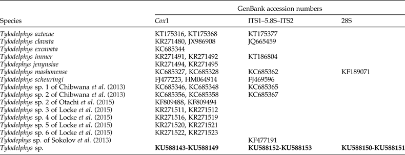
Genomic DNA was extracted from seven single ethanol-fixed metacercaria specimens of Tylodelphys sp. in 200 μl of a 5% suspension of Chelex® in deionized water containing 0.1 mg/ml proteinase K, followed by incubation at 56°C for 5 min, boiling at 90°C for 8 min, and centrifugation at 14,000 g for 10 min. The following gene regions were amplified: partial fragment of the large ribosomal subunit (28S rDNA) [1800 bp; primers U178F: 5′–GCA CCC GCT GAA YTT AAG–3′ and L1642R: 5′–CCA GCG CCA TCC ATT TTC A–3′ (Lockyer et al., Reference Lockyer, Olson and Littlewood2003)], the ITS1–5.8S–ITS2 ribosomal gene cluster (ITS) [900 bp; primers D1: 5′–AGG AAT TCC TGG TAA GTG CAA G–3′ and D2: 5′–CGT TAC TGA GGG AAT CCT GGT–3′ (Galazzo et al., Reference Galazzo, Dayanandan, Marcogliese and McLaughlin2002)] and partial fragment of the mitochondrial cytochrome c oxidase subunit I gene (Cox1) [600 bp; primers Plat-diploCOX1F: 5′–CGT TTR AAT TAT ACG GAT CC–3′ and Plat-diploCOX1R: 5′–AGC ATA GTA ATM GCA GCA GC–3′ (Moszczynska et al., Reference Moszczynska, Locke, McLaughlin, Marcogliese and Crease2009)]. Polymerase chain reaction (PCR) amplifications were performed in 25 μl reactions containing 2.5 μl of extraction supernatant, 1 × PCR buffer (16 mm (NH4)2SO4, 67 mm Tris–HCl at pH 8.8), 2 mm MgCl2, 200 μm of each deoxynucleoside triphosphate (dNTP), 0.5 μm each primer and 0.7 unit BIOTAQ™ DNA polymerase (Bioline (Aus) Pty Ltd, Alexandria, Australia). Thermocycling conditions used for amplification of the rDNA regions follow Blasco-Costa et al. (Reference Blasco-Costa, Balbuena, Kostadinova and Olson2009) for the 28S fragment and Chibwana et al. (Reference Chibwana, Blasco-Costa, Georgieva, Hosea, Nkvvengulila, Scholz and Kostadinova2013) for the ITS region. Thermocycling conditions for the Cox1 fragment were as follows: initial denaturation at 95°C for 2 min followed by 40 cycles with denaturation at 94°C for 40 s, annealing at 50°C for 30 s and extension at 72°C for 45 s; with a final extension step at 72°C for 5 min. PCR amplicons were purified prior to sequencing using exonuclease I and shrimp alkaline phosphatase enzymes (Werle et al., Reference Werle, Schneider, Renner, Volker and Fiehn1994). Amplicons were cycle-sequenced from both strands, using, besides PCR primers for the 28S and Cox1 genes, an internal primer for the 28S fragment [L1200R: 5′–GCA TAG TTC ACC ATC TTT CGG–3′ (Littlewood et al., Reference Littlewood, Curini-Galletti and Herniou2000)] and two other primers for the ITS fragment [BD1: 5′–GTC GTA ACA AGG TTT CCG TA–3′ and BD2: 5′–TAT GCT TAA ATT CAG CGG GT–3′ (Luton et al., Reference Luton, Walker and Blair1992)]. Sequencing was performed at the commercial facility Macrogen (Seoul, Korea). Contiguous sequences were assembled and edited using Geneious® (v. 8.1 Biomatters Ltd, Auckland, New Zealand) and submitted to GenBank.
Newly generated sequences for the 28S rDNA, the ITS1–5.8S–ITS2 gene cluster and the Cox1 fragment were aligned in three independent datasets together with published sequences of identified strigeids and diplostomids from GenBank. The Cox1 dataset was aligned using MUSCLE implemented in MEGA v. 6 (Tamura et al., Reference Tamura, Peterson, Peterson, Stecher, Nei and Kumar2011). The extremes of the alignments were trimmed to match the shortest sequence prior to phylogenetic analyses. The 28S and ITS datasets were aligned using MAFFT in Guidance (Sela et al., Reference Sela, Ashkenazy, Katoh and Pupko2015); for the ITS dataset, positions in the alignment with a score below 0.93 were excluded. The 28S alignment (851 bp long) included two representative sequences of Diplostomum spp. and one of T. mashonense retrieved from GenBank (table 2). The ITS dataset (779 bp long of the ITS1–5.8S–ITS2 gene cluster) included two representative sequences of Diplostomum and eight of Tylodelphys spp. from GenBank. The Cox1 dataset (413 bp long) included two representative sequences of Diplostomum and 27 of Tylodelphys spp. Sequences of Apatemon and Australapatemon spp. belonging to the Strigeidae, sister family to the Diplostomidae, were included as outgroups in all analyses. Phylogenetic analyses were run on the Cox1 and ITS datasets individually under the maximum likelihood (ML) and Bayesian inference (BI) criteria, employing the model of nucleotide evolution GTR + Γ (estimated using jModelTest 2.1.1 (Guindon & Gascuel, Reference Guindon and Gascuel2003; Darriba et al., Reference Darriba, Taboada, Doallo and Posada2012)). ML analyses were conducted using the program RAxML v. 7.3 (Stamatakis, Reference Stamatakis2006; Stamatakis et al., Reference Stamatakis, Hoover and Rougemont2008). All model parameters and bootstrap support values (1000 repetitions) were estimated using RAxML. BI trees were constructed using MrBayes v. 3.2 (Ronquist et al., Reference Ronquist, Teslenko, van der Mark, Ayres, Darling, Hohna, Larget, Liu, Suchard and Huelsenbeck2012), running two independent MCMC runs of four chains for 20 million generations and sampling tree topologies every 2000 generations. Burn-in periods were set to the first 2500 generations. A consensus topology and nodal support estimated as posterior probability values (Huelsenbeck et al., Reference Huelsenbeck, Ronquist, Nielsen and Bollback2001) were calculated from the remaining trees. All MrBayes and RAxML analyses were performed on the computational resource CIPRES (Miller et al., Reference Miller, Pfeiffer and Schwartz2010). Genetic divergences were calculated as uncorrected p-distances for each gene region/species using MEGA.
GenBank accession numbers for Tylodelphys species/lineages used as ingroup in the phylogenetic analyses.

In bold, accession codes for the newly obtained sequences for this study.
Diplostomidae Poirier, 1886; Tylodelphys Diesing, 1850; Tylodelphys sp.
References: Tylodelphys sp. in Lagrue & Poulin (Reference Lagrue and Poulin2015)
Description of metacercaria
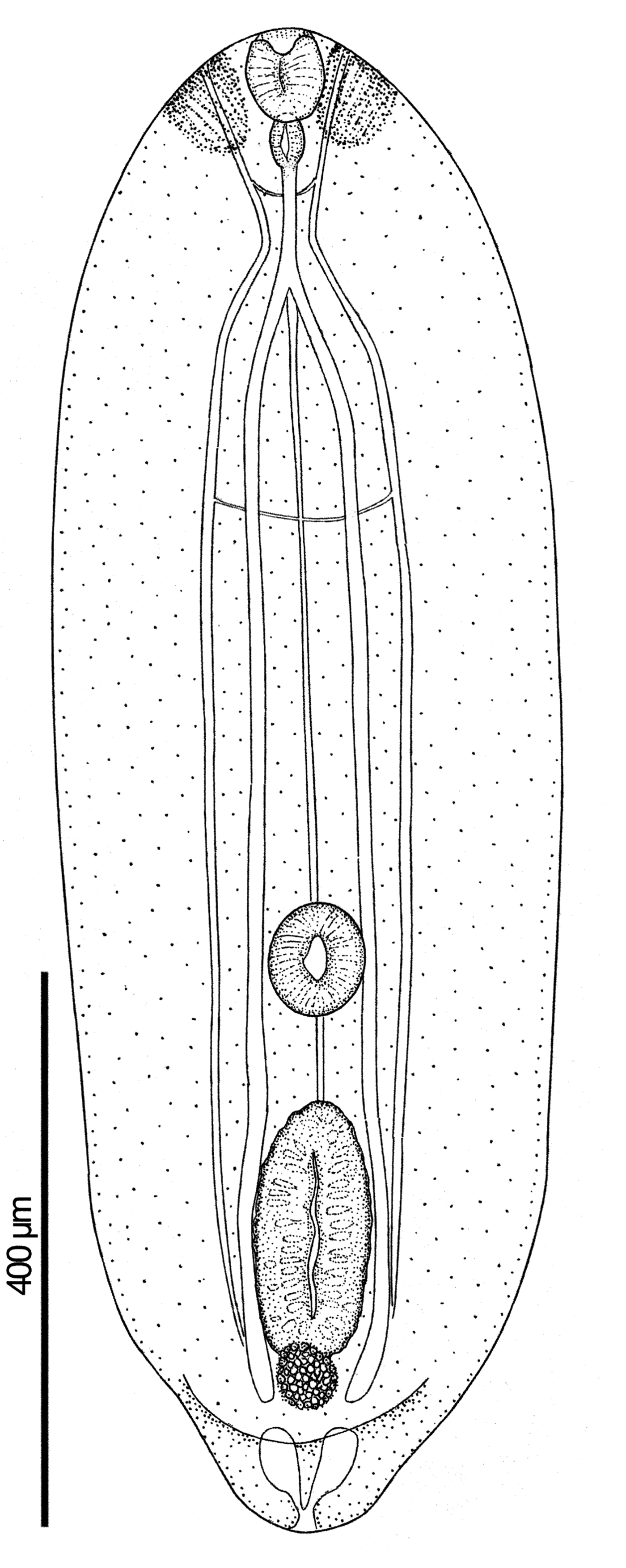
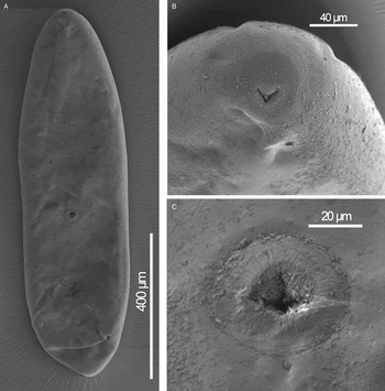
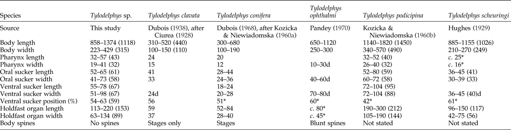
Based on 30 stained and mounted specimens and two specimens for SEM (figs 1, 2). Body linguiform, slightly concave ventrally, anterior end rounded, posterior end bluntly pointed, conical; length 858–1374 (1118 ± 167), width at level of holdfast organ 223–429 (315 ± 54). Tegument devoid of spines or papillae (fig. 2). Hindbody small and not well differentiated, only discernable on one specimen: length 143 (13% of total body length), width at widest (anterior margin) 161. Oral sucker sub-terminal, well-developed, smaller than ventral sucker (VS:OS width ratio 1:0.7–1.1 (1:0.9); 52–65 (61 ± 5) × 41–73 (55 ± 7). Pseudosuckers (lappets) either side of oral sucker, barely differentiated, only discernable on one specimen; 45–46 × 35–37. Ventral sucker sub-round, protrusive in lateral view; 55–78 (67 ± 7) × 51–98 (68 ± 10), and 464–756 (597 ± 80) from oral sucker. Ventral sucker situated 54–63 (59 ± 2)% length of body. Prepharynx absent. Pharynx longitudinal oval; 32–57 (43 ± 7) × 19–49 (32 ± 7). Oesophagus long; length 8–77 (46 ± 22). Intestinal caeca terminate posterior to holdfast organ. Holdfast organ strongly muscular, longitudinal oval, located midway between ventral sucker and posterior end of body; 113–220 (153 ± 26) × 63–134 (89 ± 20). Primordia of gonads directly posterior to holdfast organ within hindbody. Excretory bladder V-shaped with pore at posterior tip of body; three main excretory ducts united in region of pharynx by transverse duct about midway between oral sucker and ventral sucker. Minor ducts and flame cells not observed. Forebody of metacercaria filled with oval granular inclusions, average 10 × 5 in size.
Tylodelphys sp. metacercaria ex Gobiomorphus cotidianus.

Tylodelphys sp. metacercaria, scanning electron micrographs. Ventral view of the metacercaria (A), detail of the oral sucker (B) and detail of the ventral sucker (C).

Second intermediate host: common bully, Gobiomorphus cotidianus McDowall (Actinopterygii: Eleotridae).
Site of infection in second intermediate host: vitreous and aqueous humour of eye (see supplementary video V1).
Locality: Lake Hayes, South Island, New Zealand (44°59′S, 168°48′E, freshwater, elevation 332 m).
Prevalence in second intermediate host: 80%.
Mean intensity in second intermediate host: 2.6.
Material: voucher specimens (30 mounted metacercariae on slides and two mounted metacercariae for SEM); paragenophores (two specimens preserved in 70% ethanol); and molecular voucher specimens (extracted gDNA of two specimens) MHNG-PLAT-92964.
DNA sequences: Cox1, KU588143–KU588149; 28S, KU588150–KU588151; ITS1, KU588152–KU588153.
Remarks
The above-described metacercaria conforms to the description of the ‘diplostomulum’ type of metacercaria according to Niewadomska (Reference Niewadomska, Gibson, Jones and Bray2002), and it resembles all described species of Tylodelphys metacercariae. Of the 30 or so species of Tylodelphys, 18 have been described at the metacercarial stage (table 1). Of these, T. argentinus, T. barilochensis, T. cardiophilus, T. cerebralis, T. craniaria, T. crubensis, T. destructor, T. excavata, T. grandis, T. jenynsiae and T. xenopi occur in the cranial cavity, brain, spinal cord, pericardial sac or body cavity.
The remaining species, T. clavata, T. conifera, T. ophthalmi, T. podicipina and T. scheuringi, in common with the above-described species, inhabit the eye of their second intermediate fish host. Of these, T. clavata, T. conifera and T. scheuringi are smaller in all, or nearly all, metrics than the present species, and T. podicipina is larger in body size, ventral sucker and holdfast sizes (table 3). Tylodelphys ophthalmi is reported to have a comparatively very small pharynx and tegumental spination. It is likely, therefore, that the metacercaria reported here from bullies in New Zealand is a hitherto undescribed species, the full description and naming of which will be completed either experimentally, or as and when the adult is discovered. This represents the first record for a species of Tylodelphys and for a member of the Diplostomidae in New Zealand.
Comparative metrical data for metacercariae of Tylodelphys species parasitizing the eye of fish. The range is followed by the mean in parentheses when available.

* Data inferred from illustrations in original descriptions or redescriptions. Blank cells are missing data and not possible to infer from literature.
Abbreviation: d, diameter.
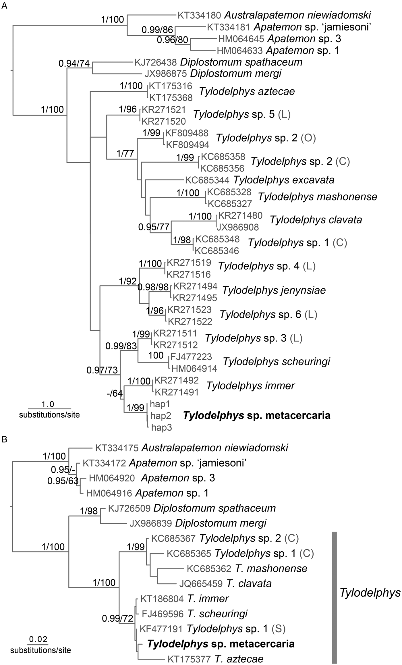
At the molecular level, three unique Cox1 haplotypes were retrieved from sequences of seven metacercariae of Tylodelphys sp. from New Zealand (NZ). The three haplotype sequences formed a separate reciprocally monophyletic lineage, supporting the distinct species status of these metacercariae. Phylogenetically, Tylodelphys sp. from NZ appeared closely related to species from North America, sister to T. immer in a clade together with T. scheuringi and Tylodelphys sp. 3 of Locke et al. (Reference Locke, Al-Nasiri, Caffara, Drago, Kalbe, Lapierre, McLaughlin, Nie, Overstreet, Souza, Takemoto and Marcogliese2015) (fig. 3a ). The relationships among Tylodelphys species/lineages were generally poorly supported. Phylogenetic analyses of the ITS region included sequences for eight Tylodelphys species besides our newly obtained sequences from NZ and depicted two clades with strong support (fig. 3b ). One clade included sequences representative of American species together with that of our metacercariae and one sequence of Tylodelphys sp. metacercaria of Sokolov et al. (Reference Sokolov, Lebedeva and Yadrenkina2013) from West Siberia. The other clade comprised sequences of the European and African species/lineages.
Phylogenetic relationships inferred by maximum likelihood analysis for representatives of Tylodelphys based on Cox1 (A) and ITS (B) sequence data; with posterior probability values followed by bootstrap percentages above the branches (posterior probabilities <0.90 and bootstrap values <60 not reported). Species in bold were newly sequenced in this study. Abbreviations: hap, haplotype (followed by a number); (L), Locke et al. (Reference Locke, Al-Nasiri, Caffara, Drago, Kalbe, Lapierre, McLaughlin, Nie, Overstreet, Souza, Takemoto and Marcogliese2015); (C), Chibwana et al. (Reference Chibwana, Blasco-Costa, Georgieva, Hosea, Nkvvengulila, Scholz and Kostadinova2013); (O), Otachi et al. (Reference Otachi, Locke, Jirsa, Fellner-Frank and Marcogliese2015); and (S), Sokolov et al. (Reference Sokolov, Lebedeva and Yadrenkina2013).

Intraspecific genetic divergence in the Cox1 fragment varied between 0.2 and 1.2% within Tylodelphys species; whereas Tylodelphys sp. from NZ showed 0.8% intraspecific variation. No intraspecific variability was detected in the ITS or 28S sequences of the two metacercariae of Tylodelphys sp. newly sequenced. Mean interspecific genetic divergence for the Cox1 sequences of Tylodelphys spp. showed a range of variation of 8–16.5%. Cox1 sequences of Tylodelphys sp. from NZ diverged 8.6–14.2% from the sequences of other Tylodelphys species/lineages. Mean interspecific genetic divergence for the ITS sequences of Tylodelphys species/lineages ranged from 0.7 to 8.3%, whereas Tylodelphys sp. from NZ diverged between 1.1–7.7% from other Tylodelphys species. Comparison of the partial sequences for the 28S rRNA gene of our metacercariae with the only available 28S sequence of Tylodelphys in GenBank (T. mashonense, referred to as T. mashonensis in GenBank; KF189071), revealed a genetic divergence of 3.1%; while divergence between the sequences for Tylodelphys spp. and those for Diplostomum spp. was 5.3–5.9%. Molecular information altogether confirms the generic affiliation of the newly sequenced specimens to Tylodelphys and their distinct status from all Tylodelphys species and lineages molecularly characterized so far.
Note on the taxonomic status of Tylodelphys spp.
Tylodelphys ophthalmi was first described as Diplostomulum ophthalmi Pandey, 1970, although it was cited as Pandey (Reference Pandey1968) (an abstract) in Pandey & Tewari (Reference Pandey and Tewari1984). However, the name D. ophthalmi was used prior to the description of the species in Chakrabarti (Reference Chakrabarti1968). Therefore, D. ophthalmi should have been considered a nomen nudum. Pandey & Tewari (Reference Pandey and Tewari1984) redescribed T. ophthalmi and proposed T. cerebralis as a junior synonym of the former, based on the similarity of its morphological features. We do not question the synonymy of the two forms; however, since D. ophthalmi should have been considered as nomen nudum and, attending to the Principle of Priority, the name T. cerebralis should prevail over T. ophthalmi, and the latter should be considered as its junior synonym (table 1). Additionally, Yamaguti (Reference Yamaguti1971) considered T. strigicola a nomen nudum because it lacked a description. However, Odening (Reference Odening1962) proposed T. strigicola for the material described as Tylodelphys sp. by Odening & Bockhardt (Reference Odening and Bockhardt1961), for which type specimens were also deposited. Thus, we consider T. strigicola as a valid species for the time being notwithstanding the remarks regarding its validity made by Niewiadomska (Reference Niewiadomska1963b).
Up to 14 reciprocally monophyletic lineages (putative species) of Tylodelphys have been characterized genetically so far (Moszczynska et al., Reference Moszczynska, Locke, McLaughlin, Marcogliese and Crease2009; Chibwana et al., Reference Chibwana, Blasco-Costa, Georgieva, Hosea, Nkvvengulila, Scholz and Kostadinova2013; Georgieva et al., Reference Georgieva, Soldánová, Pérez-del-Olmo, Dangel, Sitko, Sures and Kostadinova2013; García-Varela et al., Reference García-Varela, Sereno-Uribe, Pinacho-Pinacho, Hernández-Cruz and Pérez-Ponce de León2015; Locke et al., Reference Locke, Al-Nasiri, Caffara, Drago, Kalbe, Lapierre, McLaughlin, Nie, Overstreet, Souza, Takemoto and Marcogliese2015), but only half of them have been matched to already described species on the basis of either the adult or metacercarial stage (see table 1). Recently, García-Varela et al. (Reference García-Varela, Sereno-Uribe, Pinacho-Pinacho, Hernández-Cruz and Pérez-Ponce de León2015) proposed to consider as incertae sedis all South American species known only from their metacercaria form. We agree with these authors that the relationships between these forms and described species from adults cannot be established with the available morphological information. Thus, we propose to consider all named species on the sole basis of their larval metacercaria stage as incertae sedis. We exclude, however, those named metacercariae for which molecular data have been provided (i.e. T. jenynsiae and T. scheuringi), since evaluation of their phylogenetic relationship with other named species, either adults or metacercariae, is possible. Nonetheless, the adult stages of these recognized metacercaria species await formal description upon discovery or molecular matching to a known species when such data are obtained. Similarly, unnamed species/lineages of Tylodelphys, with molecular data supporting their uniqueness (at the molecular level at least), should be considered valid, especially when supplemented with morphological descriptions. Notwithstanding the incertae sedis status, these species should be taken into account in future morphological comparisons of metacercariae in order to eventually uncover their life cycle and clarify their status. Therefore, we recognize 21 nominal species of Tylodelphys and eight additional genetic lineages that we consider as valid.
Almost certainly, Tylodelphys species diversity is very different from what we know today, given that one-third of the known Tylodelphys spp. are distinguished solely based on the morphology of the metacercaria larval stage. Often a higher number of morphospecies than genetically identified units (lineages) has been found when morphological variation of metacercaria forms is assessed in combination with molecular data (Chibwana & Nkwengulila, Reference Chibwana and Nkwengulila2010; Chibwana et al., Reference Chibwana, Blasco-Costa, Georgieva, Hosea, Nkvvengulila, Scholz and Kostadinova2013; Otachi et al., Reference Otachi, Locke, Jirsa, Fellner-Frank and Marcogliese2015). Host-induced variation in the metacercariae of other diplostomids has also been documented (Niewiadomska & Szymanski, Reference Niewiadomska and Szymanski1991, Reference Niewiadomska and Szymanski1992). This leads us to think that the two subspecies of T. excavata and T. podicipina recognized by Dubois (Reference Dubois1964, Reference Dubois1969) may not be valid; in particular T. excavata spinnata (Gupta, 1962), which was already suggested as a likely junior synonym of T. excavata excavata (Rudolphi, 1903) by Dubois (Reference Dubois1964). We refrain from proposing a change in taxonomy until molecular sequence data are obtained from the original host and geographical region of the above subspecies. Nevertheless, we strongly recommend no further description and naming of new Tylodelphys species on the basis of morphological differences of the metacercarial forms alone.
Supplementary material
Video V1. Live movement of Tylodelphys sp. metacercariae in the vitreous humour of the eye of Gobiomorphus cotidianus immediately after eye removal. Observation made from a dissecting microscope.
To view supplementary material for this article, please visit http://dx.doi.org/10.1017/S0022149X16000298
Acknowledgements
We are thankful to Dr Carlos Rouco (University of Cordoba, Spain) and Dr Clement Lagrue (University of Otago, New Zealand) for their assistance in the field, and Dr André Piuz (Natural History Museum of Geneva, Switzerland) for his help with scanning electron microscopy. We are also grateful to two anonymous reviewers for their helpful suggestions and detailed corrections.
Financial support
This work has been supported indirectly by a University of Otago Research Grant and through a Marie Curie Outgoing International Fellowship for Career Development (IB-C, grant number PIOF-GA-2009–252124) within the 7th Framework Programme (FP7/2007–2013) of the European Commission.
Conflict of interest
None.
Ethical standards
The sampling in this paper complies with the current laws and animal ethics regulations of New Zealand.