Introduction
With increased capacity for mechanical ventilation in intensive care facilities, the incidence of iatrogenic tracheal stenosis is on the rise. The inflammatory changes that occur during prolonged ventilation lead to tracheal stenosis in 20 per cent of patients, among whom less than 1 per cent become symptomatic.Reference Pearson and Andrews1,Reference Brichet, Verkindre, Dupont, Carlier, Darras and Wurtz2
Open airway surgical procedures, though potentially safe, can have grave complications, such as sudden massive haemorrhage, shock and even death, when anatomical variations of the blood vessels are overlooked.Reference Silen and Spieker3 Here, we report two cases of intra-operative complications associated with the anomalous course of the brachiocephalic arteries. This report highlights the anatomical variations that can be encountered, and the vigilance required prior to and during airway surgical procedures to avoid such incidents.
Case reports
Case one
A 60-year-old female presented to our out-patient department with an inability to phonate or be decannulated for 3 years. She had been tracheostomised for grade IV tracheal stenosis following ventilation for post-traumatic brain injury.
After evaluation, she underwent tracheal resection and anastomosis. The first six tracheal rings had to be excised because of unhealthy mucosa, tracheomalacia, and disfigured cartilages that included the stenotic segment. On extubation, she developed stridor due to supraglottic oedema, for which a nasotracheal stent was inserted and kept in place. She did not tolerate stent removal given the persistent supraglottic oedema; hence, an emergency tracheostomy was performed.
During the emergency tracheostomy procedure, an inadvertent arterial injury occurred. Temporary haemostasis was achieved by manual compression until help arrived. The vascular surgeons identified a tear at the origin of the common carotid artery from the brachiocephalic trunk, which involved two-thirds of its circumference. This was repaired by primary suturing with size 5-0 Prolene,® and the tracheostomy was completed.
The patient had an uneventful post-operative period. She was subsequently decannulated and continues to be followed up.
On reviewing the pre-operative computed tomography (CT) scan, it was found that the origin, as well as the bifurcation of the brachiocephalic artery, was way to the left of the midline. It was seen arising from the arch of the aorta approximately 11 mm to the left of the midline, running tortuously for a length of 2 cm, before bifurcating in the midline. Hence, the right common carotid artery was seen originating 17 mm to the left of the right sternoclavicular joint traversing to the right, adherent to the anterior tracheal wall just inferior to the tracheostoma, before coursing cranially and laterally to reach the right side of the neck (Figure 1).
Computed tomography images: (a) coronal section showing right brachiocephalic trunk bifurcating at the left side of the trachea (red arrow); and (b) axial section showing right common carotid artery coursing cranially and laterally to reach the right side of the neck (yellow arrow).

Case two
A 47-year-old male manual labourer was scheduled to undergo tracheal resection and anastomosis. He had undergone an emergency tracheostomy earlier, following presentation to the out-patient department with severe stridor. He developed tracheal stenosis after prolonged ventilation for the management of organophosphorus poisoning. Repeated balloon dilatations were unsuccessful; hence, he was scheduled to undergo tracheal resection and anastomosis. Contrast-enhanced CT of the neck showed tracheal stenosis with deformed tracheal cartilages.
During the surgery, while dissecting laterally and above the tracheostoma to expose the trachea, there was a torrential bleed from below. This was temporarily controlled by manual pressure and the vascular surgeons were called in. The injury could not be identified through the cervical exposure; hence, a sternotomy had to be performed.
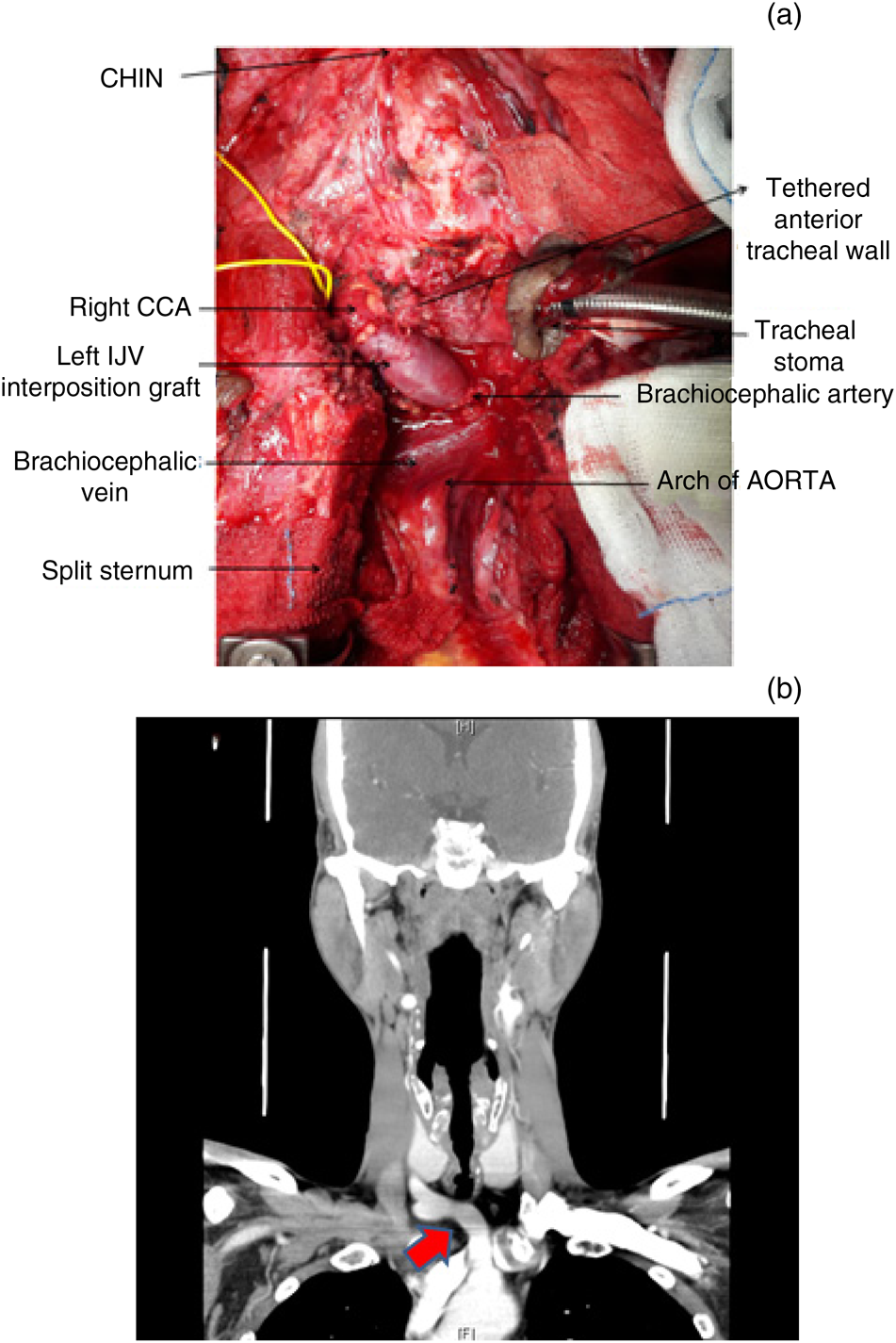
Subsequently, in the superior mediastinum, an irregular laceration was identified on the inferior aspect of brachiocephalic trunk, and its superior surface was found to be adherent to the anterior tracheal wall. The tear was repaired using the left internal jugular vein as an interposition graft, which connected the brachiocephalic trunk with the right common carotid artery, and this was reinforced with a strap muscle sling (Figure 2a). The anterior tracheal wall that was breached while freeing it from the adherent vessel wall to facilitate the grafting had to be repaired. The tracheoplasty performed was overlaid with strap muscle. The tracheal resection had to be abandoned. However, the immediate post-operative period was uneventful and there were no neurological deficits.
(a) An intra-operative photograph showing a left internal jugular venous (IJV) graft connecting the injured brachiocephalic trunk and the right common carotid artery (CCA); and (b) computed tomography coronal section showing an unbranched brachiocephalic artery trunk in the lower region of the neck (broad red arrow). H = head; F = feet

While revisiting the patient's CT neck scans, it was noted that the brachiocephalic trunk, which measured 4.2 cm, had an anomalous horizontal course. It was observed to be hugging the anterior wall of the trachea in the suprasternal region in the anterior region of the neck, bifurcating at the right lateral border of the trachea, 1.5 cm above the right sternoclavicular joint (Figure 2b).
Six weeks later, the patient presented with spontaneous moderate bleeding from the tracheostomy site, which subsided with inflation of the tracheostomy cuff. A CT angiogram was performed, which showed a tortuous brachiocephalic trunk in close proximity to the tracheostomy. In addition, there was extravasation of contrast from the graft site into the tracheostoma on release of the cuff tamponade of the tracheostomy tube, confirming a tracheo-innominate fistula. This was managed by two covered stents (10 mm × 59 mm) in the brachiocephalic artery, extending across the right subclavian artery and distally into the right common carotid artery (Figure 3).
Digital subtraction angiography images showing: (a) contrast enhancement within the tracheostoma, at a 6–9’o clock position close to the graft site, suggestive of tracheo-innominate fistula (red arrow); and (b) two well-deployed endovascular stents in the brachiocephalic artery extending across the right subclavian artery, distally into the right common carotid artery (yellow arrow).

The patient continues to have a tracheostomy. There have been no further complications and he receives regular follow up.
Discussion
Haemorrhage is one of the most dreaded complications that can occur during or following anterior neck surgery. It can result in death by exsanguination from the injured vessel, or haematoma leading to airway obstruction.Reference Klemm and Nowak4 Although large blood vessels lying in close proximity to the trachea are few and are not commonly encountered in the neck, the risk is higher in surgical procedures for tracheal stenosis. Individuals with tracheal stenosis often have to undergo low tracheostomy in an emergency life or death situation, to bypass the diseased segment.Reference Praveen and Martin5,Reference Upadhyaya, Bertellotti, Laeeq and Sugimoto6
The brachiocephalic artery, the first and the largest branch from the aortic arch, is the most common artery encountered and is also at risk during these procedures.Reference Silen and Spieker3 It originates on the right anterior aspect of the trachea behind the manubrium sternum, and runs superiorly from left to right over the right anterolateral portion, before dividing at the level of right sternoclavicular joint into the right common carotid artery and right subclavian artery.Reference Furlow and Mathisen7 The possibility of an anomalous course of the brachiocephalic trunk is of cardinal importance in surgical procedures of the neck, and even more so in percutaneous dilatational tracheostomy.Reference Mukadam and Hoskins8
Although developmental changes are known to occur in the anatomy of brachiocephalic trunk, it usually progresses to lie more towards the midline with increasing age.Reference Fawcett, Gomez, Hughes and Set9 A lot of other variations have been observed and reported based on cadaveric dissections and angiographic studies.Reference Namita and Sharma10,Reference Lale, Toprak, Yagiz, Kaya and Uyanık11 These variations may not occur in isolation and are of profound relevance, as accidental injury during surgical or interventional procedures can potentially lead to fatal haemorrhage or irreversible brain damage.
• Anatomical variations in the major blood vessels around the trachea can complicate potentially safe tracheal surgery
• The first variation reported was early bifurcation of the brachiocephalic trunk, leading to the right common carotid artery from the left side of the trachea
• The second variation reported was an undivided brachiocephalic trunk in the lower neck region with a horizontal pretracheal course
• Peritracheal vascular assessment is as important as endoluminal airway assessment, to avoid primary haemorrhage during tracheal surgery
The reported variations have occurred at the origin, along the course of the artery and at the level of branching. The origin can include the brachiocephalic trunk, arising from the aortic arch in front of the trachea, before crossing over to the right side.Reference Iterezote, de Medeiros and Petrella12 The brachiocephalic trunk can have a common origin with the left common carotid artery, referred to as the bovine aortic arch variant.Reference Layton, Kallmes, Cloft, Lindell and Cox13 It is also reported to arise from the descending aorta, crossing the midline posterior to the oesophagus, referred to as the retroesophageal aberrant innominate artery.Reference Bein, Saba, Patel, Reinhartz and Hanley14 Other variations in the brachiocephalic artery course include the main trunk and the bifurcating branches positioned higher up in the neck, called the high-riding variant. At times, the artery can even reach the left lobe of the thyroid along the left side of trachea, and then cross to the right side pretracheally, referred to as the sub-isthmic innominate artery.Reference Namita and Sharma10,Reference Dave and Upadhya15
The anomaly in the first case was the bifurcation of the brachiocephalic trunk much earlier on the left side of the trachea; hence, the right common carotid artery had an oblique pretracheal course, as reported in a cadaveric study.Reference Shankar, Raveendranath, Ravindranath and Manjunath16 In the second case, the main trunk of the brachiocephalic artery was abnormally coursing and bifurcating in the neck above the clavicle, with a horizontal pretracheal course from the left side of the trachea to the right.
Cross-sectional imaging of the anterior cervical region prior to any surgical intervention has been advocated, to decrease the possibility of inadvertent catastrophic haemorrhage.Reference Lale, Toprak, Yagiz, Kaya and Uyanık11 A major vessel may be found anterior to the trachea, and this is most likely in the suprasternal notch.Reference Weightman and Gibbs17 However, imaging may not always be possible in acute emergencies; thus, an ENT surgeon, and an airway surgeon in particular, should be aware of the various possible vascular anatomical variations of the great vessels of the neck.
Conclusion
Assessment of peritracheal vascular structures via imaging conducted prior to surgery is as important as endoluminal airway assessment to achieve the best outcome. A thorough knowledge of the anatomical structures, diligent assessment and anticipation of anatomical variations at the time of dissection will help to prevent complications.
Acknowledgement
We would like to acknowledge the vascular surgery, thoracic surgery and interventional radiology departments for their timely interventions.
Competing interests
None declared