Introduction
Azoospermia is considered as one of the common causes of male infertility and results from spermatogenesis disruption (Esteves, Reference Esteves2015). Approximately 1% of all men suffer from azoospermia that accounts for 10–15% of male infertility (Jarow et al., Reference Jarow, Espeland and Lipshultz1989). Azoospermia is divided into obstructive and non-obstructive groups, therefore the development of an appropriate treatment plan for men with non-obstructive azoospermia is a challenging task for embryologists (Jarow et al., Reference Jarow, Espeland and Lipshultz1989; Cocuzza et al., Reference Cocuzza, Alvarenga and Pagani2013). Given the fact that round spermatids are chromosomally similar to haploid sperms (Ogura et al., Reference Ogura, Ogonuki, Miki and Inoue2005), round spermatid injection (ROSI) has been proposed as a therapeutic option to overcome the defects of spermatogenesis in patients with late maturation arrest (Vloeberghs et al., Reference Vloeberghs, Verheyen and Tournaye2013; Tanaka et al., Reference Tanaka, Nagayoshi, Takemoto, Tanaka, Kusunoki, Watanabe, Kuroda, Takeda, Ito and Yanagimachi2015). Although fertilization rate and ROSI-generated offspring have been reported following the injection of spermatids in various mammalian species (Ogura et al., Reference Ogura, Matsuda and Yanagimachi1994; Tesarik et al., Reference Tesarik, Mendoza and Testart1995; Hirabayashi et al., Reference Hirabayashi, Kato, Aoto, Ueda and Hochi2002; Haigo et al., Reference Haigo, Yamauchi, Yazama, Yanagimachi and Horiuchi2004; Tanaka et al., Reference Tanaka, Nagayoshi, Takemoto, Tanaka, Kusunoki, Watanabe, Kuroda, Takeda, Ito and Yanagimachi2015), the success rate of fertilization and embryo development following ROSI is markedly lower than that of intracytoplasmic sperm injection (ICSI) (Levran et al., Reference Levran, Nahum, Farhi and Weissman2000; Vicdan et al., Reference Vicdan, Isik and Delilbaşi2001; Urman et al., Reference Urman, Alatas, Aksoy, Mercan, Nuhoglu, Mumcu, Isiklar and Balaban2002; Benkhalifa et al., Reference Benkhalifa, Kahraman, Biricik, Serteyl, Domez, Kumtepe and Qumsiyeh2004).
In addition to the limited success rate, genetic concerns attributed to ROSI have hampered its extensive use in ART. Preimplantation embryo development is a complicated process that is regulated using epigenetic reprogramming that includes DNA methylation, genomic imprinting, RNA silencing, and covalent modifications of histones, without any alteration in DNA sequence (Shi and Wu, Reference Shi and Wu2009; Rivera and Ross, Reference Rivera and Ross2013). Epigenetic reprogramming, in turn, controls the expression of early embryonic genes, the establishment of cellular memory and differentiation during embryo development. Therefore, it is believed that the ineffectiveness of ROSI may be related to abnormal epigenetic changes that disrupt subsequent zygotic gene expression. Kishigami and colleagues observed a highly remethylated DNA in a ROSI-derived zygotic paternal genome prior to the first mitosis after initial demethylation, and that was rather different from the normal paternal genome following mature spermatozoa injection (Kishigami et al., Reference Kishigami, Van Thuan, Hikichi, Ohta, Wakayama, Mizutani and Wakayama2006b).
Histone deacetylase inhibitors (HDACI) such as trichostatin A (TSA) can dramatically reduce abnormal DNA methylation through increasing acetylated histones and DNA demethylation in animal cloning using somatic cells (Kishigami et al., Reference Kishigami, Mizutani, Ohta, Hikichi, Thuan, Wakayama, Bui and Wakayama2006a; Salehi et al., Reference Salehi, Kato and Tsunoda2014). One research group succeeded in achieving 2–5 times higher success rates for therapeutic cloning when they treated embryos with TSA (Rybouchkin et al., Reference Rybouchkin, Kato and Tsunoda2006). As the pattern of DNA hypermethylation is similar between cloned and ROSI-derived embryos, TSA as an HDAC inhibitor is expected to improve epigenetic reprogramming in ROSI (Rybouchkin et al., Reference Rybouchkin, Kato and Tsunoda2006).
To date, previous studies have incubated ROSI-derived embryos with TSA. However, the probable effect of TSA-treated spermatid in the subsequent developmental competency following ROSI has not been addressed. Therefore, due to the increased risk of imprinting defects following ROSI, accurate evaluation of this method and provision of an effective strategy for improving the development and reduction of epigenetic defects seems necessary. Here, we treated spermatid with TSA before injection into oocytes to determine whether it can promote efficient pre-embryonic development and epigenetic changes.
Materials and methods
The Ethics Committee of Shahid Beheshti University of Medical Sciences, Cellular and Molecular Research Center approved all animal experiments described in this project. All procedures were carried out in compliance with the Committee’s standards for animal care and use. All chemicals and reagents used in the present study were obtained from Sigma Chemical Co. (St. Louis, MO, USA) unless stated otherwise.
Study design
Round spermatids were incubated with 100 nM TSA for 45 min prior to injection into the oocyte. Our three experimental groups consisted of test group: blastocysts derived from round spermatids incubated with TSA (ROSI/+TSA); sham group: blastocysts derived non-treated spermatids (ROSI/−TSA); and control group: blastocysts derived from sperm injection (ICSI). Matured oocytes, round spermatids and spermatozoa were collected from BDF1 (C57BL/6 × DBA/2) mice aged 6–8 weeks.
Mouse oocyte collection
To maximize the number of matured oocytes, ovarian stimulation of female BDF1 mice was performed as previously described (Hosseini et al., Reference Hosseini, Dehghani-Mohammadabadi, Ghafarri Novin, Haji Molla Hoseini, Arefian, Mohammadi Yeganeh and Salehi2017). Briefly, mice were sacrificed by cervical dislocation, the oviducts were separated and placed in a dish containing pre-warm flushing holding medium (FHM) supplemented with 4% bovine serum albumin (BSA). The oocytes with their surrounding cumulus cells (COCs) were released by puncturing the ampulla of the oviduct using a fine needle. To remove the cumulus cells, we put COCs into hyaluronidase enzyme (80 IU/ml) for 30 s. Denudated oocytes were transferred into a microdrop of potassium simplex optimization medium (KSOM) + 4% BSA and placed in an incubator for at least 1 h prior to microinjection.
Spermatozoa preparation for piezo-actuated ICSI
Spermatozoa were collected from the caudae epididymides of male BDF1 mice (at least 10 weeks old). The epididymides were separated and placed in human tubal fluid (HTF) supplemented with 4% BSA. Sperm was isolated by dicing epididymes with a fine needle and placed at 37°C under 5% CO2. To perform ICSI, we mixed sperm with polyvinyl pyrrolidone (PVP) 10% and put small drops of PVP and FHM on the bottom of a microinjection dish that had been covered with mineral oil. The sperm head was aspirated into the microinjection pipette and the head was separated from the midpiece following applying several pulses with the piezo to neck region. A single head was injected per matured oocyte. After 10 min of recovery of injected oocytes at room temperature, the oocytes were cultured in KSOMaa + 4% BSA at 37°C under 5% CO2 in air for 96 h.
Spermatid preparation for piezo-actuated ROSI
To collect round spermatids, the testes of the same male BDF1 mouse were separated and placed in HTF + 4% BSA medium. At first, the tunica albuginea from the testicles was removed. Next, seminiferous tubules were minced with fine scissors to obtain spermatogenic cells. After that, the spermatogenic cell suspension was treated with TSA so that the final concentration of TSA was 100 nM and this was incubated at 37°C for 45 min (Bui et al., Reference Bui, Wakayama, Kishigami, Kim, Van Thuan and Wakayama2008) prior to ROSI. ROSI was conducted in accordance with a protocol described previously (Kimura and Yanagimachi, Reference Kimura and Yanagimachi1995). Briefly, spermatogenic cells with or without TSA were added to separated drops of 10% PVP. We distinguished spermatids from other round cells according to their small diameter (∼10 μm) and a nuclear chromatin granule that is located centrally. Only one spermatid was injected into each oocyte. All ROSI-fertilized oocytes were subjected to activation medium containing 10 mM SrCl2 for 6 h and then cultured in KSOM + 4% BSA medium. Embryo development was monitored for 96 h following injection and compared among experimental groups.
cDNA synthesis and quantitative real-time PCR (qRT-PCR) analysis
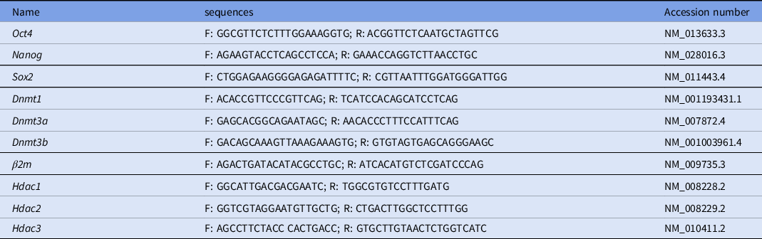
A reverse transcription reaction was performed based on a previous study (Hosseini et al., Reference Hosseini, Dehghani-Mohammadabadi, Ghafarri Novin, Haji Molla Hoseini, Arefian, Mohammadi Yeganeh and Salehi2017). Each reaction tube contained two blastocysts. cDNA synthesis was followed by qRT-PCR using the Master SYBR Green I mix (Yekta Tajhiz, Tehran, Iran) and a StepOne™ Real-Time PCR instrument (Applied Biosystems, USA) to assess quantification of Oct4, Sox2, Nanog, Dnmt1, Dnmt3a, Dnmt3b, Hdac1, Hdac2 and Hdac3. The transcript levels of qRT-PCR data were normalized relative to that of the beta2m transcript as an endogenous control. Data were analyzed using the Relative Expression Software Tool (REST, version 2009). Table 1 presents the primer sequences used for qRT-PCR.
Primer sequences used for qRT-PCR

Immunocytochemistry (ICC)
Immunofluorescence analysis of ROSI and ICSI-derived blastocysts was carried out to detect OCT4 protein expression. Briefly, the blastocysts were fixed in 4% paraformaldehyde for 20 min and permeabilized with 0.2% Triton X-100 in phosphate-buffered saline (PBS) at room temperature. Next, the fixed blastocysts were blocked with 3% BSA in PBS for 40 min and incubated overnight at 4°C with rabbit polyclonal anti-OCT4 antibody (Abcam, CA, USA, 1:200). The blastocysts were rinsed three times with PBS (each time 10 min) and incubated with goat anti-rabbit IgG, FITC conjugated antibody (1:50 dilution) (Razibiotech, AF8035) for 45 min at 37°C. Nuclei were counterstained with DAPI and visualized using a fluorescence microscope. The fluorescence intensity of each nucleus for OCT4 was measured using ImageJ software (National Institutes of Health, Bethesda, MD, USA).
Differential staining of blastocysts
Trophoblast cells and the inner cell mass of blastocysts were evaluated using a double staining technique (Salehi et al., Reference Salehi, Kato and Tsunoda2014). Briefly, embryos (n = 20) reaching the blastocyst stage were ‘zona freed’ using acidic Tyrode’s solution and were transferred into rabbit anti-mouse serum for 30 min. Next, blastocysts were washed in FHM and placed in 30% guinea pig complement supplemented with propidium iodide (PI; 10 μg/ml) and Hoechst stain (H33342; 10 μg/ml) for 30 min. After incubation, embryos were immediately fixed in a solution containing 70% ethanol. Fixed blastocysts were mounted on glass slides in a drop of glycerol, and flattened with coverslips. Cell counting was performed using a fluorescence microscope.
Statistical analysis
One-way analysis of variance (ANOVA) was applied for statistical analysis using Statistical Package for Social Sciences (SPSS) v.21 software (SPSS, Chicago, IL, USA). Relative gene expression levels of different genes among the studied groups were analyzed using REST 2009 Software. All data were expressed as mean ± standard deviation (SD) and a P-value < 0.05 was considered to be statistically significant.
Results
ROSI with TSA-treated spermatid enhanced the developmental competence of preimplantation embryos
We compared preimplantation development among the three studied groups. Our results showed no statistically significant differences in fertilization rate following ROSI/+TSA (89.28 ± 1.8) compared with the non-treated-ROSI and ICSI groups (87.18 ± 2.5% and 95.41 ± 4.3, respectively). As can be seen in Table 2, the cleavage rate in treated spermatid-derived embryos was higher than that of non-treated-ROSI, even though it was not significantly statistically. Importantly, blastocyst formation dramatically increased when TSA-treated spermatids were injected into an oocyte when compared with the non-treated-ROSI. However, the percentage of embryos that reached the blastocyst stage in the ROSI/+TSA group was lower than for ICSI.
Comparison of developmental competency among treated and non-treated spermatid injection and ICSI

Non-similar letters in each row show significant differences. (*P < 0.001).
Relative expression level of Oct4, Sox2 and Nanog increased in ROSI/+TSA-derived embryos
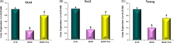
The relative expression levels of Oct4, Sox2 and Nanog genes at the blastocyst stage were measured in TSA-treated ROSI, non-treated ROSI and ICSI groups to identify the effect of TSA on critical developmentally related genes. Differential gene expression analysis indicated that the gene expression levels of the Oct4, Nanog and Sox2 in non-treated ROSI embryos were significantly lower than those of ICSI embryos. The expression levels of Oct4, Nanog and Sox2 were upregulated in the TSA-treated group compared with the non-treated-ROSI group, while they were similar to the expression levels observed in the ICSI-derived embryos (Figure 1).
Relative expression levels of Oct4 (A), Sox2 (B) and Nanog (C) genes in blastocyst-derived ICSI, ROSI/+TSA and ROSI/−TSA. Non-similar letters, in each bar chart, show significant difference (P < 0.05).

Higher protein expression level of OCT4 was achieved following TSA-treated spermatid injection
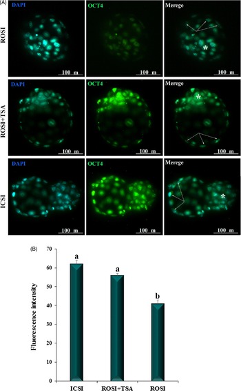
The protein level of OCT4 in blastocysts was assessed by immunofluorescence staining. ICC analysis indicated that the intensity of OCT4 was significantly lower in ROSI-derived blastocysts compared with TSA-treated and ICSI groups, whereas TSA treatment increased OCT4 protein expression levels to be almost identical to that of ICSI-derived blastocysts (P < 0.05). (Fig.2).
Protein expression of OCT4 at the blastocyst stage. (A) Immunofluorescent of OCT4 in blastocysts (green), nuclei were visualized with DAPI (blue). Asterisk shows ICM and arrows show trophoblast cells. (B) Comparison of OCT4 fluorescence intensity among the studied groups. Non-similar letters, in each bar chart, show significant difference (P < 0.05).

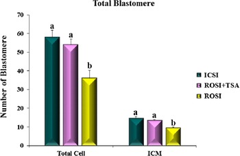
We counted the inner cell mass (ICM) and total blastomere to evaluate the quality of ROSI-derived blastocysts. We performed differential staining of blastocysts for ICM and trophectoderm cell counting. As can be seen from Figure 3, there was no significant difference either in total blastomeres or ICM at the blastocyst stage between ICSI and ROSI/+TSA groups, while these notably declined in blastocysts produced by ROSI compared with blastocysts derived from ICSI and ROSI/+TSA.
Average number of total blastomeres and inner cell mass (ICM) in blastocysts of each group. Non-similar letters, in each bar chart, show significant difference (P < 0.05).

Relative expression levels of Dnmt and Hdac genes in ROSI/+TSA-derived embryos
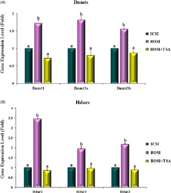
To determine the quantitative expression levels of Dnmt and Hdac genes we utilized qRT-PCR. Figure 4 depicts that the expression levels of evaluated genes in ROSI/+TSA-derived embryos declined compared with non-treated embryos. These results showed no significant difference between ICSI and ROSI/+TSA-derived embryos.
Relative mRNA expression of Dnmt1, Dnmt3a and Dnmt3b (A) and Hdac1, Hdac2 and Hdac3 (B) genes in blastocysts derived from ICSI, ROSI/+TSA and ROSI/−TSA. Non-similar letters, in each bar chart, show significant difference (P < 0.05).

Discussion
ROSI was introduced as a therapeutic approach in azoospermic men more than 20 years ago (Ogura et al., Reference Ogura, Matsuda and Yanagimachi1994; Tesarik et al., Reference Tesarik, Mendoza and Testart1995). However, the percentage fertilization and subsequent pregnancy rate have remained low. Aberrant DNA methylation of ROSI-derived embryos is believed to be a probable reason for lower success rates following spermatid injection. Accordingly, TSA, a histone deacetylase inhibitor that either reduces the abnormal DNA hypermethylation or improve epigenetic reprogramming following somatic cell nuclear transfer may increase the success rate of ROSI. In this study, we treated round spermatid with TSA to examine whether it had the ability to improve developmental competency of ROSI-derived embryos.
In terms of preimplantation development, our results showed a similar fertilization rate in non-treated spermatid-derived embryos and TSA-treated spermatids. Surprisingly, the fertilization rate was comparable with that of the ICSI group in contrast with other studies that have reported marked low fertilization rates following ROSI compared with the injection of spermatozoa (Levran et al., Reference Levran, Nahum, Farhi and Weissman2000; Vicdan et al., Reference Vicdan, Isik and Delilbaşi2001; Sousa et al., Reference Sousa, Cremades, Silva, Oliveira, Ferraz, Teixeira da Silva, Viana and Barros2002; Benkhalifa et al., Reference Benkhalifa, Kahraman, Biricik, Serteyl, Domez, Kumtepe and Qumsiyeh2004). Another study, in agreement with our findings, reported a fertilization rate of 89% in mouse spermatid injection (Kong et al., Reference Kong, Yin, Chen, Li, Li, Xing, Jiang, Fang, Lyu and Chen2017). The discrepancy between our results and other studies is most probably related to artificial oocyte activation after ROSI. It is proposed that fertilization failure following ROSI is attributed to premature chromatin condensation (PCC) and reduced oocyte-activating capacity. An immature spermatid may not be able to activate the oocyte because of low oocyte-activating ability (Yazawa et al., Reference Yazawa, Yanagida and Sato2007). Here, TSA increased blastocyst formation rate (63%). This is in agreement with the Kong and colleagues study (Kong et al., Reference Kong, Yin, Chen, Li, Li, Xing, Jiang, Fang, Lyu and Chen2017) in which embryos derived from ROSI were incubated with Scriptaid as HDACI. Given the enhanced blastocyst development rate in our study, we suggested that TSA could epigenetically manipulate round spermatid nuclei and this led to increased round spermatid reprogramming.
To confirm the role of TSA in epigenetic improvement in ROSI-derived embryo development, Dnmt expression levels and subsequently core components of pluripotency genes including Oct4, Sox2 and Nanog were assessed. DNA methylation regulates either induction or repression of genes involved in embryo development (Bartolomei and Ferguson-Smith, Reference Bartolomei and Ferguson-Smith2011). As previously described, a high level of remethylation occurs in the ROSI-derived zygotic paternal genome. In our study, incubation of spermatid with TSA decreased the expression levels of Dnmt1, Dnmt3a and Dnmt3b in blastocysts derived from ROSI compared with the non-treated group. Notably, Dnmt gene expression levels were similar to that of blastocyst-derived mature spermatozoa. DNMT controls the epigenetic mechanism through DNA methylation and regulation of early developmentally related genes. Kishigami and colleagues demonstrated that TSA-treated zygotes reduced DNA methylation in the spermatid-derived paternal genome.
There was a significant increase in both mRNA and protein expression levels of OCT4 prior to the 8-cell stage and through to the blastocyst stage (Szczepańska et al., Reference Szczepańska, Stańczuk and Maleszewski2011). OCT4 is critically involved either in the self-renewal of undifferentiated embryonic stem cells (ESCs) or differentiation of ESCs into trophoblast cells (Wu and Schöler, Reference Wu and Schöler2014). Here, we demonstrated a higher expression level of Oct4, Sox2 and Nanog in ICSI-derived embryos than in ROSI-derived embryos at the blastocyst stage. Notably, Oct4, Sox2 and Nanog gene expression levels in TSA-treated ROSI-derived embryos were substantially increased and were similar to the expression levels in embryos derived from ICSI. Immunofluorescence analysis of OCT4 protein confirmed that higher numbers of cells in the ICM expressed OCT4 protein following TSA-treated spermatid injection compared with non-treated ROSI-derived embryos. Histone acetylation is one of the mechanisms responsible for activating chromatin and the subsequent gene expression. Cervoni and Szyf (Reference Cervoni and Szyf2001) revealed that histone acetylation directed methylation activity and led to gene demethylation.
The histone acetylation state following TSA treatment relaxes the chromosome structure. Consequently, the transcriptionally permissive state of chromosomes results in increased chromatin decondensation and DNA replication. As a result, this improves somatic cell nuclei reprogramming through histone modification (Zlatanova et al., Reference Zlatanova, Caiafa and Van Holde2000; Bui et al., Reference Bui, Wakayama, Kishigami, Park, Kim, Thuan and Wakayama2010). Additionally, it has been reported that embryo treatment with TSA inhibits histone deacetylase, exposes DNA active binding sites and decreases DNA methylation, which is important for gene activation (Cervoni and Szyf, Reference Cervoni and Szyf2001). Moreover, we confirmed the efficiency of TSA in development using different methods, reflected by a dramatically higher success rate.
Overall, the present study showed that TSA enhanced the success rate of development to the blastocyst stage and the proportion of pluripotent cells in the ICM (as evidenced by Oct4 expression) that led to the higher efficiency of ROSI. More importantly, our findings would provide a new efficient approach for the practical improvement of development and reduction of epigenetic defects by ROSI.
Acknowledgements
This study was supported by the Molecular and Cellular Research Center, Shahid Beheshti University of Medical Sciences, Tehran, Iran.
Conflict of interest
All authors declare they have no conflict of interest.