Introduction
There are numerous factors affecting reproductive efficiency in cattle and one of them is body condition. This factor has been correlated with reproductive performance both phenotypically (Buckley et al., Reference Buckley, O’Sullivan, Mee, Evans and Dillon2003) and genetically (Berry et al., Reference Berry, Buckley, Dillon, Evans, Rath and Veerkamp2003) and supports the premise that nutritional status affects reproductive functions. Cows with low BCS at calving, or that suffer excess BCS loss early post-partum, are less likely to ovulate, have a reduced submission rate to artificial insemination and decreased conception rate (Moreira et al., Reference Moreira, Risco, Pires, Ambrose, Drost, Lorenzo and Thatcher2000; Santos et al., Reference Santos, Thatcher, Pool and Overton2001; Berry et al., Reference Berry, Buckley and Dillon2007; Roche et al., Reference Roche, Friggens, Kay, Fisher, Stafford and Berr2009).
Dietary intake can affect morphology (O’Callaghan et al., Reference O’Callaghan, Yaakub, Hyttel, Spicer and Boland2000), quality (Lozano et al., Reference Lozano, Lonergan, Boland and O’Callaghan2003) and developmental capacity of oocytes and embryo development in vivo and in vitro (McEvoy et al., Reference McEvoy, Robinson, Aitken, Findlay, Palmer and Robertson1995, Reference McEvoy, Robinson, Aitken, Findley and Robertson1997; Negrao et al., Reference Negrao, Nibart and Humblot1997). It was shown that the embryo cleavage and blastocyst formation rates were greater for oocytes from cows with a high BCS (3.3 to 4.0) than a low BCS (1.5 to 2.5) (Snijders et al., Reference Snijders, Dillon, O’Callaghan and Boland2000; Adamiak et al., Reference Adamiak, Mackie, Watt, Webb and Sinclair2005, Reference Adamiak, Powell, Webb and Sinclair2006). With regards to embryo developmental potency at in vitro embryo production (IVP), the percentage of zygotes initiating division and the percentage of forming blastocysts were higher in cows with a BCS of 3.3–4.0 than with a BCS of 1.5–2.5 (Snijders et al., Reference Snijders, Dillon, O’Callaghan and Boland2000). Concerning embryo quality, embryo cell number was greater for low BCS-derived oocytes compared with moderate BCS heifers; however TUNEL index did not differ between BCS groups (Adamiak et al., Reference Adamiak, Powell, Webb and Sinclair2006).
The changing seasons is another important factor. Climate changes such as air temperature, relative humidity, air movement and solar radiation affect fertility of beef and dairy cows (De Rensis & Scaramuzzi, Reference De Rensis and Scaramuzzi2003). Some seasonal studies have shown that quality of bovine oocytes and rate of embryo development deteriorate in the summer compared with winter months (Rocha et al., Reference Rocha, Randel, Broussard, Lim, Blair, Roussel, Godke and Hansel1998; Rutledge et al., Reference Rutledge, Monson, Northey and Leibfried-Rutledge1999). It was found that the Bos indicus cows exhibited better fertility during the spring and summer months, compared with the autumn and winter (Zeitoun et al., Reference Zeitoun, Rodriquez and Randel1996). In contrast, other studies showed that cattle fertility and conception rates decreased significantly in a hot summer compared with in a cool season (Badinga et al., Reference Badinga, Collier, Thatcher and Wilcox1985; Monty & Racowski, Reference Monty and Racowski1987; Takuma et al., Reference Takuma, Sakai, Ezoe, Ichimaru, Jinnouchi, Kaedei, Nagai and Otoi2010), and which can be caused by compromised oocyte quality and by altered patterns of follicular development, steroid production and gene expression (Hansen, Reference Hansen2007).
To date, little information is available on the cell quality of IVP embryos in relation to the body condition of donor cows and the season of the year. Therefore, the aim of our study was to investigate the effect of body condition and season on the development and quality of in vitro produced bovine preimplantation embryos.
Our observations bring additional data on the effect of body condition and season on the yield of IVP embryos as well as on their quality, using fluorescence assays for embryo cell number, apoptosis and quality of actin cytoskeleton.
Materials and methods
Animals
Donors of oocytes were Holstein-Friesian cows which were examined for body condition score (BCS) before slaughter at a local slaughterhouse. BCS was determined by the same operator using visual estimation and palpation according to a five-point scale (Edmonson et al., Reference Edmonson, Lean, Weaver, Farver and Webster1989) of 1 (emaciated) to 5 (obese). For BCS1, BCS2 and BCS 3 groups, respectively, average ages were 6.2, 4.1, and 5.7 years and milk yields were 8815, 7937, and 7744 kg. For our experiments only BCS 1 cows (n = 18), BCS2 (n = 50) and BCS3 cows (n = 60) were available.
Oocyte recovery and quality evaluation
Ovaries were collected immediately after slaughter, tagged with labels according to the BCS of donor cows and transported to the laboratory within 3–3.5 h in isothermic boxes at ambient temperature. Connective tissue was removed from the ovaries and they were washed with sterile saline and 70% ethanol solution (to avoid possible bacterial contamination on the ovarian surface, Stringfellow & Seidel, Reference Stringfellow and Seidel1998). Ovarian follicles (2–8 mm) were aspirated with a syringe with HEPES-TALP medium and a 20G needle. Follicular fluid (with oocytes) was transferred to a Petri dish and the oocytes (cumulus–oocyte complexes, COCs) were collected, washed in HEPES-TALP medium and classified according to their morphological characteristics (Gordon, Reference Gordon1994) into four qualitative categories: quality 1 (Q1): compact multilayered cumulus, homogenous cytoplasm; quality 2 (Q2): compact multilayered cumulus, homogenous cytoplasm, but with darker zone at the periphery of the oocyte; quality 3 (Q3): less compact cumulus, irregular cytoplasm, with darker clusters; quality 4 (Q4): expanded cumulus (overmatured oocytes), cumulus cells scattered in dark clamps, irregular cytoplasm with dark clusters, ‘nude oocytes’.
Only Q1 and Q2 grade oocytes were considered to be of suitable quality for in vitro maturation and were assigned as ‘maturation-grade’ oocytes.
Procedure of in vitro embryo production (IVP)
Selected oocytes were washed twice in maturation medium TCM199 with glutaMAX (Gibco), sodium pyruvate (0.25 mmol.l−1), gentamycin (0.05 mg.ml−1), fetal bovine serum 10% (FBS, BioWhitaker, Verviers, Belgium) and FSH/LH (1/1 I.U., Pluset, Lab. Calier, Barcelona, Spain) and maturated for 24 h at 38.5°C in a humidified atmosphere with 5% CO2 in air. Matured oocytes were identified according to the expansion of cumulus cells surrounding the oocyte. Oocytes that were either naked or contained dark granules in the ooplasm were considered to be immature. Following maturation, the presumptive metaphase II oocytes were washed twice and placed into 100-μl drops of fertilization medium IVF-TALP under mineral oil for 20–22 h at 38.5°C in a humidified atmosphere with 5% CO2 in air and incubated with 1 × 106.ml−1 spermatozoa in the presence of 10 μg/ml heparin (Sigma-Aldrich, Germany). Following in vitro fertilization the presumptive zygotes were picked up in the drop, cleaned of excessive sperm and remaining cumulus cells by vortexing, washed twice in medium and cultured in Upgraded INRA B2 Menezo (CCD, Vernouillet, France) embryo medium in co-culture with a Buffalo Rat Liver (BRL) cell monolayer in a humidified atmosphere at 38.5°C. Embryo development was examined on the basis of embryo cleavage rate (day 2) and blastocyst rate (day 7).
Analysis of embryo quality (embryo cell number, apoptotic index, actin cytoskeleton)
The embryos were processed for apoptosis detection using the In Situ Cell Death Detection Kit (Roche Slovakia Ltd, Bratislava, Slovak Republic) according to the product manual. For labelling of actin filaments the embryos were incubated in phalloidine–TRITC conjugate solution (Chemicon International, Temecula, CA, USA) for 45 min and then covered with a drop of Vectashield anti-fade medium containing DAPI stain (cell number; Vector Laboratories, Burlingame, CA, USA). The samples were mounted between coverslip and microslide and stored at –20°C until fluorescence analysis. The actin cytoskeleton was classified as belonging to three quality grades (Makarevich et al., Reference Makarevich, Kubovičová, Hegedušová, Pivko and Louda2011): grade I – best quality; grade II – fair quality and grade III – poor quality.
Stained embryo samples were checked under a Leica fluorescence microscope using specific filters with wavelengths for green, red and blue fluorescence and a ×20 magnification objective.
Statistical analysis
Oocyte yield, embryo cleavage and blastocysts rate were analyzed by chi-squared test. One-way analysis of variance (ANOVA) and Tukey test were used to analyse differences in the TUNEL index between groups. The treatment effect on actin cytoskeleton quality was tested using the Mann–Whitney U-test. All calculations were performed using the SAS software package (SAS Institute, 2001).
Results
Oocytes used for IVP were recovered from the BCS 1–3 cows within the spring, summer and autumn periods. The rate of good quality oocytes (Q1 and Q2) was significantly higher (P < 0.05) in the BCS 2 and BCS 3 groups, compared with the BCS 1 group. Conversely, there were no differences in cleavage and blastocyst rates among BCS groups (Table 1).
Effect of the BCS on oocyte quality and in vitro fertilization (IVF) outcome

a versus b at P < 0.05 (chi-squared test).
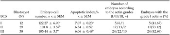
IVP blastocysts were analysed for cell number, apoptotic index and actin cytoskeleton quality. The average embryo cell number was significantly higher (P < 0.05) in the BCS 1 group compared with BCS 2 and BCS 3 groups (Table 2). Significantly higher cell numbers in the BCS 1 group corresponded to the highest apoptotic index in this group. It was slightly higher compared with BCS 2 group and significantly higher (P < 0.05) compared with the BCS 3 group (Table 2). There were no differences in the number of embryos with grade I actin (highest quality) between the BCS groups, although the highest, but not significant, proportion of embryos with grade I actin was in the BCS 2 group (Table 2).
Effect of the body condition score (BCS) on embryo quality
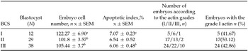
a versus b at P < 0.05 (embryo cell number: t-test; apoptotic index: one-way ANOVA; actin quality: Mann–Whitney U-test). SEM, standard error of the mean.
Evaluation of the effect of season (Table 3) on oocyte quality (Q1 + Q2) showed that the proportion of good quality oocytes was the lowest during the spring months (49.45%), then significantly increased during the summer (P < 0.05; 63.26%) and decreased again during the autumn months (50.07%).
Effect of season on oocyte quality and IVF outcome

a versus b at P < 0.05 (chi-squared test).
Conversely, the proportion of matured oocytes was highest during the summer months (85.00%), then the maturation rate decreased during the autumn (P < 0.05; 73.42%) and increased again (80.55%) during the spring months. Although the maturation rate was highest during the summer months, the proportion of cleaved embryos was highest during autumn months (76.20%), then slightly decreased during spring months (68.27%) and significantly decreased (42.00%; P < 0.05) during the summer months. Similarly, the blastocyst yield during the autumn (25.00%) was significantly higher compared with spring (12.12%; P < 0.01) and summer (16.66%; P < 0.05) months (Table 3). Significant differences (P < 0.05) in total cell numbers between the embryos produced during the spring (99.38), autumn (110.10) or summer (108.96) months were observed (Table 4). The proportion of dead cells (TUNEL index) was not significantly different in embryos produced from oocytes recovered within different season months (6.45, 6.13 and 6.18, respectively, Table 4). The highest (although not significant) dead cell index was determined during spring months (6.45 ± 0.47), and was corresponded to the lowest embryo cell number (99.38 ± 3.90).
Effect of season on embryo quality

a versus b at P< 0.05; c versus d at P < 0.001 (embryo cell number: t-test; apoptotic index: one-way ANOVA; actin quality: Mann–Whitney U-test). SEM, standard error of the mean.
Evaluation of actin cytoskeleton quality showed that a significantly higher (P < 0.001) proportion of embryos with grade 1 actin quality (75.00%) was obtained during the summer months compared with the autumn (35.56%) or spring months (28.57%).
Discussion
Quality of oocytes recovered from ovarian follicles may be affected by the high genetic value, milk yield, lactation rank, feeding level and body condition of cows (Dominguez, Reference Dominguez1995; Jaškowski, Reference Jaškowski2001). Cows that lose a lot of body condition in early lactation (Snijders et al., Reference Snijders, Dillon, O’Callaghan and Boland2000) and those with high hepatic triacylglycerol contents, which were indicative of excessive fat mobilisation at this time (Kruip et al., Reference Kruip, Wensing and Vos2001), produced oocytes of inferior quality as measured by reduced ability to develop in vitro. Decreased BCS may be associated with decreased oocyte quality measured by rates of in vitro fertilization and early embryonic development (Snijders et al., Reference Snijders, Dillon, O’Callaghan and Boland2000).
The effect of the cow's body condition on the number of ovarian follicles and the number and quality of oocytes have been analyzed in many studies (Dominguez, Reference Dominguez1995; Drion et al., Reference Drion, Beckers, Ectors, Hanzen, Hontain and Lonergen1996; Snijders et al., Reference Snijders, Dillon, O’Callaghan and Boland2000; Fihri et al., Reference Fihri, Klakhdissi, Derqaoui, Hajji, Naciri and Goumari2005; Nowak et al., Reference Nowak, Kowski, Olechnowicz and Bukowska2009). In our study, the body condition of cows affected the proportion of good quality oocytes; it was significantly higher in the BCS 2 and BCS 3 groups, compared with the BCS 1 group. These results confirm our former observations that cows tended to emaciation (BCS 1) have significantly lower prematuration oocyte quality, and that the cows with average body condition (BCS 3) have an optimal oocyte quality suitable for IVP (Kubovičova et al., Reference Kubovičova, Makarevič, Hegedušová, Slezáková and Bezdíček2012). Our results are in accordance with the results of Nowak et al. (Reference Nowak, Jaškowski, Szczepankiewicz and Olechnowicz2011), who aspired more oocytes from cows with moderate body condition (BCS 3), while lower numbers were collected from emaciated cows. This is in concert with the suggestion of Snijders et al. (Reference Snijders, Dillon, O’Callaghan and Boland2000) and Fihri et al. (Reference Fihri, Klakhdissi, Derqaoui, Hajji, Naciri and Goumari2005) that the number of follicles, oocyte yield and the quality of oocytes increase as the BCS increases and that the low oocyte yield of cows with a low body condition may be related to the energy level of the diet.
There were no differences between the cow oocyte donors with BCS 1–3 in the cleavage and blastocysts rates. This is contrary to the results of Snijders et al. (Reference Snijders, Dillon, O’Farrell, Diskin, Wylie, O’Callaghan, Rath and Boland2001), who reported that the percentage of zygotes initiating division and the percentage of forming blastocysts were higher in cow oocyte donors with BCS 3.3–4.0 than in cows with a BCS of 1.5–2.5.
The changing seasons are another important factor that affects reproductive performance in cows. In our study, the best oocyte quality (maturation grade) was noted during summer months (63.26%) compared with autumn (50.07%) and spring (49.45%) months. The best results on embryo cleavage and blastocyst rates were obtained during the autumn period. On the basis of these results we can state that there was a variable effect of the season on oocyte and embryo quality.
In most cited works (Badinga et al., Reference Badinga, Collier, Thatcher and Wilcox1985; Monty & Racowski, Reference Monty and Racowski1987; Takuma et al., Reference Takuma, Sakai, Ezoe, Ichimaru, Jinnouchi, Kaedei, Nagai and Otoi2010), conditions during the summer months were hyperthermic, whilst in our ambient temperature conditions the summer season was not characterised by the long-term high temperature. Therefore, we do not consider our summer season as a hyperthermia period.
In mammals, programmed cell death has been also identified as the mechanism underlying the depletion of oocytes from the ovarian pool throughout life (Morita & Tilly, Reference Morita and Tilly1999; Tilly, Reference Tilly2001). Presumably, apoptosis begins in the follicle and then affects the oocyte. It is also known, that IVP embryos exhibit relatively high levels of apoptosis (with varying degrees of cytoplasmic fragmentation) and a high incidence of developmental arrest during culture (Hardy, Reference Hardy1999; Meier et al., Reference Meier, Finch and Evan2000). Incidence of apoptosis serves as a quality evaluation marker in preimplantation embryo development and is measured by apoptotic index. In our study, significant difference in the apoptotic index was observed between the BCS 1 and BCS 3 embryos. Generally, maximal apoptotic index was noted in the lowest BCS1 embryos and then decreased with the elevation in BCS embryos. We can suppose that the significantly higher apoptotic rate in the BCS1 embryos compared with the BCS 3 ones, can be explained by less cell numbers in the BCS3 group. However, an increase in the apoptotic index in BCS 1 embryos could be also caused by suboptimal conditions in which the oocytes developed in vivo before recovery. These observations were also confirmed by the fact that in the BCS 1 group a lower proportion of oocytes was selected for IVM.
Actin is a major component of cytoskeleton. During oocyte maturation, fertilization, and embryo development, polymerization and depolymerization of actin filaments are important processes (Sun & Schatten, Reference Sun and Schatten2006). Actin microfilaments play an important role in cell cleavage during embryo development (Bassell et al., Reference Bassell, Taneja, Kislaukis, Sandelt, Pouters, Singer, Estes and Higgins1994; Volderrama et al., Reference Volderrama, Babia, Ayala, Kok, Renaupiqueras and Egea1998). It was found in IVP embryos of hamster (Barnett et al., Reference Barnett, Clayton, Kimura and Bavister1997), rat (Matsumoto et al., Reference Matsumoto, Shoji, Umezu and Sato1998), pig (Wang et al. Reference Wang, Abeydeera, Han, Prather and Day1999; Kidson et al., Reference Kidson, Schoevers, Tharasanit, Hazeleger, Duero-Steverink, Colenbrander, Dieleman and Kidson2004) and horse (Tremoleda et al., Reference Tremoleda, Schoevers, Stout, Colenbrander and Bevers2001, Reference Tremoleda, Stour, Lagutina, Lazzari, Bevers, Colenbrander and Galli2002a, Reference Tremoleda, Colenbrander, Stout and Beversb, Reference Tremoleda, Stout, Lagutina, Lazzari, Bevers, Colenbrander and Galli2003), that the organization of actin filaments can be affected by environmental conditions such as temperature, pH, and ion concentration in culture medium, that finally affect embryo cleavage or compromise further development. The disruption of microfilaments can adversely affect the structural integrity of cells, with serious consequences for embryo cleavage. Staining of microfilaments of the cytoskeleton enabled the identification of differences in its quality between the embryos. In the present study, the distribution of embryos according to actin cytoskeleton quality was significantly different between the autumn and spring months compared with the summer months. During the summer months the proportion of grade I actin embryos was significantly higher compared with autumn and spring months. It was reported that intracellular actin filament distribution is directly related to the 2-cell block in hamster (Barnett et al., Reference Barnett, Clayton, Kimura and Bavister1997) and in rat (Matsumoto et al., Reference Matsumoto, Asano and Endo1997) embryos. How actin filaments modulate embryo development is poorly understood. It is clear that abnormal microfilament distribution results in abnormal cell function, because the actin cytoskeleton is required for normal function of mitochondria, Golgi complex and other cell organelles (Boldogh et al., Reference Boldogh, Vojtov, Karmon and Pon1998). A direct association between actin quality and the morphology of the blastocyst was shown (Kidson et al., Reference Kidson, Schoevers, Tharasanit, Hazeleger, Duero-Steverink, Colenbrander, Dieleman and Kidson2004). It was demonstrated that good quality blastocysts contained grade I actin, whereas the presence of any anomalies in gross morphology of the blastocyst was correlated to a large extent with poor actin quality (grade II actin).
Knowledge about current body condition of dairy cows may help in the control of nutrition status and optimizing the feeding efficiency of cows, health and reproduction status, longevity and general breeding economy. An important requirement for successful reproduction is an adequate body condition of cows and heifers, expecially during parturition. In commercial practice, body condition scoring should be carried out regularly and satisfactorily in circumstances where weighing may be impractical. The technique is easy to learn and is useful when practiced by the same person in the same herd over several years. Breeders should monitor the body status of dairy cows regularly to avoid drastic changes in the body condition.
Our results indicate that body condition affects initial quality of in vivo recovered oocytes. The rate of good quality oocytes was significantly higher in BCS 2 and BCS 3 groups, compared with the BCS 1 group. Conversely, BCS does not affect cleavage and blastocyst rates and embryo quality, which may suggest that further development in vitro can mask the influence of BCS on preimplantation development. A variable effect of a season on the oocyte and embryo quality was observed. The more favorable season for in vitro embryo development is the autumn.
Acknowledgements
This work was supported by the Slovak Research and Development Agency from the grants APVV-0137–10 and APVV-0556–011 and by the Ministry of Education (Slovak Republic) under the contract KEGA No. 011UPJŠ-4/2014 and VEGA No. 1/0415/12. The research leading to these results has received funding from the European Community under project no 26220220180: Building Research Centre ‘Agro Biotech’ and LAGEZ No. 26220120051 supported by the Operational Programme Research and Development funded from the European Regional Development Fund.