Implications
Osteoporosis is a widespread health and welfare issue in laying hens as it causes bone damage and pain. Besides modification of housing systems, nutritional manipulation is also a common practice to improve skeletal heath. Present research indicates that dietary probiotic supplementation improves bone mass of hens and reduces unmarketable egg production without deterioration of physiological homeostasis and eggshell quality. Dietary probiotic supplementation could be an alternative management practice for improving poultry skeletal health and production.
Introduction
Osteoporosis is a widespread skeletal damage in laying hens, increasing bone weakness and frequency of fractures that subjects hens to chronic pain (Nasr et al., Reference Nasr, Nicol and Murrell2012). It is caused by an imbalance of bone remodeling between bone formation and resorption under the influence of estrogen. At the onset of sexual maturity, the level of estrogen markedly increases and is in favor of medullary bone deposition, providing a labile source of calcium for eggshell formation (Whitehead and Fleming, Reference Whitehead and Fleming2000). However, continuous deposition of medullary bone with age results in deterioration of structural bone. Consequently, age-related loss of structural bone over the course of the production cycle eventually leads to osteoporosis (Whitehead and Fleming, Reference Whitehead and Fleming2000), being most severe at the end of lay. Osteoporosis is most prevalent in caged layers, while currently ~85% of all eggs in the United States (and 90% around the world) are laid by hens kept in the cage systems (UEP, 2017).
Recent studies demonstrate that supplementation of probiotics improves skeletal health in humans and rodents (Scholz-Ahrens et al., Reference Scholz-Ahrens, Ade, Marten, Weber, Timm, Acil, Gluer and Schrezenmeir2007; McCabe et al., Reference McCabe, Britton and Parameswaran2015). Limited studies have been conducted in poultry, especially in laying hens, to evaluate the effects of probiotics on bone health (Mutus et al., Reference Mutus, Kocabagli, Alp, Acar, Eren and Gezen2006; Panda et al., Reference Panda, Rao, Raju and Sharma2006; Houshmand et al., Reference Houshmand, Azhar, Zulkifli, Bejo, Meimandipour and Kamyab2011; Ziaie et al., Reference Ziaie, Bashtani, Torshizi, Naeeimipour, Farhangfar and Zeinali2011; Abdelqader et al., Reference Abdelqader, Irshaid and Al-Fataftah2013b; Fuentes et al., Reference Fuentes, Orozco, Vicente, Velasco, Menconi, Kuttappan, Kallapura, Latorre, Layton, Hargis and Téllez2013; Sadeghi, Reference Sadeghi2014). The objective of the present experiment was to investigate the effects of a dietary probiotic inclusion on performance traits and bone mineralization, including the tibia, femur, humerus and keel bone, in laying hens as well as its possible mechanisms.
Materials and methods
Birds, management and diets
Ninety-six 56-week-old White Leghorn laying hens of the Hy-Line W-36 strain were provided by a commercial egg farm (Creighton Brothers Farm, Atwood, IN, USA). Hens were assigned to 24 cages with four hens per cage based on their BW so that each cage had similar mean BW. The cage dimensions were 38×51×48 cm (length×width×height) providing 484.5 cm2 of floor space per hen. Each cage contained two nipple drinkers and one feeder providing 10.3 cm feeder space per hen. A piece of cardboard was installed between the feeders to ensure that hens were not able to consume feed from the adjacent feeders. Each cage had a lined under tray for collecting manure; and the liners were replaced daily. Hens were housed in one room; and average room temperature was 20°C throughout the experimental period. The photoperiod was 16 light (0400 to 2000 h) : 8 dark. The experimental protocol had been approved by the Purdue Animal Use and Care Committee (PACUC Number: 1111000262).
Before the start of the experiment, all hens were given 4 weeks (56 to 59 weeks of age) to adapt to their housing environment and fed a regular layer diet (Table 1). The egg production and BW were monitored during the pre-trail period. Hens were transferred among the cages as necessary to ensure egg production and BW evenly distributed among the cages at the end of 59 weeks of age. At 60 weeks of age, the 24 cages were randomly assigned to 1 of 4 dietary treatments: a layer diet mixed with a commercial probiotic product (PoultryStar, BIOMIN America, Inc., San Antonio, TX) at 0, 0.5, 1.0 or 2.0 g/kg feed (Control; 0.5×10⁶ cfu/g; 1.0×2×106 cfu/g; and 2.0×, 4×10⁶ cfu/g of feed, respectively) for 7 weeks. The dose of 1.0 g/kg feed was recommended by the company for laying hens. The probiotic consists of four microbial strains (Enterococcus faecium, Pediococcus acidilactici, Bifidobacterium animalis and Lactobacillus reuteri). Feed and water were provided ad libitum. Diets were mixed at the Purdue University feed mill using a step-increase procedure. Feed samples were collected at the beginning and the end of this study for analysis of microorganism recovery by the manufacturer’s proprietary assay (Biomin America Inc., San Antonio, TX, USA).
Composition and nutrient analysis of the laying hen diet

1 Myco curb Dry (Carl S. Akey, Inc., Lewisburg, OH, USA): propionic acid, sodium hydroxide, calcium hydroxide, amorphous silicon dioxide, sorbic acid, benzoic acid, propylparaben, methylparaben and BHA.
2 Ethoxyquin (Carl S. Akey, Inc.).
3 The premix supplied per kg of diet: vitamin A, 12 320 IU; vitamin D3, 4620 IU; vitamin E, 15.4 IU; vitamin K, 3.08 mg; riboflavin, 6.16 mg; niacin, 46.2 mg; vitamin B12, 23.1 mg; pantothenic acid, 15.4 mg; folic acid, 0.31 mg; choline, 401 mg; iron, 50.4 mg; zinc, 71 mg; manganese, 90 mg; copper, 7 mg; iodine, 0.7 mg and selenium, 0.25 mg.
At the end of this study, starting from 0900 h, blood sample (8 ml) was collected from each hen via cardiac puncture following sedation with sodium pentobarbital (i.v., 30 mg/kg BW) within 2 min after removal from the cage. Each sample was placed into an ice cooled ethylenediamine tetraacetic acid-coated tube (Becton, Dickinson and Company, Franklin Lakes, NJ, USA). Duplicate blood smears on glass slides were made per hen. Plasma was collected by centrifuging the whole blood at 700×g for 15 min. The plasma samples collected from hens of the same cage were pooled by drawing the same amount of plasma from each hen and then frozen at −80°C until assayed. Following blood collection, hens were euthanized by cervical dislocation. Cecal content was collected from the lumen of both ceca of each hen, and stored at −80°C. Cecal tonsil was randomly collected from one hen per cage and frozen at −80°C until assayed. The left wing, thigh, drum, and breast were collected from each hen in the study and frozen at −20°C until analysis.
Production performance
Eggs were collected daily and classified as normal (intact egg with clean eggshell and without visual cracks), dirty (intact egg with blood spots or feces on its shell) and unmarketable (visually cracked or shell-less egg). The egg collection area, inside the cages and the trays under the cages were checked carefully for shell-less eggs. Hen-day egg production and % dirty eggs were calculated on a weekly basis. The productions of cracked, shell-less and unmarketable eggs were calculated on a weekly as well as on a cumulative basis. Within a cage, calculation for weekly cumulative hen-day production was by the following formula. Weekly cumulative hen-day production=(total unmarketable eggs, cracked eggs or shell-less eggs laid up to the given week)/(four hens×total days up to the given week)×100%.
In all, 24 intact hard-shelled eggs per treatment were collected randomly over a 2-day period every other week for eggshell quality analysis. Briefly, the proportion of eggshell was calculated by dividing shell weight by egg weight and multiplying the quotient by 100. Eggshell strength was measured using an egg force reader (Orka Food Technology, Bountiful, UT, USA). Eggshell thickness was determined at three different places along the egg’s equator using a digital micrometer (Coolant Proof Micrometer Series 293; Mitutoyo America Corp., Aurora, IL, USA).
Hens were weighed individually at 60, 63 and 66 weeks of age. Mortality was recorded daily. Feed intake and total egg weight during a 3-day period were determined by cage at 60, 62, 64 and 66 weeks of age. Feed conversion ratio was calculated as kg of eggs laid per kg of feed.
Bone mineralization and breaking force
The left wing, thigh, drum, and breast were thawed and then scanned using dual-energy X-ray absorptiometry (Norland Medical Systems Inc., Fort Atkinson, WI, USA) with muscle, skin and feathers intact (except the keel bone) to quantify bone mineral density, bone mineral content and bone area of the humerus, femur, tibia/fibula and keel. The muscles were removed from the keel bone to allow the bone to be oriented laterally in a similar manner among all samples. After scanning, soft tissue was removed from the tibia. The tibia bones were placed in plastic bags and refrozen until bone-breaking strength analysis.
Bone-breaking strength was determined using a shear testing method that entailed a load frame with the MTS TestSuite TW Elite Software (MTS Criterion Model 43; MTS Systems Corp., Eden Prairie, MN, USA) at Purdue engineering laboratory. Before the test, bones were brought to room temperature. Bones were sheared at midshaft using a crosshead speed of 5.0 mm/min to minimize splintering.
Bone remodeling
Commercial ELISA kits (MyBioSource, San Diego, CA, USA) were used for detecting plasma levels of osteocalcin and c-terminal telopeptide of type I collagen (CTX) following the manufacturer’s instructions, respectively. All samples and related controls for each parameter were analyzed with the same kit. The within-assay CV was less than 15%.
Cecal microbial analysis
Microbial analysis of the cecal content was conducted within 24 h following collection. One gram of the cecal content from each hen was diluted with 9 ml of buffered peptone water (Neogen Corp., Lansing, MI, USA) and then homogenized in a snap-cap tube. Each homogenized sample was serially diluted from 10−1 to 10−5. Each diluted sample (10 µl) was plated on Rogosa agar and Bifidus selective medium agar to determine the presentation of Lactobacillus spp. and Bifidobacterium spp., respectively. Both plates were incubated anaerobically at 37°C and counted for bacterial colonies after 48 h of incubation. The results were expressed as log10 cfu/g of fresh sample.
Immune cytokines
Commercial ELISA kits were used for measuring plasma cytokine levels of interleukin (IL)-1β (Lifeome BioLabs, Oceanside, CA, USA), IL-6 (MyBioSource), IL-10 (MyBioSource), tumor necrosis factor-α (TNF- α; MyBioSource), and interferon-γ (IFN- γ; MyBioSource) according to the manufacturer’s instructions. All samples and related controls for each parameter were analyzed with the same kit. The within-assay CV was less than 15%.
Cecal tonsil mRNA expression of IL-6, IL-1β, and lipopolysaccharide-induced TNF factor (LITAF) was detected by real-time PCR using primers and probes (Table 2) developed elsewhere (Strong et al., Reference Strong, Hester, Eicher, Hu and Cheng2015). Glyceraldehyde 3-phosphate dehydrogenase was used as a housekeeping gene. The cecal tonsils were homogenized using a tissue homogenizer, and RNA was extracted using the RNeasy Mini Kit (Qiagen, Valencia, CA, USA), and then quantified by using the GeneQuantTM 100 Spectrophotometer (GE Healthcare, Chicago, IL, USA). Adjusted volume of each RNA sample and RNase-free water (Ambion Inc., Austin, TX, USA) were added to 61.5 μl of reverse transcription mix for a total of 100 μl. The reverse transcription mix consisted of 2.5 μl of Multi-Scribe reverse transcriptase, 22 μl of 25 mM MgCl, 5 μl random hexamers, 2 μl RNase inhibitor, 20 μl dNTPs and 10 μl of TaqMan reverse transcription buffer provided in the TaqMan Reverse Transcription Reagent Pack (Applied Biosystems, Foster City, CA, USA). The mixture was reversely transcribed using Techne TC-3000G PCR Thermal Cycler (Bibby Scientific Limited, Stone, Staffordshire, UK) and amplified using StepOnePlusTM System (Applied Biosystems). Followed the reverse transcription, 2.5 μl cDNA was mixed with the PCR mixture which contained 1.625 μl of TaqMan probe, 2.25 μl of gene-specific TaqMan forward and reverse primers each, 12.5 μl of PCR Mastermix (Applied Biosystems), and 3.875 μl RNase-free water. The cycling conditions were 50°C for 2 min and 95°C for 10 min of the holding stage, followed by 40 cycles of 95°C for 15 s, 60°C for 1 min. Results were quantitated by using of standard curve method. Standards were measured in duplicates with a standard deviation of less than 2.0 and a CV less than 2.0%.
Taqman primers and probes used for chickenFootnote 1

IL-1β=interleukin 1 beta mRNA; IL-6=interleukin-6 mRNA; LITAF=lipopolysaccharide-induced TNF-α factor mRNA; f=forward primer; r=reverse primer; p=probe.
1 Gene expression reported in relative abundance to glyceraldehyde 3-phosphate dehydrogenase.
Corticosterone and heterophil to lymphocyte ratio
Plasma samples were analyzed for corticosterone concentrations using a commercial kit (Arbor Assays LLC, Ann Arbor, MI, USA) as previously reported (Borsoi et al., Reference Borsoi, Quinteiro-Filho, Calefi, Ferreira, Astolfi-Ferreira, Florio and Palermo-Neto2015). All samples were analyzed with the same kit, and the within-assay CV was less than 15%.
Blood smears were stained with Camco three-step staining reagents (Cambridge Diagnostic, Inc., Fort Lauderdale, FL, USA). The heterophils and lymphocytes were counted at 1000×(oil immersion lens) until a total of 100 cells per slide was reached (i.e., 200 cells per hen). The heterophil to lymphocyte ratio was calculated by dividing the number of heterophils by the number of lymphocytes (Yan et al., Reference Yan, Hester, Enneking and Cheng2013). The ratios from the two slides per pullet were averaged and the mean was used for the statistical analysis.
Statistical analysis
Body weight, egg production, % unmarketable egg, % cracked egg, % shell-less egg, % dirty egg, egg weight, eggshell strength, % eggshell, eggshell thickness, feed intake and feed conversion ratio were analyzed using a two-way ANOVA with repeated measures over the hen’s age. The mixed model procedure of SAS 9.4 software was used (SAS Institute Inc., Cary, NC, USA). Cage was used as the experiment unit, and fixed effects included the probiotic supplementation and the hen’s age. A one-way ANOVA was used for all other measures. Body weight was used as a covariate for bone mineralization and bone area (Steel et al., Reference Steel, Torrie and Dickey1997). Transformation of data was performed for normality when variances were not homogeneous (Steel et al., Reference Steel, Torrie and Dickey1997). Logarithmic transformation was performed for cecal microbial count, and arcsine square root transformation was performed for % unmarketable egg, % cracked egg, % shell-less egg, and % dirty egg. Statistical trends were similar for both transformed and untransformed data; therefore, the untransformed least square means and SEM were presented except for microbial analysis. The Tukey–Kramer test was used to partition differences among means. Statistical significance was set at P<0.05.
Results
The concentrations of microorganisms in the probiotic-supplemented diets at the beginning and the end of the experiment were similar to the targeted doses (Supplementary Material Table S1).
There was no probiotic by age interaction for any parameter measured in the study. In addition, there were no probiotic effect on BW, feed intake, feed conversion ratio, egg weight, egg production, % of dirty eggs, % of cracked eggs and eggshell quality traits (Table 3). No mortality occurred in the study. An age effect was found on BW only. As expected, hens weighed more as they aged (1.47, 1.48 and 1.51 kg at 60, 63 and 66 weeks of age, respectively; P<0.0001).
The effects of dietary probiotic supplementation on performance traits of White Leghorn hens

a,bLeast square means within a row lacking a common superscript differ (P<0.05).
1 A regular laying hen diet was mixed with the probiotic at 0, 0.5, 1.0 or 2.0 g/kg feed (Control, 0.5×, 1.0× and 2.0×).
2 Values for BW represent the least square means averaged over 3 ages of the hen at 60, 63 and 66 weeks of age. The number of observations per least square mean was 72.
3 Values for feed intake, feed conversion ratio, and egg weight were determined over a 3-day period and averaged by cage over 4 ages of the hen at 60, 62, 64 and 66 weeks of age. The number of observations per least square mean was 24.
4 Values for hen-day egg production and the proportion of dirty egg, shell-less egg, and cracked egg were calculated on a weekly basis. Unmarketable eggs are the sum of cracked and shell-less eggs. The number of observations per least square mean was 42.
5 Values for % shell, eggshell thickness and strength were determined over 4 ages of the hen at 60, 62, 64, and 66 weeks of age. The number of observations per least square mean was 96.
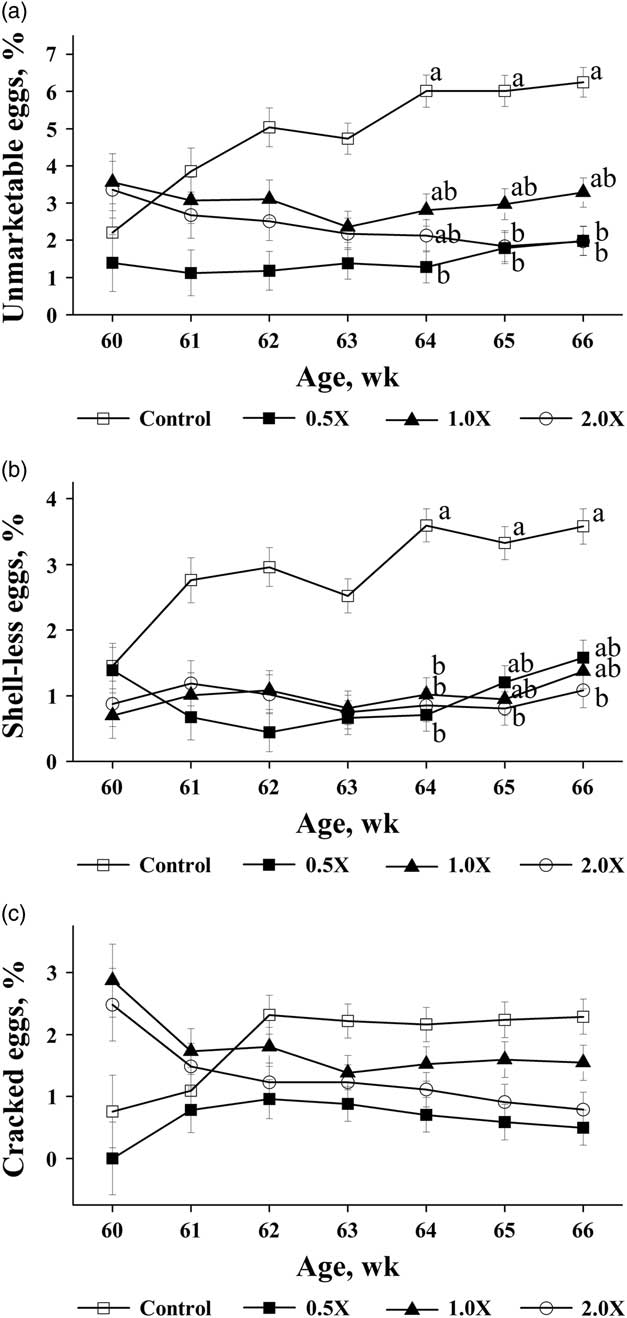
There were probiotic effects on the egg quality. The % of unmarketable eggs (a sum of cracked and shell-less eggs) was decreased in hens from both the 0.5× and 2.0× groups as compared with the control group (P=0.02) with the reduction of % of shell-less eggs (P=0.05) rather than cracked eggs (P>0.05, Table 3). The % of unmarketable eggs laid by hens in the 1.0× group was intermediate in the value, but without differences from the other groups (Table 3). Although there was no significant age effect, the % of the weekly cumulative unmarketable eggs (Figure 1a) and shell-less eggs (Figure 1b) were continuously increased from week 60 to 66 in control group; while it was relatively stable in the probiotic-fed groups. Compared with the control group, consequentially, the % of unmarketable eggs was reduced in the 0.5× group from week 64 to 66 (P<0.05. Figure 1a) and from week 65 to 66 in the 2.0× group (P<0.05. Figure 1a). While, the % of the weekly cumulated shell-less eggs, compared with the controls, was reduced at week 64 in the both 0.5× and 1.0× groups (P<0.05) and from week 64 to 66 in the 2.0× group (P<0.05, Figure 1b). There was no difference in the weekly cumulative unmarketable eggs among treatments before 64 weeks of age.
The effects of a dietary probiotic supplementation on weekly production of unmarketable eggs (a) the sum of shell-less (b) and cracked eggs (c) in laying hens from 60 to 66 weeks of age. A regular laying hen diet was mixed with the probiotic at 0, 0.5, 1.0 or 2.0 g/kg feed (Control, 0.5×, 1.0× and 2.0×). Least square means±the SEM within the age of a hen lacking common superscripts differ (P<0.05). The number of observations per least square mean was 6.

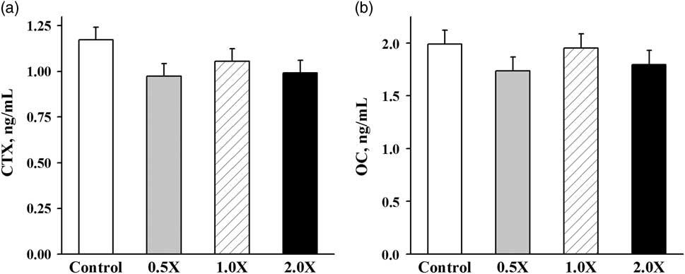
The probiotic feeding increased tibial and femoral mineral density and femoral mineral content (P=0.04, 0.03 and 0.02, respectively, Table 4), with a concomitant trend for increases in humerus mineral density and tibial mineral content (P=0.07 and 0.08, respectively, Table 4), occurred in the 2.0× group as compared with the control group. Bone mineralization values in the 0.5× and 1.0× groups were intermediate between the control and 2.0× groups. Keel mineralization, bone area, bone-breaking force (Table 4) and the bone remodeling indicators of circulating OC and CTX were similar among treatments (Figure 2).
The effects of dietary probiotic supplementation on plasma concentrations of bone remodeling indicators, CTX (a) and osteocalcin (b), in laying hens at 66 weeks of age. A regular laying hen diet was mixed with the probiotic at 0, 0.5, 1.0 or 2.0 g/kg feed (Control, 0.5×, 1.0× and 2.0×). The number of observations per least square mean was 6. CTX=c-terminal telopeptide of type I collagen.

The effects of dietary probiotic supplementation on bone mineral density, mineral content, area and breaking force of bones retrieved from 66-week-old White LeghornsFootnote 1

a,bLeast square means within a row lacking a common superscript differ (P<0.05).
1 The number of observations per least square mean was 24.
2 A regular laying hen diet was mixed with the probiotic at 0, 0.5, 1.0 or 2.0 g/kg feed (Control, 0.5×, 1.0× and 2.0×).
3 BW was used as a covariate except for keel bone area and tibia breaking force.
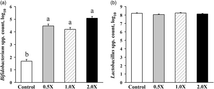
In comparison to the control group, the counts of cecal Bifidobacterium spp., but not Lactobacillus spp., were increased in all probiotic-fed groups (P<0.001, Figure 3) at 66 weeks of age.
The effects of dietary probiotic supplementation on cecal microbial count of Bifidobacterium (a) and Lactobacillus (b) in laying hens at 66 weeks of age. A regular laying hen diet was mixed with the probiotic at 0, 0.5, 1.0 or 2.0 g/kg feed (Control, 0.5×, 1.0× and 2.0×). Least square means±the SEM lacking common superscripts differ (P<0.05). The number of observations per least square mean was 24.

As the 2.0× group exhibited the greatest bone accrual effects compared with the control group, it was chosen for further investigation if the overdose, 2.0×, of dietary probiotic causes negative reactions in immunity. No differences were observed for plasma concentrations of cytokines, including pro-inflammatory IL-1β, IL-6, IFN-γ and TNF-α as well as anti-inflammatory IL-10 (Table 5). In line with the plasma cytokine levels, similar mRNA expressions of IL-1β, IL-6, and LITAF in the cecal tonsil were observed between the 2.0× group and control group (Table 5). Plasma levels of IgM, IgY and IgA in the probiotic group were also comparable with the control group (Table 5). With respect to the treatment response, the probiotic supplementation did not affect the heterophil to lymphocyte ratio and plasma corticosterone levels (Table 5).
The effects of dietary probiotic supplementation on immunology and stress indicators in laying hens at 66 weeks of ageFootnote 1

IL=interleukin; TNF-α=tumor necrosis factor-α; IFN-γ=interferon-γ; LITAF=lipopolysaccharide-induced tumor necrosis factor-α factor.
1 The number of observations per least square mean was 6.
2 Control: a regular layer diet; 2.0×: the regular layer diet plus probiotic at 2.0 g/kg feed.
Discussion
The current data showed that a dietary probiotic inclusion has beneficial in decreasing unmarketable eggs (the sum of cracked eggs and shell-less eggs) in laying hens without detectable negative effects up to 2.0× level. Similar to the current findings, reduced unmarketable and shell-less eggs have been reported in multiple strains of laying hens fed various probiotics at different ages (Mikulski et al., Reference Mikulski, Jankowski, Naczmanski, Mikulska and Demey2012; Zhang et al., Reference Zhang, Xie, Ji, Yang, Wu, Li, Ma and Bi2012; Abdelqader et al., Reference Abdelqader, Irshaid and Al-Fataftah2013b). For example, the laying hens fed diet mixed with Pediococcus acidilactici for 24 weeks showed reduced production of unmarketable eggs as compared with the control hens fed without the relative probiotic (Mikulski et al., Reference Mikulski, Jankowski, Naczmanski, Mikulska and Demey2012). The authors additionally found a significant increase in eggshell thickness, specific gravity and % of eggshell in hens fed probiotic. On the other hand, Zhang et al. (Reference Zhang, Xie, Ji, Yang, Wu, Li, Ma and Bi2012) noticed reduced production of unmarketable eggs but failed to find any difference in eggshell thickness when a multiple-strain based probiotic was fed to Lohman Pink hens for 8.6 weeks, similar to the current results. The short duration of the current study (7 weeks) along with the study of Zhang et al. (Reference Zhang, Xie, Ji, Yang, Wu, Li, Ma and Bi2012) could be one reason for the lack of effects of the probiotic on eggshell traits. In the current study, the cumulative production of unmarketable eggs did not show a reduction until the last 3 weeks of the 7-week study (Figure 1a) may suggest that the beneficial bacteria of the probiotic supplementation require time to establish their colonies in the intestinal lumen before the probiotic effects occurred. In addition to the duration of probiotic supplementation, other multiple factors, including microbial strain and dose, the genetic background and age of the hens, and environmental conditions such as ambient temperature and humidity, are affecting the study outcomes.
Previous studies have showed that increases of tibial size, density and mass occurred in broiler chickens fed various probiotics (Mutus et al., Reference Mutus, Kocabagli, Alp, Acar, Eren and Gezen2006; Panda et al., Reference Panda, Rao, Raju and Sharma2006; Houshmand et al., Reference Houshmand, Azhar, Zulkifli, Bejo, Meimandipour and Kamyab2011; Ziaie et al., Reference Ziaie, Bashtani, Torshizi, Naeeimipour, Farhangfar and Zeinali2011; Fuentes et al., Reference Fuentes, Orozco, Vicente, Velasco, Menconi, Kuttappan, Kallapura, Latorre, Layton, Hargis and Téllez2013; Sadeghi, Reference Sadeghi2014). For instance, a multi-species based probiotic significantly improved tibia weight, length, bone wall thickness and tibiotarsi weight/length index (bone density indicator) in male Ross 308 broilers fed for 42 days (Ziaie et al., Reference Ziaie, Bashtani, Torshizi, Naeeimipour, Farhangfar and Zeinali2011). However, a Bacillus subtilis-based probiotic improved tibia bone ash and calcium content of Ross 308 broilers only at 21 days of age rather than 42 days of age (Sadeghi, Reference Sadeghi2014). In laying hens, one study reported increased tibia weight, density and ash content following fed Bacillus subtilis for 10 weeks (Abdelqader et al., Reference Abdelqader, Irshaid and Al-Fataftah2013b). Our study provided further evidence of bone promoting effects of probiotic in laying hens as the probiotic feeding significantly enhanced tibial and femoral bone mineral density in the 2.0× group compared with the control group. The current findings, together with the results from Abdelqader et al. (Reference Abdelqader, Irshaid and Al-Fataftah2013b), indicated that probiotics have positive effects on skeletal health in laying hens, which is not at the expense of reducing egg production or eggshell quality.
In the current study, cecal microbiota composition was modified in hens as indicated by the increase in Bifidobacterium spp. population in probiotic-fed groups compared with the control group. However, the population of Lactobacillus spp. was not affected by the probiotic supplementation. Our results were comparable with the studies conducted on broilers using the same probiotic product (Giannenas et al., Reference Giannenas, Papadopoulos, Tsalie, Triantafillou, Henikl, Teichmann and Tontis2012; Mountzouris et al., Reference Mountzouris, Palamidi, Tsirtsikos, Mohnl, Schatzmayr and Fegeros2015); although some studies reported an increase in the levels of both Bifidobacterium spp. and Lactobacillus spp. (Mountzouris et al., Reference Mountzouris, Tsirtsikos, Kalamara, Nitsch, Schatzmayr and Fegeros2007 and Reference Mountzouris, Tsitrsikos, Palamidi, Arvaniti, Mohnl, Schatzmayr and Fegeros2010). Bifidobacterium spp. synthesize and secret a wide range of biochemical and metabolites in the intestines including short chain fatty acids (SCFA) (Wang et al., Reference Wang, Shoji, Sato, Nagata, Ohtsuka, Shimizu and Yamashiro2007). Short chain fatty acids can regulate bone mass directly through inhibiting the bone-absorption of osteoclasts and activating the bone-forming of osteoblasts (Iwami and Moriyama, Reference Iwami and Moriyama1993) or indirectly through increased intestinal absorption of minerals (Legette et al., Reference Legette, Lee, Martin, Story, Campbell and Weaver2012). Although the levels of cecal SCFA were not measured in the current study, increased concentrations of cecal propionate, butyrate and total SCFA have been reported in broiler chickens fed the same probiotic product (Murugesan and Persia, Reference Murugesan and Persia2015). However, the lack of probiotic effects on the bone remodeling indicators, circulating OC and CTX, did not support the direct mode in this study. In the current study, increased Bifidobacterium spp. may lead to increased Ca bioavailability and retention as reported elsewhere (Abdelqader et al., Reference Abdelqader, Al-Fataftah and Das2013a), which may in turn lead to the improvement of bone mass.
Numerous evidence demonstrates the significant role of cytokines in regulating bone mass (Lorenzo et al., Reference Lorenzo, Horowitz and Choi2008), making it becomes one of the main themes of the proposed mechanisms for the intestinal microbiome influencing bone metabolism. In the current study, we failed to find significant changes of cytokines in both plasma and the cecal tonsil between the 2.0× and control groups. Whether cytokines were altered in the bone of probiotic-fed hens remained to be determined. Most researches detecting the biological functions of probiotics regarding to bone physiology and health were conducted in rats and mice (McCabe et al., Reference McCabe, Britton and Parameswaran2015), and several research groups have attributed the bone accrual function to cytokine regulation under different pathophysiological conditions. For example, oral Saccharomyces cerevisiae administration, alone or with standard therapy, led to reduced alveolar bone loss, associated with decreased pro-inflammatory cytokine levels of TNF-α and IL-1β as well as increased anti-inflammatory cytokine IL-10 levels (Garcia et al., Reference Garcia, Knoll, Longo, Novaes, Assem, Ervolino, de Toledo and Theodoro2016). In a type 1 diabetes-mediated bone loss model, Lactobacillus reuteri inhibited the bone loss and rescued the TNF-α induced down-regulation of Wnt10b in both bone and osteoblasts (Zhang et al., Reference Zhang, Motyl, Irwin, MacDougald, Britton and McCabe2015).
Excessive level of corticosterone is well known to decrease bone mass through inhibition of osteoblastogensis, increasing osteoblast and osteocyte apoptosis, and promoting osteoclast survival (O’Brien et al., Reference O’Brien, Jia, Plotkin, Bellido, Powers, Stewart, Manolagas and Weinstein2004; Jia et al., Reference Jia, O’Brien, Stewart, Manolagas and Weinstein2006). Probiotics alleviate the stress response along the hypothalamus–pituitary–adrenal axis by reducing plasma or brain levels of corticotropin releasing hormone, adrenocorticotropic hormone and corticosterone (Sohail et al., Reference Sohail, Ijaz, Yousaf, Ashraf, Zaneb, Aleem and Rehman2010; Ait-Belgnaoui et al., Reference Ait-Belgnaoui, Colom, Braniste, Ramalho, Marrot, Cartier, Houdeau, Theodorou and Tompkins2014). In these studies, stressors such as heat or psychological stimuli were applied. The laying hens in the current study were raised under normal management condition without subjecting to known stressors, so it is not surprising that plasma corticosterone levels and heterophil to lymphocyte ratios were unaffected by the probiotic treatment.
In conclusion, the present study demonstrated that the dietary probiotic inclusion improves bone mineralization in aged White Leghorn laying hens without detrimental effects on egg production and eggshell quality. Instead, probiotic supplementation concomitantly reduces unmarketable egg production. The possible modes of the action may be associated with the regulation of gut microbial composition, but do not appear to be through the modulation of bone remodeling, immune cytokines, and corticosterone. Further studies are needed to investigate other cellular mechanisms such as regulating brain functions in regulating bone health via the microbiota-gut-brain and brain-bone axes.
Acknowledgments
The hens were donated by the Creighton Brothers Farm (Atwood, IN). The authors thank G. L. Nowling, J. Y. Hu, U. T. Mahmoud and M. G. Erickson of the USDA-ARS Livestock Behavior Research (West Lafayette, IN, USA) for technical assistance. Bone-breaking force was performed in Dr. Richard Stroshine’s lab of the College of Engineering at Purdue University. The study was financially supported by the grant award of the NIFA-AFRI, USDA. Mention of trade names or commercial products in this article is solely for the purpose of providing specific information and does not imply recommendation or endorsement of the USDA. The USDA is an equal opportunity provider and employer.
Declaration of interest
The authors declare that there is no conflict of interest.
Ethics statement
The experimental protocol had been approved by the Purdue Animal Use and Care Committee (PACUC Number: 1111000262).
Software and data repository resources
No software, data or models were deposited in official repositories.
Supplementary Material
To view supplementary material for this article, please visit https://doi.org/10.1017/S175173111800109X