Introduction
Supracricoid laryngectomy with cricohyoidoepiglottopexy or with cricohyoidopexy is an organ-preserving surgical procedure designed to treat laryngeal cancers. Since 1997, we have performed laryngectomy with cricohyoidoepiglottopexy and supracricoid laryngectomy with cricohyoidopexy for tumour (T) stage T2 and T3 laryngeal cancer cases and some T4 cases. This technique involves removal of the entire thyroid cartilage, including the bilateral true and false vocal folds, except for one or two arytenoid regions; reconstruction is then performed by fixing the cricoid cartilage to the hyoid bone by a three heavy chronic catgut stitches.
Amongst our patients undergoing these operations, two cases manifested laryngeal chondritis due to physical contact between the cricoid cartilage and cervical vertebrae osteophytes. The clinical symptoms and management of these two cases are reported.
Case reports
Case one
This case involved a 67-year-old man whose chief complaint was hoarseness. The patient had a two-month history of hoarseness before presenting to our hospital. He did not have any notable past history. He had smoked approximately 15–20 cigarettes/day for 30 years and consumed alcohol occasionally.
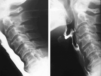
On laryngoscopy, a tumour was found involving the right vocal fold, with an extension to the laryngeal ventricle. The movement of the right arytenoid was not fixed. Tumour extension towards the paraglottic space was observed by target computed tomography (CT) scan. Histopathological analysis of a biopsy from the laryngeal tumour revealed a squamous cell carcinoma, and tumour–node–metastasis (TNM) staging was T3 N0 M0. Excessive osteophyte formation was found at the level of the C3–4 vertebral body during the pre-operative barium swallowing test (Figure 1). The posterior wall of the hypopharynx and cervical oesophagus was irregularly shaped because of the osteophyte. The patient, however, did not suffer from dysphagia before surgery.
Pre-operative barium swallow test showing excessive osteophyte formation at the level of the C3–4 vertebral body.

We conducted a laryngectomy with cricohyoidoepiglotto-pexy on the second day of hospitalisation. The bilateral arytenoids were preserved following tumour removal.
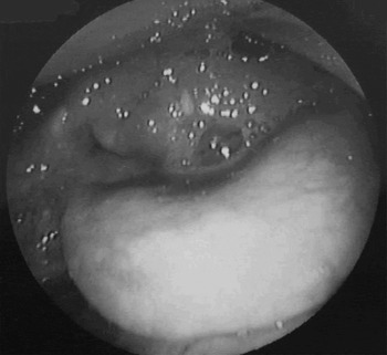
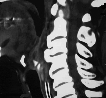
The early post-operative course was uneventful, with no particular symptoms indicating wound infection. However, the patient was unable to breathe from the neoglottis because of persistent oedema of the arytenoids (Figure 2). A sagittal view of the target CT scan revealed that the posterior end of the upper elevated cricoid cartilage directly contacted the osteophyte at the C3–4 level (Figure 3). Because of this, the posterior wall of the pharynx was also swollen.
Laryngoscopic view showing neoglottic stenosis caused by persistent oedema of the arytenoids.

Sagittal target computed tomography scan, showing the posterior end of the upper elevated cricoid cartilage directly contacting the C3–4 level osteophyte.

Approximately two months after the laryngectomy with cricohyoidoepiglottopexy procedure, laryngeal microlaryngoscopy was performed under general anaesthesia. This revealed white, necrotic tissue surrounded by granulation tissue located in the posterior wall of the pharynx. The posterior region of the cricoid cartilage contacting the pharynx also showed surface necrosis. It was clear that the persistent oedema of the arytenoids and the posterior pharyngeal wall was due to contact between both tissues.
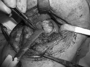
An external exploration was performed a few days later. A whitish, oval, 2 × 3 cm area covered with necrotic mucosa was found in the anterior region of the vertebrae (Figure 4). Beneath this whitish area, an osseous malformation was found and excised. The defect was covered by suturing the adjacent prevertebral mucosa. This osteophyte was presumed to be the osseous change observed at the C3–4 level. Laryngeal chondritis was also noted, involving the upper edge of the cricoid cartilage with ulceration and necrosis. Excision of the necrotic part of the cricoid and arytenoid cartilages was performed; the defect was covered by suturing the adjacent laryngeal mucosa. This exploration revealed that the chondritis was caused by physical contact between the elevated laryngeal cartilage and the C3–4 osteophyte.
At operation, a whitish, oval, 2 × 3 cm area covered with necrotic mucosa was found in the anterior region of the vertebrae.

The post-operative course was uneventful, with the laryngeal and pharyngeal oedema subsiding rapidly. A lateral view of pharyngeal X-ray revealed that the C3–4 osteophyte had completely resolved.
Two months after the second operation, the patient had achieved satisfactory swallowing and vocal functions.
Case two
The second patient was a 69-year-old man whose chief complaint was also hoarseness. This patient had a three-month history of hoarseness before presenting to our hospital. He had had pulmonary tuberculosis at the age of 20 years and was currently being treated for hypertension and hepatitis associated with hepatitis C virus. He had smoked 10–15 cigarettes/day for 50 years and was a heavy alcohol drinker.
Laryngoscopy revealed a tumour involving the right vocal fold, with an extension to the laryngeal ventricle. Movement of the right arytenoid was good. Biopsy of the laryngeal tumour and histopathological analysis revealed a squamous cell carcinoma, and the tumour was staged as T2 N0 M0. Excessive osteophyte formation was found at the C3–4 vertebral body during the pre-operative barium swallowing test. The posterior wall of the hypopharynx and cervical oesophagus was irregularly shaped by the osteophyte. However, the patient had not suffered dysphagia prior to surgery.
Laryngectomy with cricohyoidoepiglottopexy was undertaken on the fourth day of hospitalisation. The anterior two-thirds of the right arytenoid were removed.
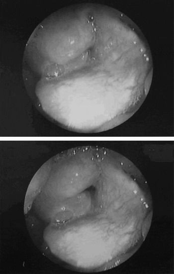
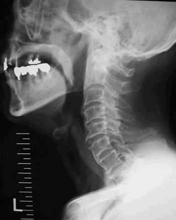
During the early post-operative course, the surgical wound became infected, with methicillin-resistant Staphylococcus aureus amongst other microbes, and the residual right arytenoid showed persistent oedema (Figure 5). A lateral view of pharyngeal X-ray revealed that the posterior end of the upper elevated cricoid cartilage directly contacted an osteophyte at the C3–6 level osteophytes (Figure 6).
Laryngoscopic views during the early post-operative course, showing infection of the surgical wound (which involved methicillin-resistant Staphylococcus aureus) and persistent residual right arytenoid oedema.

Lateral view of pharyngeal X-ray showing the posterior end of the upper elevated cricoid cartilage directly contacting the C3–6 level osteophytes.

One month after the initial procedure, a microlaryngoscopy exploration was performed. Laryngeal chondritis was noted, involving the upper edge of the cricoid cartilage, with a scarred neoglottis. Excision of the necrotic cricoid and laser resection of the scarred neoglottis was performed. This exploration revealed that the chondritis was caused by physical contact between the elevated laryngeal cartilage and a C3–4 osteophyte.
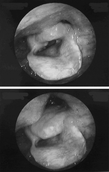
The post-operative course was uneventful, with the laryngeal and pharyngeal oedema subsiding rapidly (Figure 7). A further lateral view of pharyngeal X-ray revealed that the C3–4 osteophyte had been completely removed.
Laryngoscopic views taken during the later, uneventful post-operative course, showing rapid reduction of the laryngeal and pharyngeal oedema.

Two months after the second operation, the patient had achieved good swallowing and vocal functions.
Discussion
Ankylosing vertebral hyperostosis is a disease characterised by osteophyte formation and bony change of the anterior longitudinal ligament at the vertebral body. In 1950, Forestier and Rotes-Querol first reported as ankylosing spondylitis hyperostosis the case of a patient with excessive bone formation.Reference Forestier and Rotes-Querol1 In 1971, Resnick et al. redefined the bony malformation as diffuse idiopathic skeletal hyperostosis, after observing a tendency towards increased new bone formation which involved the general articulation joints.Reference Resnick, Shaul and Robins2
Although most of the observed patients were asymptomatic, dysphagia might occur if the osteophyte grew to a massive size.Reference Resnick, Niwayama, Resnick and Niwayama3 In a further publication, Resnick et al. noted that ankylosing spondylitis hyperostosis was observed in 6 to 12 per cent of autopsy cases.Reference Resnick and Niwayama4 This fact indicates that the surgeon must be aware of this cervical condition when considering laryngectomy with cricohyoidoepiglottopexy or supracricoid laryngectomy with cricohyoidopexy.
In our department, we have observed nine patients with a large osteophyte of the anterior cervical vertebrae, among 31 patients receiving laryngectomy with cricohyoidoepiglottopexy or supracricoid laryngectomy with cricohyoidopexy. However, only the two cases presented above manifested clinical problems post-operatively. In case one, laryngeal oedema persisted, resulting in difficulty in closing the tracheostoma. Laryngeal chondritis and necrosis of the pharyngeal wall were detected via laryngomicroscopy, and subsequently managed via external exploration. The patient was able to retain his laryngeal function. In case two, laryngeal chondritis induced by a C3–4 osteophyte resulted in stricture of the neoglottis. We were able to manage the stricture by laser excision of the scar tissue, thus preserving the patient's laryngeal function.
Based on laryngeal X-ray analyses of our cases, we observed that the cricoid cartilage was shifted approximately 0.9 cm posteriorly and 3.3 cm superiorly, on average, after laryngectomy with cricohyoidoepiglottopexy or supracricoid laryngectomy with cricohyoidopexy.Reference Satoru, Meijinn and Makito5 A cervical osteophyte at the level of C3–4 is likely to be the closest surface to form physical contact with an elevated laryngeal structure. Osteophytes can be located at a lower level, such as C5–7, but this is unlikely to create a problem.
Following laryngectomy with cricohyoidoepiglottopexy, severe complications involving the neoglottis can include a ruptured pexis and laryngeal stenosis. Ruptured pexis occurs at a rate of 0.8 per cent, and might occur immediately after the procedure or several months later.Reference Laccourreye, Brasnu, Laccourreye and Weinstein6 In order to avoid rupture, tension-free sutures must be placed after sufficient dissection of the mediastinal trachea, an adequate distance must be allowed between the sutures, and impaction needs to be performed by properly aligning the anterior border of the cricoid cartilage and the hyoid bone. Laryngeal stenosis resulting in unsuccessful decannulation has been reported to occur at a rate of 3.7 per cent.Reference Diaz, Laccourreye, Veivers, Garcia, Brasnu and Laccourreye7 Technical error might result in stricture of the neoglottis, resulting in delayed decannulation after the procedure. Laryngeal chondritis due to an osteophyte, occurring following supracricoid laryngectomy, has not previously been reported; our report is the first to present this complication.
• Supracricoid laryngectomy with cricohyoidoepiglottopexy and with cricohyoidopexy has been described for T2 and T3 laryngeal cancer cases and some T4 cases
• This paper describes two cases of laryngeal chondritis following laryngectomy with cricohyoidoepiglottopexy. This complication was caused by C3–4 cervical osteophytes physically contacting the cricoid cartilage
• When encountering a patient with excessive osteophyte formation at the level of C3–4, the surgeon must take extra precautions when undertaking laryngectomy with cricohyoidoepiglottopexy or cricohyoidopexy
The surgeon may need to consider a different approach when observing excessive osteophyte formation at the level of the C3–4 cervical vertebrae. Such an alterative approach may include radical excision of the osteophyte following the main laryngectomy with cricohyoidoepiglottopexy or cricohyoidopexy procedure; however, precautions regarding local infection may be needed. Adequate informed consent is also necessary in order to safely manage this type of case.
Conclusion
We present two cases of laryngeal chondritis caused by C3–4 cervical osteophytes following a laryngectomy with cricohyoidepiglotto-pexy procedure. This is the first paper reporting this type of complication following such a procedure. When encountering a patient with excessive osteophyte formation at the level of C3–4, one needs to take extra precautions, during the laryngectomy with cricohyoidoepiglottopexy or cricohyoidopexy procedure, in order to avoid this type of complication.