Introduction
Cryopreservation of mammal embryos has been technically feasible for many years, but morphological injuries still persist in fish embryos during cryopreservation (Ahammad et al., Reference Ahammad, Bhattacharyya and Jana1988; Ninhaus et al., 2008). The impediments to develop a safety protocol have been: the lower permeability of the embryonic membranes; embryo size with low surface-to-volume ratio; large-sized cells; and the sensitivity of the eggs to the subfreezing temperatures (Hagedorn et al., Reference Hagedorn, Hsu, Kleinhans and Wildt1997; Zhang & Rawson, Reference Zhang and Rawson1998).
Cryopreservation consists of storing quiescent embryos in liquid nitrogen for long periods (Neves, Reference Neves2008). Consequently, a successful protocol can have profound effects on farming and fishery management, preservation of rare species (Wildt, Reference Wildt, Seal, Rall, Cloud and Thorgaard1993), offspring production of rheophilic species during all the year and the safe storage of diverse gene pools in minimal space (Janik et al., Reference Janik, Kleinhans and Hagedorn2000).
Cryoprotectants are the substances required to submit fish embryos to subzero temperatures (Harvey, Reference Harvey1983). These chemicals are classified according to their site of action. Methanol, ethylene-glycol and dimethyl sulphoxide (DMSO), for example, have intracellular activities (Reichenbach et al., Reference Reichenbach, Oliveira, Lima, Santos-Filho, Andrade, Gonsalves, Figueiredo and Freitas2001), and sucrose, glucose and trehalose have extracellular activities (Niemann, Reference Niemann1991; Denniston et al., Reference Denniston, Michelet, Godke, Tiersch and Mazik2000). Thus, Streit Jr et al. (Reference Streit, Digmayer, Ribeiro, Sirol, Moraes and Galo2007) tested several combinations of cryoprotectants, and suggested cooling the embryos of P. mesopotamicus with 17.0% sucrose plus 9.0% methanol.
The methods for preserving bovine embryos are based on the standard freezing curve developed by Willadsen et al. (Reference Willadsen, Polge and Rowson1976, Reference Willadsen, Polge and Rowson1978), who recommended decreases in thermal rates of between 3 and 1°C/min. Thus, the embryos are induced to seeding after stabilization at −7°C, which is followed by freezing at −33 or −35°C, they are then stored in liquid nitrogen. However, these methods have often been detrimental to the embryo viability because cryopreservation is still resulting in morphological injuries to these embryos (Neves, Reference Neves2008; Ninhaus-Silveira, 2008).
The investigation of many morphological modifications by using the stereo- or light microscopy to describe and evaluate the morphological structures of fish embryos is more cost-effective. Nevertheless, electron micrographic images have been the best way to characterize abnormalities, deformities and injuries (Galman & Avtalion, Reference Galman and Avtalion1989; Shardo, Reference Shardo1995; Ninhaus-Silveira et al., Reference Ninhaus-Silveira, Foresti, Azevedo and Agostinho2007; Neves, Reference Neves2008).
In Brazil, researchers have been developing technological on-farm systems to raise P. mesopotamicus since the mid-1970s (Urbinati & Gonçalves, Reference Urbinati, Gonçalves, Baldisseroto and Gomes2005) because the pacu has excellent farming characteristics for large acreages (Castagnolli & Zuim, Reference Castanholli and Zuim1985; Lima et al., Reference Lima, Bernardino, Val-Sella, Fava-De-Moraes, Schemy and Borella1991; Silva et al., Reference Silva, Barroso and Benevides1997).
The pacu is also an excellent model for the present investigation because of its rheophilic behaviour. Thus, the objective of the present experiment was to investigate the effects of different concentrations of sucrose with 9.0% methanol on the morphological structures of P. mesopotamicus embryos.
Materials and methods
The experiment was conducted in the Estação de Hidrologia e Aqüicultura of the DUKE ENERGY INTERNATIONAL–Geração Paranapanema, Salto Grande (SP) in collaboration with the PeixeGen/UEM and Aquam/UFRGS research groups.
Parental fish were randomly selected at the gonad stage, which was visually detected through the enlarged abdomen and the reddish urogenital pore in the females, and the presence of milting after a gentle pressure along the male body (Woynarovich & Horváth Reference Woynarovich and Horváth1983). Thereafter, they were transferred to an aquarium with 1000 l of clean-flowing water.
The females received injections of carp pituitary at 5.5 mg kg−1 of body weight divided into 10% at the first application, and the remained 90% at the second application 12 h later. When this second injection was applied to the females, one dose of carp pituitary at 2.5 mg kg−1 of body weight was also applied to the males. Thereafter, the water temperature and the reproductive signals from the females were monitored at 1-h intervals because the pacu requires about 240 h to spawn. The gametes and semen were both expelled into the same beaker after applying a gentle pressure along the fish body. After the addition of water for the fertilizing, the eggs were transferred into 7-l capacity incubators.
The fertilization rate was evaluated by counting the number of live embryos and dead eggs from three samples randomly collected soon after blastopore closure. The hatching rate of the live embryos that were incubated without any cryoprotectant (control 1) was evaluated from three replicates of 100 individuals.
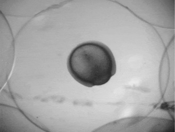
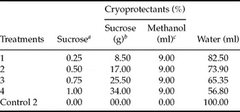
Fish embryos developed under 27.4 ± 0.2°C for 6 h were selected according to their viability, health and normal chorion, which was visualized at blastopore closure (Fig. 1). Groups of 15 individuals collected using Pasteur pipettes were submitted to the treatments described in Table 1.
Fish embryo image of Piaractus mesopotamicus at the post-gastrula stage, 95% epiboly–blastopore closure (digital image at 12 megapixels taken with a stereomicroscope, ×25).

Sucrose solutions for freezing the embryos of Piaractus mesopotamicus.

aMolar concentration of sucrose; bPer cent sucrose; cPer cent methanol.
In the 0.5 ml palettes, 15 fish embryos were submitted to the freezing curve regime using a programmable cooler Biocom, model Dominum K, in which the decrease in temperature was 0.5°C/min until stabilization at −7°C for manual seeding. Thereafter, the temperature was reduced to −33°C and the embryos were stabilized for 6 min. Next, the embryos were stored in liquid nitrogen.
The frozen embryos were thawed at 45°C for 15 s according to the protocol of Streit Jr et al. (Reference Streit, Benitis, Moraes, Ribeiro, Sakaguti and Caldieri2006). Soon after thawing, these embryos were immersed in 10% sucrose to reduce osmotic pressure. Next, they were transferred ito incubators with 1.5 l of flowing water. Samples were randomly taken before fixing these embryos with 2% glutaraldehyde in 0.2 M phosphate buffer.
The fixed samples were first rinsed once with alcohol at 30, 50 and 70% for 30 min, twice in 90% alcohol and then in 100 ethanol for 15 min. Next, the samples were submitted to the Critical Point Dryer (BAL-TEC CDP 030), which uses liquid and gaseous CO2 for complete dehydration. Finally, they were coated with a gold–palladium alloy in the metal stubs of the Metal Desk II Denton vacuum, and scanning electron micrographic images were used to detect normal development or injuries, photographed using a JEOL (JSM-540) scanning electron microscope at the UFRGS, Porto Alegre, Rio Grande do Sul State, Brazil.
Results
In the control 1 group, the fertilization rate was 88.60 ± 8.2% and the hatching rate was 93.40 ± 2.14%. These responses indicated the use of high quality biological material. All the thawed embryos, however, died during the first few hours in the incubators, although some were well rehydrated and presented good morphological conformation.
Despite the apparent normality of some morphological components treated with sucrose at 8.50, 17.00 and 25.50%, the chorion was only visualized after rehydration of those embryos submitted to 17.0% sucrose plus 9% methanol.
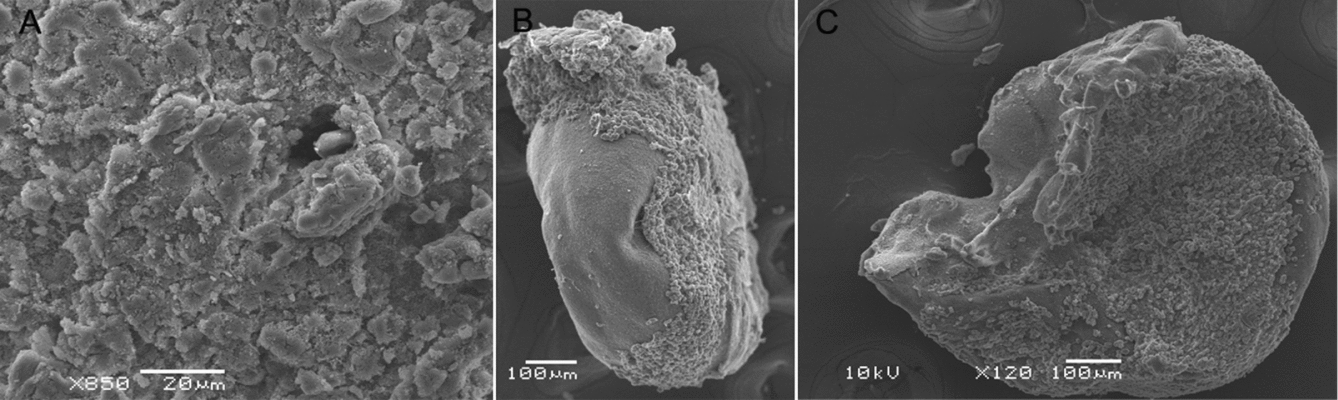
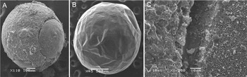
In the control 2 group, the images showed fish embryos with damaged blastoderm cells and disfigured vitello. The blastoderm was incapable of overcoming the freezing process, and the micropore, which is the chorion microstructure that controls fluid exchanges (Rawson et al., Reference Rawson, Zhang, Kalicharan and Jongebloed2000), was completely damaged (Fig. 2).
Scanning electron micrographic images after the freezing/thawing process, and taken from fish embryos treated with no cryoprotectant. (A) The injuries in the blastoderm cells. (B) Fish embryo with a disfigured vitello and completely damaged blastoderm. (C) The destroyed vitello and the damaged blastoderm.
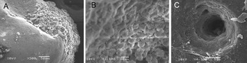
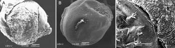
The use of 8.5% sucrose led to some injuries, but in most of the embryos there were no vitello destruction (Fig. 3). The embryos treated with 17.0% sucrose had no apparent injuries in the chorion, and the vitelline membrane had no structural damage. Furthermore, the blastoderm had damage, and the syncytial vitelline layer showed deformities (Fig. 4).
Scanning electron micrographic images of fish embryos (Piaractus mesopotamicus) after the freezing process using 8.5% sucrose plus 9.0% methanol. (A) Injuries in the blastoderms cells and the modifications in the vitello. (B) The embryonic cells with structural modifications in the blastoderm. (C) The chorion micropore with injuries caused by the flux/influx of cryoprotectant and water.

Scanning electron micrographic images of fish embryos (Piaractus mesopotamicus) after the freezing process using 17.5% sucrose plus 9.0% methanol. (A) Fish embryo with intact vitello and injuries in the blastoderm. (B) The hydrated fish embryo showing the intact chorion. (C) Apparent damages in the syncytial vitelline layer.

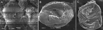
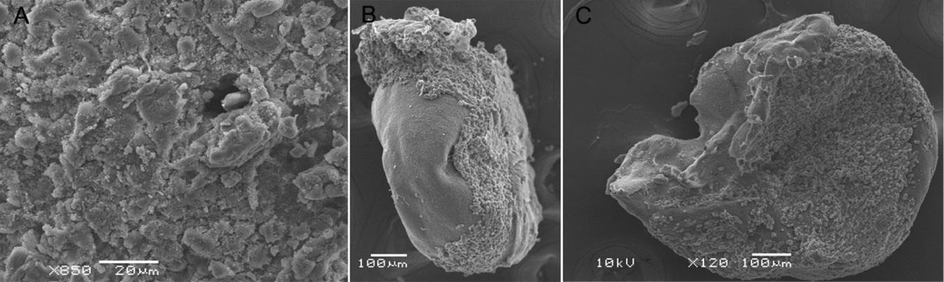
Under 25.6% sucrose the fish embryos presented structural modifications that characterized the presence of injuries, and a reduced and damaged syncytial layer. The chorion was disrupted and all the vitellos presented structural modifications (Fig. 5). In 34.0% sucrose, the vitellos of all fish embryos were destroyed and the blastoderms were injured (Fig. 6).
Scanning electron micrographic images of fish embryos after the freezing process using 25.6% sucrose plus 9.0% methanol. (A) The damaged blastoderm and the destroyed chorion. (B) The disfigured vitello and the destroyed blastoderm. (C) Apparent damages in the syncytial vitelline layer.

Scanning electron micrographic images of fish embryos (Piaractus mesopotamicus) after freezing using 34.2% sucrose plus 9.0% methanol. (A) The destruction of the blastoderm cells. (B) The destroyed vitello and the remains of the blastoderm. (C) Vitellos completely destroyed.

These scanning electron micrographic images, therefore, showed some of the inherent difficulties in maintaining the integrity of the fish embryos after use of sucrose and methanol.
Discussion
In the control 1 group, the fertilization rate reflected the healthy conditions of the embryos and, consequently, they were perfectly capable of resisting cryopreservation treatments (Lahnsteiner, Reference Lahnsteiner2008); the high viability (Bobe & Labbé, 2009) was important when comparing the efficiency of these cryoprotectants.
The presence of whitish embryos after the thawing process indicated the formation of ice crystals (Zhang & Rawson, Reference Zhang and Rawson1996). These whitish embryos were different to the translucent embryos, which had good conformation. Ice crystals were also reported by Neves (Reference Neves2008) who also studied P. mesopotamicus and Ninhaus-Silveira et al. (2008) who studied the corimbatá (P. lineatus). Injuries in the blastoderm cells and the disfigured vitello were probably caused by these ice crystals, as reported by Ninhaus-Silveira et al. (Reference Ninhaus-Silveira, Foresti, Azevedo and Agostinho2007). These ice crystals may have also been responsible for chorion absence and the destruction of the embryonic and vitelline membranes, as reported by Neves (Reference Neves2008).
Although most of the fish embryos were severely injured by these cryoprotectant solutions, the responses from every concentration showed different levels of intensity. Sucrose at 8.5%, for example, induced injuries, but the vitello was not destroyed in most embryos (Fig. 3). The blastoderm, however, was incapable of overcoming the freezing process, and the micropore was completely damaged (Fig. 2C). These injuries, which should have been prevented by the use of sucrose solutions (Woelders, Reference Woelders1997), may have been caused by the flux/influx of water under a sucrose concentration of 8.5%. Sucrose at 25.5 or 34.0% can disrupt the vitello because of osmotic pressure (Ahammad et al., Reference Ahammad, Bhattacharyya and Jana2003). This fact can be associated with the present treatments because, under 34.0% sucrose all vitellos were destroyed and the blastoderms were injured (Fig. 6).
The successful cryopreservation of oocytes and fish embryos has numerous constraints associated with injuries throughout the freezing process. The failure may have been due to one of the following four causes: first, most fish eggs have a diameter more than 1 mm, which represents a low area-to-volume ratio that reduces flux of both water and cryoprotectant during the cryopreservation; the second cause is the quantity of vitello; third, the numerous morphological structures of fish embryos; and, finally, the complex system of membranes, which is characterized by low levels of permeability and high sensitiveness to freezing conditions (Billard & Zhang, 2000).
Therefore, in conclusion, in the cryopreservation of P. mesopotamicus, the different concentration levels of sucrose plus 9.0% methanol were unable to avoid the formation of ice crystals during the freezing and thawing processes. Ice crystals were responsible for these injuries, but the reduced level of injury observed in the morphological structures of fish embryos treated with 17.0% sucrose plus 9.0% methanol is a compelling argument for additional studies.
Acknowledgements
The authors thank the Estação de Hidrologia e Aqüicultura of the DUKE ENERGY INTERNATIONAL–Geração Paranapanema for providing the facilities where the present experiment was carried out, the Universidade Federal do Rio Grande do Sul (UFRGS) for the scanning electron microscope, the Universidade Estadual de Maringá (UEM) and the Research Group PeixeGen for human resources, the CAPES (Coordenação de Aperfeiçoamento de Pessoal de Nível Superior) and CNPq (Conselho Nacional de Pesquisa) for the economic support.