Osteoporosis is a common skeletal disease characterised by low bone mineral density (BMD) and defects in the microarchitecture of bone, with a consequent increase in bone fragility and susceptibility to fracture (Ref. 1). The burden of osteoporosis on the healthcare system is increasing with the ageing of society (Ref. Reference Burge2), and current direct costs exceed $19 billion per year in the United States alone (Ref. Reference Organization3).
Osteoporosis is a common complex disease caused by the interplay of genetics and environmental factors, with adverse environmental exposures acting on a genetically susceptible individual to produce disease (Ref. Reference Chakravarti and Little4). Many factors influence the risk of osteoporosis, including diet, physical activity, medication use and coexisting diseases, but one of the most important clinical risk factors is a positive family history, emphasising the importance of genetics in its pathogenesis (Refs Reference Sigurdsson5, Reference Torgerson6). The allelic architecture of osteoporosis is likely to be multifactorial, with each imparting a relatively small effect, but their combined effect, as well as their interaction with environmental factors, probably leads to clinical disease (Ref. Reference Manolio and Collins7).
In the past decade, much progress has been made in reproducibly identifying genes that influence osteoporosis. Since 2007, we have experienced an acceleration in the identification of osteoporosis loci with the advent of large-scale, high-density genome-wide association studies (GWASs). GWASs, in which several hundred thousand to more than a million single-nucleotide polymorphisms (SNPs) are assayed in thousands of individuals, have been successfully used thus far to identify common genetic variants (those with a minor allele frequency greater than ~5%) that are associated with common human diseases (Ref. Reference Hindorff8).
Osteoporosis is defined clinically through the measurement of BMD, which is primarily evaluated by a dual-emission X-ray absorptiometry scan. Other osteoporosis-related phenotypes such as bone geometry and quantitative ultrasonography can also provide information on fracture prediction (Ref. Reference Kaptoge9) and assessment of the response to osteoporosis treatment (Refs Reference Greenspan10, Reference Uusi-Rasi11).
BMD is a highly heritable trait, with heritability estimates of 0.6–0.85 (Ref. Reference Ng12). Twin and family studies have shown that peak BMD, as well as BMD loss, is genetically determined (Refs Reference Slemenda13, Reference Smith14, Reference Gueguen15, Reference Krall and Dawson-Hughes16). Heritability studies have also confirmed that other phenotypes relevant to the pathogenesis of osteoporosis, such as ultrasound properties of bone and bone geometry, have significant heritable components (Ref. Reference Arden17). Segregation analysis in families has shown that regulation of BMD and other osteoporosis-related phenotypes is polygenic and determined by the effects of several genes, each with relatively small effects, rather than a small number of genes with large effects (Ref. Reference Gueguen15).
This article reviews the results of linkage and association studies for osteoporosis, with particular focus on the new findings revealed by GWASs since 2007, and discusses future directions for the dissection of the genetic determinants of osteoporosis in the post-GWAS era.
Linkage studies
Linkage analysis has long been used to identify disease-associated loci in inherited diseases. This approach relies on the principle of identity by descent combined with phenotypic information to identify loci conferring disease susceptibility. Linkage analysis is a powerful methodology to detect the causative gene in monogenic bone diseases; however, for complex diseases, the identification of susceptibility genes located within the linkage peak region has proved difficult, and the identified loci have not often been replicated for osteoporosis (Ref. Reference Richards18). More than a dozen genome-wide linkage scans have been performed on BMD and other osteoporosis-related phenotypes, including femoral neck (FN) geometry (Ref. Reference Peacock19), ultrasound properties of bone (Ref. Reference Wilson20) and bone loss. However, even a very large-scale meta-analysis of nine genome-wide linkage studies (combined n = 11 842) did not yield any genome-wide significant loci for BMD (Ref. Reference Ioannidis21). This probably reflects the fact that common variants that regulate BMD have modest effects that are difficult to detect reproducibly by conventional linkage analysis.
Candidate gene association studies
Before GWASs, candidate gene association studies (CGASs) were widely used to identify genes that plausibly have an important role in the disease process, and well-designed CGASs, in combination with fine mapping of known osteoporosis loci, remain an attractive and efficient way of identifying new susceptibility genes or variations until the costs of high-throughput sequencing decrease. Although CGASs are attractively simple in methodology, they have often produced spurious and nonreplicated results. This may be because the statistical power to detect these associations is limited unless causal SNPs or very good surrogates have been typed. Additionally, other factors such as differences in sample characteristics, including ethnicity, sex, age and menopausal status, might produce different results in different studies.
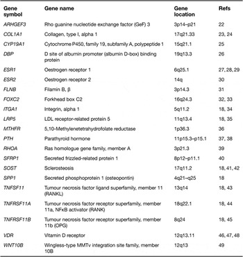
A recent CGAS attempted to overcome these difficulties by standardising genotyping and phenotype definitions across 19 195 European subjects in five international, multicentre population-based studies. This study (Ref. Reference Richards18) used genome-wide genotyping data, but the authors restricted their analysis to 150 candidate genes and 36 016 SNPs within these loci. They found evidence for association in only 9 of 150 (6%) candidate genes previously tested for their association with osteoporosis. Details of these loci can be found on the HUgeNet website (http://www.hugenavigator.net/). These results suggest that candidate genes tested for BMD rarely replicate in large consortia with standardised phenotyping and genotyping. Table 1 lists the candidate genes for osteoporosis.
Promising candidate genes in osteoporosis

Genome-wide association studies
GWASs represent a powerful and efficient study design for investigating the genetic architecture of complex diseases arising from common base-pair variants (Refs Reference Hardy and Singleton50, Reference Manolio51). GWASs use dense maps of common SNPs (minor allele frequency >~5%) that cover the human genome to look for allele-frequency differences between cases and controls (diseases study design) or genotype variation, which can explain trait variation (quantitative traits study design). A significant observation in the variant site is taken to indicate that the corresponding region of the genome contains functional DNA-sequence variants that influence the disease or trait in question (Ref. Reference Kruglyak52). GWASs have proved to be a powerful approach in screening the susceptibility genes (loci) for common diseases. An updated list of published GWASs can be found at the National Cancer Institute–National Human Genome Research Institute catalogue of published GWASs (http://www.genome.gov/26525384). A major advantage of GWASs over candidate gene studies is that they offer the possibility of identifying novel susceptible genes and pathways.
In 2007, Kiel et al. (Ref. Reference Kiel53) published the first GWAS of osteoporosis. In this study, they used the early and low-density commercial Affymetrix GeneChip and genotyped ~100 000 SNPs in 1141 Framingham Heart Study subjects, and identified 40 SNPs that could potentially be associated with several bone phenotypes. However, owing to sample size limitations and other factors, none of the P-values exceeded the threshold of genome-wide significance. In 2008, two GWASs (Refs Reference Richards54,Reference Styrkarsdottir55) reported nine genome-wide significant hits for BMD. In these two studies, more than 300 000 SNPs were genotyped in much larger cohorts. The following 2 years (2009 and 2010) witnessed a deluge of GWASs conducted on osteoporosis and related phenotypes (Refs Reference Guo38, Reference Zhao56, Reference Paternoster57, Reference Kung58, Reference Koller59, Reference Karasik60, Reference Hsu61, Reference Guo62, Reference Xiong63, Reference Timpson64, Reference Styrkarsdottir65, Reference Rivadeneira66, Reference Liu67, Reference Cho68, Reference Liu86, Reference Duncan92, Reference Kou93).
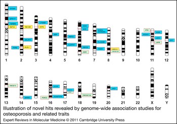
GWASs for osteoporosis and related phenotypes validated several candidate genes for osteoporosis, including LRP5 (Refs Reference Richards54, Reference Rivadeneira66), ESR1 (Refs Reference Styrkarsdottir55, Reference Styrkarsdottir65, Reference Rivadeneira66), SOST (Ref. Reference Styrkarsdottir65), TNFRSF11B (Refs Reference Richards54, Reference Styrkarsdottir55, Reference Rivadeneira66), TNFSF11 (Refs Reference Styrkarsdottir55, Reference Paternoster57, Reference Styrkarsdottir65, Reference Rivadeneira66, Reference Cirulli and Goldstein91), TNFRSF11A (Refs Reference Styrkarsdottir55, Reference Rivadeneira66), PTH (Ref. Reference Guo38) and FOXC2 (Ref. Reference Rivadeneira66). Novel loci that have been identified as being associated with osteoporosis and related phenotypes by recent original GWASs and subsequent meta-analysis of GWAS data are discussed in this section (Fig. 1). Note that the SNPs from each study are the reported SNPs either near or within the genes; however, they probably do not represent the causal variants.
Illustration of novel hits revealed by genome-wide association studies for osteoporosis and related traits. Novel bone mineral density loci are in blue, new loci for other osteoporosis-related phenotypes are in yellow and candidate loci validated by GWASs are in green. Please refer to Table 1 and the main text for full gene names and references.

BMD loci revealed by GWASs
JAG1
rs2273061, located near the JAG1 (jagged 1) locus on 20p12.2, showed significant association with spine BMD (P = 5 × 10⁻⁸) in a GWAS carried out on Southern Chinese subjects in Hong Kong and replicated in other ethnic groups (Ref. Reference Kung58). The study also confirmed association of the high BMD-related allele G of rs2273061 with higher JAG1 mRNA expression in both human bone-derived cells and peripheral blood mononuclear cells. The function of JAG1 seems related to an increase in bone mineral deposition (Ref. Reference Nobta69). JAG1 protein was detected in trabecular osteoblasts and endosteal osteoblasts, and its expression was increased with intermittent parathyroid hormone treatment (Ref. Reference Weber70).
IL21R
Guo et al. (Ref. Reference Guo38) performed a GWAS for FN BMD in a discovery sample consisting of 983 unrelated white subjects and replicated the top 175 SNPs (with P < 5 × 10⁻⁴) in a family-based sample of 2557 white subjects. Three IL21R (interleukin 21 receptor) SNPs, rs8057551, rs8061992 and rs7199138, showed consistent association with FN BMD in discovery and replication samples, with combined P-values of 2.31 × 10⁻⁶, 8.62 × 10⁻⁶ and 1.41 × 10⁻⁵, respectively, in the total sample. IL21R is a cytokine receptor that is important to bone biology and has been identified negatively to be correlated with the destruction of cartilage and bone (Ref. Reference Jungel71).
ALDH7A1
This susceptibility gene was first found to be associated with osteoporotic fractures (OFs) in a case–control GWAS in 700 elderly Chinese Han subjects, with a follow-up replication study in an independent Chinese sample containing 390 cases with hip OFs and 516 controls (Ref. Reference Guo62). SNP rs13182402 within the ALDH7A1 (aldehyde dehydrogenase seven family, member A1) gene on chromosome 5q31 was strongly associated with OFs (P = 2.08 × 10⁻⁹, odds ratio = 2.25). This SNP was confirmed as consistently associated with hip BMD in both Chinese subjects and Caucasians (P = 6.39 × 10⁻⁶). The ALDH7A1 gene encodes an enzyme of the acetaldehyde dehydrogenase superfamily, which degrades and detoxifies acetaldehyde generated by alcohol metabolism. Acetaldehyde has been shown to inhibit osteoblast proliferation and to decrease bone formation (Ref. Reference Giuliani72). Another member of the acetaldehyde dehydrogenase family, ALDH2, has also been found to be significantly associated with osteoporosis (Ref. Reference Yamaguchi73).
SP7
The SP7 (also known as OSTERIX) locus on 12q13 was first found to be associated with spine BMD (Ref. Reference Styrkarsdottir65). Common SNPs near the SP7 region were also associated with total body BMD in a GWAS in children (Ref. Reference Timpson64). SNPs in this region were genome-wide significant in the GEFOS (genetic factors for osteoporosis) meta-analysis (Ref. Reference Rivadeneira66). SP7 encodes a transcription factor that has an essential role in regulating osteoblast differentiation (Ref. Reference Baek74).
ADAMTS18 and TGFBR3
These two susceptible genes were identified by a GWAS performed in different ethnic groups (Ref. Reference Xiong63). One thousand unrelated white US subjects and 1972 subjects from white US pedigrees were genotyped, along with a Chinese hip fracture sample comprising 350 cases and controls, a Chinese BMD sample with 2955 subjects and a Tobago cohort of 908 males of African ancestry as a replication sample. ADAMTS18 (ADAM metallopeptidase with thrombospondin type 1 motif, 18) and TGFBR3 (transforming growth factor, beta receptor III) were significantly associated with BMD variation in the three major ethnic groups. They also provided in silico replication by using publicly available Framingham GWAS data (2953 whites). In addition, ADAMTS18 variants were also found to be associated with hip fracture.
The allele change from T to C of SNP rs16945612 in ADAMTS18 produced a new TEL2 (a member of the E26 transformation-specific family of transcription factors) binding site in ADAMTS18. TEL2 represses two genes, BMP6 and RARa, which are involved in regulating osteoblast differentiation and bone remodelling (Ref. Reference Gu75). Electrophoretic mobility-shift analysis confirmed the potential changes of TEL2 binding to ADAMTS18 caused by rs16945612. TGFBR3 appears to modulate the biological function of BMP2 (bone morphogenetic protein 2) (Ref. Reference Kirkbride76), and BMP2 has key roles in bone biology and is significantly associated with BMD and other bone phenotypes (Ref. Reference Styrkarsdottir77). Tgfbr3 double-null mice suffer severe abnormal skeleton defects (Ref. Reference Stenvers78).
ZBTB40
SNP rs7524102 in the 1p36 region, previously implicated in the genetic regulation of BMD by linkage analysis in families (Refs Reference Devoto79, Reference Devoto80), was identified as a potential locus for regulation of both hip and spine BMD by a GWAS with a significant association signal (P = 5 × 10⁻¹⁶) (Ref. Reference Styrkarsdottir55). This association was confirmed by other GWASs (Ref. Reference Styrkarsdottir65, Reference Rivadeneira66, Reference Cirulli and Goldstein91). In the region, ZBTB40 (zinc finger and BTB domain containing 40), which is located 80 kb downstream from the signal, may be the susceptible gene. ZBTB40 is expressed in bone; however, the function of this gene is as yet unknown and requires more functional studies to elucidate its role in bone physiology.
SOX6
By genotyping 380 000 SNPs in 1000 homogeneous unrelated Caucasians (female n = 501, male n = 499), Liu and colleagues (Ref. Reference Liu67) performed the first bivariate GWAS of obesity and osteoporosis. SNPs rs297325 and rs4756846 in intron 1 of the SOX6 (sex-determining region Y-box 6) gene were found to be associated with hip BMD in the male subjects. These findings were further validated in the 1370 male subjects of the Framingham Heart Study cohort. The SOX6 locus was also identified as the FN BMD locus by a meta-analysis (Ref. Reference Rivadeneira66). SOX6 is a member of the SOX gene family and was previously reported to have an important role in both cartilage formation (chondrogenesis) and obesity-related insulin resistance. Animal model studies show that Sox6 single-null mice present mild skeletal abnormalities, and Sox6-knockout mice fetuses die with a severe, generalised chondrodysplasia (Ref. Reference Smits81).
FAM3C and SFRP4
These two loci were identified in a GWAS in Asian populations (Ref. Reference Cho68). By genotyping 352 228 SNPs in 8842 subjects and involving eight quantitative traits, this study found that FAM3C (family with sequence similarity 3, member C) and SFRP4 (secreted frizzled-related protein 4) were associated with BMD. A FAM3C SNP rs7776725 on chromosome 7q31 was found to be associated with BMD at the radius (P = 1.0 × 10⁻¹¹), tibia (P = 1.6 × 10⁻⁶) and heel (P = 1.9 × 10⁻¹⁰). The function of FAM3C gene in bone biology is unclear to date. SNP rs1721400, mapping close to SFRP4, on chromosome 7p14 was consistently associated with BMD at the three sites mentioned above (P = 2.2 × 10⁻³, 1.4 × 10⁻⁷ and 6.0 × 10⁻⁴, respectively). SFRP4 is a member of the SFRP family, which acts as soluble modulators of Wnt signalling (Ref. Reference Cho82). Moreover, overexpression of Sfrp4 in mice has been associated with a reduction in bone density (Refs Reference Nakanishi83, Reference Nakanishi84).
MARK3
The MARK3 (MAP/microtubule affinity-regulating kinase 3) gene on chromosome 14q32.3 was significantly associated with total hip BMD (rs2010281, located in intron 1 of MARK3, had a P-value of 1.8 × 10⁻⁹) (Ref. Reference Styrkarsdottir65). This SNP was also significantly associated with FN BMD in a meta-analysis (Ref. Reference Rivadeneira66). The MARK3 gene encodes mitogen-activated protein/microtubule affinity-regulating kinase 3, a member of the adenosine monophosphate-activated protein kinase superfamily of proteins. MARK3 might be involved in cell cycle regulation, and alterations in MARK3 might lead to carcinogenesis. The function on the regulation of BMD needs to be explored further.
MHC region
Styrkarsdottir and colleagues (Ref. Reference Styrkarsdottir55) first found that SNP rs3130340 in the MHC (major histocompatibility complex) region was associated with spine BMD (P = 1.2 × 10⁻⁷). The SNP is located downstream of the uncharacterised chromosome 6 open reading frame 10 gene C6orf10. The association significance of this SNP was also confirmed by meta-analysis (Ref. Reference Rivadeneira66). The MHC locus on chromosome 6p21 is associated with a number of immune-related diseases; however, the precise mechanism responsible for osteoporosis in this region is unknown.
LRP4, GPR177 and CTNNB1
Three genes, all involved in the Wnt/β-catenin signalling pathway, that is, LRP4 (low-density lipoprotein receptor-related protein 4), GPR177 (G-protein-coupled receptor 177) and CTNNB1 (catenin beta 1), were found to be strongly associated with BMD. This pathway has an essential role in the regulation of bone mass. Two SNPs, rs2306033 and rs7935346, which are within or close to the LRP4 locus at 11p11.2, were found to be associated with hip BMD, but were not genome-wide significant (Ref. Reference Styrkarsdottir55). This association was further studied by a meta-analysis of five GWASs (Ref. Reference Rivadeneira66) and a collaborative meta-analysis (Ref. Reference Richards18) and confirmed that SNPs within the LRP4 locus were associated with FN BMD.
Two common SNPs, rs1430742 and rs2566755, were associated with both FN and lumbar spine (LS) BMD (Ref. Reference Rivadeneira66). The two SNPs are located within an intron of GPR177 (G-protein-coupled receptor 177) on chromosome 1p31.3. The study also showed that SNP rs87939, which is located 103 kb upstream of the CTNNB1 gene, was associated with FN BMD (Ref. Reference Rivadeneira66).
MEF2C, CRHR1, DCDC5, FLJ42280 and STARD3NL
These five novel genes were identified with genome-wide significance in a very large-scale meta analysis of five GWASs (Ref. Reference Rivadeneira66). The meta-analysis identified eight genome-wide significant loci. (In addition to the five genes mentioned above, another three genes – GRP177, CTNNB1 and SOX6 – are described earlier in this paper.) The study also reported eight known loci and four suggestive loci for BMD.
New loci for other osteoporosis-related phenotypes
TBC1D8, OSBPL1A and RAP1A
An integration of the GWAS for osteoporosis-related traits, including BMD, femoral neck-shaft angle (NSA), femoral neck length and narrow-neck width (NW), was performed in 7633 Caucasian women and 3657 men (Ref. Reference Hsu61). SNP rs2278729 located in intron 4 of TBC1D8 (TBC1 domain family, member 8) on chromosome 2q11.2, and SNP rs7227401 located in intron 4 of OSBPL1A (18q11.2, oxysterol-binding protein-like 1A) were significantly associated with NSA (P = 1.48 × 10⁻⁷) and NW (P = 4.22 × 10⁻⁷) in men. SNP rs494453, located in intron 2 of RAP1A (member of RAS oncogene family) on chromosome 1p13.2, was found to be associated with NW (P = 2.80 × 10⁻⁷) in women, and the association became more significant when analysing women and men together (P = 3.6 × 10⁻⁸). A gene expression study showed that rs2278729, allele A, is associated with smaller NSA in men and with a lower expression of TBC1D8 transcript (Ref. Reference Hsu61).
RTP3
A GWAS for FN bone geometry was performed by analysing 379 000 SNPs in 1000 Caucasians and was replicated in 1488 independent Caucasians and 2118 Chinese subjects (Ref. Reference Zhao56). SNP rs7430431 in the RTP3 (receptor transporter protein 3) gene on chromosome 3p21 was found to be significantly associated with buckling ratio and femoral cortical thickness (CT). Region 3p21 was previously reported to be linked with CT (LOD = 2.19, P = 0.0006) in 3998 subjects from 434 pedigrees (Ref. Reference Xiong85). RTP3 is a newly identified gene and its function in regulating bone geometry is still not known.
PLCL1
Four SNPs in the PLCL1 (phospholipase C-like 1) gene were found, in a GWAS performed for hip bone size (Ref. Reference Liu86), to be associated with hip bone size by testing 380 000 SNPs in 1000 homogeneous unrelated Caucasians. The association evidence of the region was validated both in an independent UK cohort comprising 1216 Caucasian females and in a Chinese sample with 403 females. The study (Ref. Reference Liu86) also suggested the association of the PLCL1 gene with hip fractures.
Perspectives
The study of common base-pair variants has provided evidence that osteoporosis is determined by a large number of common variants, each imparting a modest effect. To date, several dozen susceptibility genes have been identified. Promising genes with known function, confirmed by CGASs or GWASs, can now be classified into four biological pathways: the vitamin D endocrine pathway (VDR, DBP), the oestrogen endocrine pathway (ESR1, ESR2, CYP19A1), the Wnt–β-catenin signalling pathway (LRP5, SOST, WNT10B, SFRP1, FOXC2, LRP4, GPR177, CTNNB1) and the RANKL–RANK–OPG pathway (TNFSF11, TNFRSF11A, TNFRSF11B). New genes identified by recent GWASs are expected to reveal new biological pathways as their biological functions are carefully explained through functional studies. Although GWASs have proved to be a reliable approach for investigating the genetic basis of osteoporosis, emerging strategies, such as applications of next-generation resequencing technologies, are required to better understand the majority of heritability, which still has to be accounted for despite these advances.
Although the identification of susceptibility genes for osteoporosis by recent GWASs highlights contributions of common variants, their interacting effects have not been well described. However, note that a significant interaction between ESR1, ESR2, IGF-I and osteoporotic fracture (OF) and other phenotypes, including BMD and aspects of FN structure, has been reported in women (Ref. Reference Rivadeneira30).
Because the above-described GWASs were carried out in different populations with varying sample size, meta-analytic efforts remain an efficient way of combining signals across tens of thousands of genome-wide genotyped individuals, with follow-up genotyping in an equally well-powered sample. This is especially important when the effect of any common variant on osteoporosis risk is small and the statistical power to identify reproducible signals is insufficient in each GWAS. Such studies will yield more insights into this polygenic trait. For example, a recent meta-analysis of five GWASs of BMD with a sample size of 19 195 identified 13 novel loci and confirmed seven known loci (Ref. Reference Rivadeneira66).
The most useful aspect of GWASs is that they are able to identify entirely novel proteins that previously had no known role in bone disease. The above GWASs have identified many such loci, but an explanation of the exact mechanism whereby they exert their effect has yet to be described in most cases. Thus further functional experiments interrogating these loci are warranted.
It is now apparent that the variance explained by identified SNPs through GWASs is very low. For example, in one study, the top 15 SNPs associated with LS BMD combined explained ~2.9% of the variance of the phenotype, and the top 10 SNPs associated with FN BMD explained ~1.9% of the variance (Ref. Reference Rivadeneira66). Given the high heritability of BMD and the low variance explained by common variants, it is reasonable to assume that there are other sources of variation in the genome that account for this missing heritability.
Rare base-pair variants with a frequency less than 1% are one such source of variation. With the emergence of affordable resequencing technologies, the ability to identify and accurately measure such rare variants is now possible. Rare variants are also more likely to impart large risks for common disease (Ref. Reference Bodmer and Bonilla87). The study of the functional implications of rare causal variants has already led to an improved understanding of the aetiology of common forms of disease. For example, fine mapping of families with a rare form of localised bone remodelling a decade ago highlighted TNFRSF11A as a regulator of bone turnover (Ref. Reference Hughes88). Characterisation of this pathway led to the development of a monoclonal antibody against the protein, which when tested in large Phase III trials effectively treated osteoporosis (Ref. Reference McClung89) and prevented fractures in the general population (Ref. Reference Cummings90). We and others (Ref. Reference Cirulli and Goldstein91) suggest that the interrogation of rare base-pair variants will lead to important and biologically tractable targets for osteoporosis therapy.
Genetic variation in identified genes might also help us understand whether certain drugs are best suited for individuals with a certain genotype through pharmacogenetic studies. Although such studies are still in their infancy, they hold promise for identification of groups of individuals better suited for particular interventions. Furthermore, studies might now be undertaken to understand whether the side effects of osteoporosis therapies can be assigned to individuals harbouring certain genotypes. These two avenues of research provide potential ways of improving osteoporosis patient care through genetic information.
The final aim of genetic studies, such as those described above, is much more than the identification of susceptibility variants. First, identified genes will provide therapeutic targets after thorough functional studies. Second, efforts are also required to understand whether information on genotype profile can improve the prediction of OF risk. However, the small effects identified by GWASs arising from common variants suggest that the second aim will be difficult to fulfil. Although these studies are ongoing, the description of a large set of common base-pair variants reproducibly associated with osteoporosis represents a large step forward in understanding the genetic and biological basis of this disease.
Since submission of this manuscript, two additional GWASs have been published. Duncan and co-workers (Ref. Reference Duncan92) identified four new genes, GALNT3, IBSP, RSPO3 and SOX4, showing genome-wide significant association with BMD in a GWAS that focused on 1955 age-specific women, and with replication in 20,898 women from the general population. Kou and colleagues (Ref. Reference Kou93) conducted a GWAS with 190 osteoporosis cases and 1557 controls followed by replication of 2092 cases and 3114 controls in a Japanese population, they reported that an SNP in the FONG gene on chromosome 2q33.1 was associated with osteoporosis.
Acknowledgements and Funding
We thank the Canadian Institutes of Health Research (CIHR), the Jewish General Hospital, the Lady Davis Institute and the Fonds de la Recherche en Sante du Québec for funding this research, and the peer reviewers for their thorough and helpful review of this manuscript.