Carbon monoxide poisoning is one of the most common types of toxicological morbidity and mortality.Reference Can, Sayılı and Aksu Sayman1 It is stated that carbon monoxide poisoning results in more than 50,000 emergency department visits per year in the United States and more than 250 deaths per year in Turkey (25% of them being children).Reference Can, Sayılı and Aksu Sayman1,2 Although the annual death toll due to carbon monoxide poisoning has declined over time according to official data in Turkey, because of under-reporting and misdiagnosis, the exact number of patients suffering from carbon monoxide poisoning cannot be precisely determined.
The cardiovascular system and central nervous system are vulnerable to carbon monoxide poisoning because of their high oxygen demand.Reference Gorman, Drewry, Huang and Sames3 It has been determined that the rate of carbon monoxide-induced myocardial injury is 37–44% according to the literature.Reference Ozyurt, Karpuz, Yucel, Tosun, Kibar and Hallioglu4
CO-induced myocardial injury results from tissue hypoxia and cellular level damage. The affinity of haemoglobin for CO is 200–250 times higher than oxygen. A shift in the oxygen–haemoglobin cleavage curve, a competitive inhibition of oxygen release with low oxygen distribution, and associated tissue hypoxia occurs due to CO-Hb complex.Reference Ernst and Zibrak5 Previously, various types of cardiotoxicity such as cardiac intramural haemorrhage, myocardial necrosis, rupture, arrest/failure, mitral valve dysfunctions, increased serum heart-type fatty acid-binding protein and natriuretic peptides, ST segment elevation and depression, T-wave inversion, and first-degree atrioventricular block (AV) have been reported after acute carbon monoxide poisoning.Reference Dragelyte, Plenta and Chmieliauskas6-Reference Thomassen, Brattebø and Rostrup8 Although, the majority of those patients with carbon monoxide poisoning die due to dysrhythmias, which are caused by tissue hypoxia, the exact pathophysiology and clinical features of carbon monoxide-induced myocardial are not well understood.
Studies have been performed to detect early cardiac damage with ECG and conventional markers (CK, CKMB); however, these methods were found to not be clinically useful.Reference Al-Hadi and Fox9,Reference Aydin, Ugur, Aydin, Sahin and Yardim10 Echocardiography is an ideal tool for assessing various parameters of cardiac functions such as left ventricular systolic dysfunction and regional wall motion abnormalities in cases of carbon monoxide poisoning. It is also more sensitive than ECG changes in detecting carbon monoxide-induced myocardial injury. However, its routine use is limited due to its non-availability. CO-induced cardiac toxicity, which is highly variable and largely non-specific, may be misdiagnosed.Reference Kao and Nañagas11 The current literature suggests that the benefit is greatest if hyperbaric oxygen treatment begins as early as possible and ideally within 6 hours.Reference Huang, Ho and Chen12,Reference Bhutani and Vishwanath13
Currently, plasma brain natriuretic peptide (BNP) and N-Terminal Pro-B-Type natriuretic peptide (NT-proBNP) levels are widely used as diagnostic biomarkers for heart failure and cardiac dysfunction in adults.Reference Stienen, Salah and Moons14,Reference Zheng, Ye, Cen, Lin, Fu and Wang15 Since NT-proBNP starts to increase within 1 hour in acute carbon monoxide poisoning and the test duration lasts 15 minutes, it may also be used as a diagnostic biomarker for showing the cardiotoxicity in patients with acute carbon monoxide poisoning and it is important in choosing the best treatment modality. However, there is no paediatric study in the literature showing the association between NT-proBNP and cardiotoxicity in acute carbon monoxide poisoning. The aim of this study was to demonstrate the usefulness of NT-proBNP as an early biomarker of carbon monoxide-induced myocardial injury in children. It also aimed to identify the correlation between NT-proBNP and left ventricular systolic dysfunction findings shown by echocardiography.
Methods
Study design and setting
This prospective, observational study was conducted in a paediatric emergency department between October 2017 and April 2019. The emergency department is a tertiary-care teaching centre and one of the largest paediatric emergency departments in our country. The current study complies with the Declaration of Helsinki was approved by the local ethics committee of Ege University (18/5-38), and written informed consent was obtained from all patients or parents.
Patients
All patients who had a diagnosis of acute carbon monoxide poisoning, aged 0–18 years, presenting to the emergency department were reviewed. A total of 72 patients with acute carbon monoxide poisoning and 60 healthy controls were recruited during the study period. Congenital or valvular heart disease, pulmonary hypertension, and all conditions causing NT-proBNP elevation (sepsis, heart failure, renal failure, and obesity) were accepted as exclusion criteria. Informed consent was not given by two parents, and one patient was excluded because of CHD. The remaining 69 patients and 60 healthy controls were included in the final analysis.
Laboratory analysis
Venous blood samples and blood gas were taken upon admission to the emergency department, and the serum carboxyhaemoglobin (COHb), CK-MB Mass (CKMB-M), Troponin-T, and NT-proBNP levels were measured and recorded. The serum COHb (reference range <5%) levels were analysed (ABL800 Flex, Copenhagen, Denmark) and Troponin-T levels were measured through a one-step enzyme immunoassay with a Dimension VistaVR system (Siemens, Munich, Germany). Conventional cardiac markers such as CKMB-M (reference range <3.61 ng/ml) and Troponin-T (reference range <14 ng/L) and NT-proBNP levels (reference range <125 pg/ml) were measured in all patients and healthy controls.
Study protocol and measurements
All patients were examined by a paediatric emergency physician after the triage process and divided into three groups based on COHb levels. If the COHb level was <20%, 20–40%, and >40%, they were classified as mild, moderate, or severe, respectively. Also, although the COHb levels ≤40%, patients who has changes of ECG, elevated troponin T levels, decreased systolic function, and glasgow coma scale (GCS) <8 were included in severe groups. Since in young children, signs of carbon monoxide poisoning may be more subtle and non-specific than for those of an older age, and our study groups were also divided according to age group as follows: Group 1; less than 2 years old, group 2; 2–10 years old and group 3; 10–18 years old. The patients’ characteristics and clinical findings were recorded.
ECG and transthoracic echocardiography were performed on all patients and healthy controls. Transthoracic echocardiography was performed within the first 12 hours of admission. All ECGs and transthoracic echocardiography were reviewed and performed by a paediatric cardiologist. Carbon monoxide-induced myocardial injury diagnostic criteria were defined as Troponin-T >14 ng/L, ischaemic findings on ECG such as new ST-segment changes (elevation ≥1 mm/depression ≥0.5 mm), T-wave inversion ≥2 mm in two serial leads, or left ventricular ejection fraction <50% on transthoracic echocardiography. Length of stay in emergency department, initial treatment modality, and rate of hospitalisation were also recorded. The local ethical committee (18/5-38) approved this study.
Statistical analysis
Statistical analysis was performed using the IBM SPSS Statistics 21 (SPSS, Inc., Chicago) programme. Mean and standard deviation were calculated for laboratory tests (CKMB-M, Troponin-T, COHb, and NT-ProBNP) of the study groups, and Mann–Whitney U and Chi-square tests were used for comparison. A binary logistic regression was performed in the prediction of carbon monoxide-induced myocardial injury such as age, gender, carbon monoxide poisoning severity, clinical symptoms (syncarbon monoxide poisoninge, unconsciousness), and sinus tachycardia. Multivariable logistic regression analysis was used on parameters that were significant on the univariate analysis. Receiver operating characteristic analysis was utilised for significant variables to calculate cut-off points, with sensitivity and specificity, positive predictive value, and negative predictive value of markers. Linear regression analysis was performed for correlation analysis between NT-proBNP and COHb and left ventricular ejection fraction. A p value <0.05 was considered statistically significant.
Results
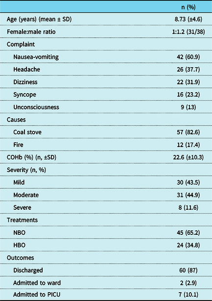
A total of 69 patients and 60 healthy controls were included in the study. The female-to-male ratio was 1:1.2 (31:38) and 1:1.1 (28/32) in the carbon monoxide poisoning patient group and the healthy control group, respectively. The mean age was 8.7 (SD±4.6) and 9.7 (SD±5.6) years in the carbon monoxide poisoning patient group and the healthy control group, respectively (Table 1). The causes of carbon monoxide poisoning were coal stove (57/69) and fire exposure (12/69).
Demographics, clinical characteristics, treatments, and outcomes of CO poisoning patients in the ED

SD=standard deviation; COHb=carboxyhaemoglobin; NBO=normobaric oxygen; HBO=hyperbaric oxygen; PICU=paediatric ICU.
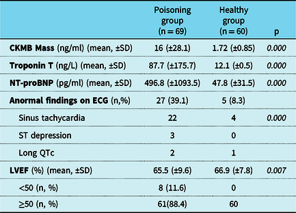
The carbon monoxide toxicity rate was found to be 43.5% mild, 44.9% moderate, and 11.6% severe. Nausea-vomiting (60.9%), headache (37.7%), dizziness (31.9%), syncarbon monoxide poisoninge (23.2%), and unconsciousness (13%) were the most common presenting symptoms (Table 1). Although headache was more likely to be present in children older than 2 (p = 0.010), no significant difference was found between the remaining presenting symptoms when compared in terms of age groups. In ECG analysis, the majority of patients (60.1%) had normal sinus rhythm, 22 (31.9%) had sinus tachycardia, 3 (4.3%) had ST depression, and 2 (2.9%) had long QT. Only in eight (11.6%) patients, reduced left ventricular ejection fraction was found (<50%) (Table 2).
Comparison of cardiac biomarkers, ECG, and LVEF on TTE between the poisoning and healthy group

ECG=electrocardiography; LVEF=left ventricular ejection fraction; SD=standard deviation; TTE=transthoracic echocardiography.
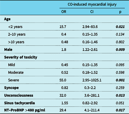
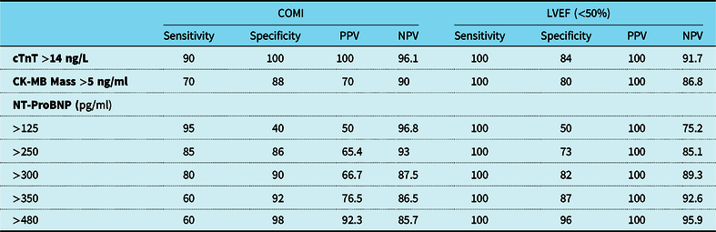
Carbon monoxide-induced myocardial injury was identified in 19 (27.5%) patients (5 had ischaemic ECG findings, 13 had high troponin T levels, and 8 had decreased transthoracic echocardiography). Multivariate logistic regression analysis was conducted to find the predictors of cardiotoxicity and revealed that male gender, younger age, COHb level (severe group), and altered mental status were found as independent predictors of carbon monoxide-induced myocardial injury (Table 3). The mean levels of COHb, CKMB-M, Troponin-T, and plasma NT-proBNP were 22.6%, 16 ng/ml, 87.7 ng/L, and 496.8 pg/ml in carbon monoxide poisoning patients, respectively (Table 2). The NT-proBNP, CKMB-M, Troponin-T, and COHb levels were significantly higher in the carbon monoxide poisoning group when compared to the healthy control group (p = 0.007, p = 0.000, p = 0.000, p = 0.000, respectively) (Table 2). NT-proBNP, CKMB-M, Troponin-T, and transthoracic echocardiography sensitivities, specificities, positive predictive values, and negative predictive values on carbon monoxide-induced myocardial injury are shown in Table 4. If the cut-off value for plasma NT-proBNP level is higher than 480 pg/ml, the sensitivity and specificity for the level of decreased left ventricular ejection fraction (<50%), which is the strongest carbon monoxide-induced myocardial injury sign, were 100 and 96%, respectively (Table 4).
Predictors of myocardial injury among poisoning children in paediatric ED

Binary logistic regression analysis and multivariate logistic regression analysis.
CI=confidence interval; OR=Odds ratio.
Sensitivity, specificity, positive predictive values (PPV), and negative predictive values (NPV) of cardiac biomarkers in COMI and LVEF

CK-MB=creatine kinase-myocardial band, COMI=CO-induced Myocardial injury, cTnT=Troponin T, LVEF=left ventricular ejection fractione, NPV=negative predictive value, NT-ProBNP=N-terminal pro-Brain natriuretic peptide; PPV=positive predictive value.
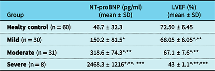
The levels of left ventricular ejection fraction were significantly decreased in the moderate and the severe groups when compared with the mild group; however, the levels of NT-proBNP were higher (p < 0.01, p < 0.01, both) (Table 5). A high negative correlation was found between NT-proBNP levels and left ventricular ejection fraction (r = −0.769, p < 0.01) in carbon monoxide poisoning group, and there was a positive correlation between the COHb and NT-proBNP levels (r = 0.583, p < 0.01).
The level of NT-proBNP and LVEF in the CO poisoning group and healty control group

LVEF=left ventricular ejection fractione; NT-ProBNP=N-terminal pro-Brain natriuretic peptide; SD=standard deviation. *p < 0.01 versus control group, **p < 0.01 versus mild group, ***p < 0.01 versus moderate group.
The most preferred treatment modality was normobaric oxygen which is administered by non-rebreathing mask (65.2%) in an emergency department and 24 children received hyperbaric oxygen. The mean follow-up time was 5.1 (SD±2.1) hours in the emergency department. In all, most of the patients (60/69) were discharged from the emergency department and only 9 patients were admitted to hospital (7 to a paediatric ICU and 2 to a ward).
Discussion
Although the major morbidity and mortality are related to the cardiotoxicity in carbon monoxide poisoning, the early detection of cardiac damage is frequently difficult. Since cardiotoxicity due to carbon monoxide poisoning is symptom free, it is often underdiagnosed. Recent adult studies showed that carbon monoxide-induced myocardial injury developed in one-third of all cases.Reference Satran, Henry, Adkinson, Nicholson, Bracha and Henry16,Reference Cha, Cha, Kim, Lee, Hwang and Kim17 Our results are consistent with recent investigations demonstrating that the carbon monoxide-induced myocardial injury occurrence rate was 27%. We did not include the CKMB-M criteria when defining carbon monoxide-induced myocardial injury; therefore, our carbon monoxide-induced myocardial injury rate may be lower than that of Satran et al, but higher than Cha et al.Reference Satran, Henry, Adkinson, Nicholson, Bracha and Henry16,Reference Cha, Cha, Kim, Lee, Hwang and Kim17
The pathophysiological mechanism of CO-induced myocardial injury results from tissue hypoxia and cellular level damage. The affinity of haemoglobin for CO is 200–250 times higher than oxygen. A shift in the oxygen–haemoglobin cleavage curve, a competitive inhibition of oxygen release with low oxygen distribution, and associated tissue hypoxia occurs due to CO–Hb complex.Reference Ernst and Zibrak5 CO binds to the cytochrome-c oxidase of the electron transport chain. As a result, the formation of developing oxygen radicals and lipid peroxidation causes cell death. In addition, high CO concentrations have been shown in the literature to induce nitric oxide-mediated cellular apoptosis.Reference Tibbles and Edelsberg18,Reference Thom, Fisher and Xu19
The predictors of carbon monoxide-induced myocardial injury were reported as male gender, altered mental status and duration of exposure in adults, a lower Glasgow coma score (≤14), and hypotension in children.Reference Satran, Henry, Adkinson, Nicholson, Bracha and Henry16,Reference Cha, Cha, Kim, Lee, Hwang and Kim17 On the other hand, many studies found no significant difference between developing carbon monoxide-induced myocardial injury and sex, age, or COHb level.Reference Cha, Cha, Kim, Lee, Hwang and Kim17,Reference Teksam, Gumus, Bayrakci, Erdogan and Kale20 In the current study, male gender, younger age, COHb level (severe group), and altered mental status were found as independent predictors of carbon monoxide-induced myocardial injury. ECG changes were observed in a minority of our patients when we exclude sinus tachycardia, but in the literature, ECG alterations in carbon monoxide poisoning were reported to be non-specific.Reference Teksam, Gumus, Bayrakci, Erdogan and Kale20,Reference Sonmez, Gunes, Saritas and Kandis21 Although sinus tachycardia is the most common finding in children, it is not reported as a sign of carbon monoxide-induced myocardial injury.Reference Teksam, Gumus, Bayrakci, Erdogan and Kale20,Reference Sonmez, Gunes, Saritas and Kandis21 Similarly, we have seen that 31.9% of our cohort had sinus tachycardia, with normal markers and transthoracic echocardiography. Although the literature showed that sinus tachycardia should not be used as a carbon monoxide-induced myocardial injury predictor, we believe that well-designed studies with different methodology such as speckle tracking echocardiography with new biomarkers are needed to demonstrate whether it is innocent or not.Reference Cha, Cha, Kim, Lee, Hwang and Kim17,Reference Teksam, Gumus, Bayrakci, Erdogan and Kale20,Reference Aslan, Uzkeser and Seven22
Carbon monoxide-induced myocardial injury is caused by tissue hypoxia and damage at the myocardial cell level.Reference Henry, Satran, Lindgren, Adkinson, Nicholson and Henry23 The early detection of carbon monoxide-induced myocardial injury may influence the method of top management modality (normobaric oxygen versus hyperbaric oxygen), and if hyperbaric oxygen is administered within 6 hours, the greatest benefit is provided.Reference Bhutani and Vishwanath13,Reference Eichhorn, Thudium and Jüttner24 There have been a few studies performed to demonstrate the positive contribution of ECG and routine cardiac markers (CK, CKMB) in the diagnosis of carbon monoxide-induced myocardial injury; however, findings on whether it is clinically useful are still uncertain.Reference Cha, Cha, Kim, Lee, Hwang and Kim17,Reference Huysal, Ustundag Budak, Aydin, Demirci, Turk and Karadag25-Reference Secilmis and Ozturk28 The total CK, Troponin-T/I, CK-MB level begin to rise 4–6 hours after the onset of the myocardial damage which is the most crucial time period for patients with carbon monoxide poisoning.Reference Teksam, Gumus, Bayrakci, Erdogan and Kale20,Reference Cardiga, Proença and Carvalho26,Reference Smiseth, Torp, Opdahl, Haugaa and Urheim27 This may be the best explanation for not using the conventional tests since good time management with earlier detection of carbon monoxide-induced myocardial injury improves the outcome.
Transthoracic echocardiography can be used to assess left ventricular ejection fraction, even in an emergency department setting. It has been used to document and monitor all the cases with myocardial damage. However, its routine use is limited due to its non-availability. There are many reports to date showing that the main sign of carbon monoxide-induced myocardial injury in echocardiography is decreased ejection fraction in acute carbon monoxide poisoning.Reference Saraçoğlu, Vuruşkan and Kılıç29,Reference Lee, Kim and Park30 In our study, left ventricular ejection fraction was reduced (<50%) in only eight patients; however, conventional cardiac markers were elevated in more patients. Discrepancies between biochemical markers and transthoracic echocardiography can be explained by a recent study conducted by Saraçoğlu et al.Reference Saraçoğlu, Vuruşkan and Kılıç29 This study found that conventional echocardiography may be insufficient for demonstrating mildly impaired left ventricular systolic function in adult carbon monoxide poisoning patients, and speckle tracking echocardiography has the potential of demonstrating subtle left ventricular systolic dysfunction.Reference Saraçoğlu, Vuruşkan and Kılıç29 They also stated that almost all individuals had left ventricular systolic dysfunction when they were evaluated by speckle tracking echocardiography. Since the basal metabolic rate and tissue oxygen demand are higher in infants and young children, myocardial cells may be more affected in these groups when carbon monoxide poisoning develops. We believe that if similar methodology such as speckle tracking echocardiography is performed in paediatric carbon monoxide poisoning, it will reveal the real rates and types of cardiac effects.
NT-proBNP levels were found to be significantly higher in carbon monoxide poisoning and its use may be recommended as a biomarker for early diagnosis of carbon monoxide-induced myocardial injury.Reference Satran, Henry, Adkinson, Nicholson, Bracha and Henry16,Reference Liu, Shen and Lv31 Also, previously published studies showed that troponin I levels increased at the 6th hour and peaked at the 12th hour in CO poisoning, while BNP levels increased in the 1st hour and reached the peak level at the 6th hour.Reference Yucel, Avsarogullari and Durukan33 In the early period of CO poisoning, although the troponin values were within normal range, NT-ProBNP levels were increased.Reference Moon, Chun, Shin and Lee34
This study is the first prospective paediatric study demonstrating the positive correlation between NT-pro-BNP and COHb and a negative correlation between NT-pro-BNP and left ventricular ejection fraction. Similar to our results, previous studies have shown that ECG and transthoracic echocardiography can be normal in acute carbon monoxide poisoning; however, increased NT-proBNP levels may be seen.Reference Davutoglu, Gunay and Kocoglu7 Mir et al have reported that the NT-proBNP levels in control children, adolescents, and adults did not show a significant age-related difference.Reference Mir, Flato and Falkenberg32 The mean plasma NT-proBNP in our healthy children was 47.8 pg/ml. Studies have shown that if the level of NT pro-BNP was higher than 100 pg/ml, then the sensitivity and specificity were 80 and 68%, respectively. Also, a positive correlation was found between NT-ProBNP and COHb levels.Reference Davutoglu, Gunay and Kocoglu7,Reference Liu, Shen and Lv31 Only 42.1% of carbon monoxide-induced myocardial injury patients in our group had abnormal transthoracic echocardiography findings, and NT-ProBNP levels were significantly increased in carbon monoxide-induced myocardial injury, and if NT-ProBNP >250 pg/ml, the sensitivity and specificity were 85 and 86% for carbon monoxide-induced myocardial injury. However, if the cut-off value for the NT-ProBNP level is set as >480 pg/ml, then the sensitivity and specificity were found as 100 and 98% on reduced left ventricular ejection fraction. We strongly believe that this is the most significant result of our study demonstrating that the early cardiotoxic effect of carbon monoxide can be predicted (correlated with reduced ejection fraction) by NT-ProBNP level, and therefore, the benefit will be greatest since hyperbaric oxygen treatment will be administered.
The current study has several limitations. The first limitation is paediatric emergency department condition such as crowded and low-experienced medical staffs during night shifts. These conditions induced misdiagnosed and disrupted patients’ evaluation. This report consists of single-centre experience and small sample patients group, and these are the second limitation. In most common fire situations, a few combustion gases other than CO and CO2 such as hydrogen cyanide (HCN), hydrogen chloride (HCl), nitrogen dioxide (NO2), and other oxides of nitrogen would be expected to act as contributory to mortality and morbidity. Since these gases detection is not easy and routinely performed in our laboratory, we cannot rule out patients’ exposure to these toxic gases.
Conclusion
Myocardial damage may develop in paediatric acute carbon monoxide poisoning despite normal ECG, standard markers, and transthoracic echocardiography findings. Male gender, younger children (<2 years), higher COHb (>40%), altered mental status, and increased NT-proBNP >480 pg/ml were predictors of carbon monoxide-induced myocardial injury. An increased level of NT-proBNP (>480 pg/ml) may be useful as an ideal biomarker for the early detection of carbon monoxide-induced myocardial injury and reduced left ventricular ejection fraction.
Acknowledgements
The authors would like to express their sincere gratitude to all members who worked together to provide technical and writing assistance, and to the departmental heads for their general support.
Financial support
This research received no specific grant from any funding agency, commercial or not-for-profit sectors.
Conflict of interest
None.
Ethical standards
The authors assert that all procedures contributing to this work comply with the ethical standards of the relevant national guidelines on human experimentation and with the Helsinki Declaration of 1975, as revised in 2008, and has been approved by the institutional committees (Ege University 18/5-38).