INTRODUCTION
Various pathogens of the intestinal tract, and their products, have been shown to interfere with the different pathways of intestinal absorption, often resulting in diarrhoea and other related symptoms. For example, the zonula occludens toxin produced by Vibrio cholerae modulates paracellular absorption in vitro and in vivo (Salama et al. Reference Salama, Fasano, Lu and Eddington2003, Reference Salama, Fasano, Thakar and Eddington 2004 ), while rotavirus infections in mice can induce increased transcellular absorption of macromolecules such as horse radish peroxidase (HRP). The same pathogens/toxins can interfere with normal intestinal ion transport.
The intestinal parasite, Giardia duodenalis, is an important pathogen worldwide and infects a range of mammals, including humans. Symptoms of giardiasis include diarrhoea, nausea, headache, flatulence, stomach cramps and weight loss. In most cases the disease is self-limiting or easily treated. However, the parasite can also cause more chronic disease, including retarded growth in children and in experimental animal models (Al-Mekhlafi et al. Reference Al-Mekhlafi, Al-Maktari, Jani, Ahmed, Anuar, Moktar, Mahdy, Lim, Mahmud and Surin2013; Bartelt et al. Reference Bartelt, Roche, Kolling, Bolick, Noronha, Naylor, Hoffman, Warren, Singer and Guerrant2013), different nutritional deficiencies (Astiazaran-Garcia et al. Reference Astiazaran-Garcia, Lopez-Teros, Valencia, Vazquez-Ortiz, Sotelo-Cruz and Quihui-Cota2010; Orden et al. Reference Orden, Apezteguia, Ciarmela, Molina, Pezzani, Rosa and Minvielle2014), development of inflammatory bowel syndrome (Hanevik et al. Reference Hanevik, Dizdar, Langeland and Hausken2009; Beatty et al. Reference Beatty, Bhargava and Buret2014) and chronic fatigue syndrome (Morch et al. Reference Morch, Hanevik, Rivenes, Bodtker, Naess, Stubhaug, Wensaas, Rortveit, Eide, Hausken and Langeland2013). Pathogenesis in giardiasis depends on both parasite and host factors. As recently reviewed by Cotton et al. (Reference Cotton, Beatty and Buret2011), these factors include mechanisms for attachment to epithelial cells and immune system evasion. During acute and chronic giardiasis, various changes are seen in the intestinal epithelium and surrounding tissue, the most important being increased epithelial cell apoptosis, loss of cell–cell contact and shortened microvilli (Maia-Brigagao et al. Reference Maia-Brigagao, Morgado-Diaz and De Souza2012; Fisher et al. Reference Fisher, Estrano and Cole2013; Koh et al. Reference Koh, Geurden, Paget, O'Handley, Steuart, Thompson and Buret2013). These changes lead to reduced intestinal barrier function, leakage of water and ions and malabsorption of nutrients. Despite increasing knowledge on the pathogenesis of giardiasis, there is still a lack of knowledge on disease mechanisms in giardiasis, and why and how different genotypes or isolates result in different symptoms and pathogenicity. In particular, we lack detailed knowledge on which transport mechanisms (transcellular or paracellular) are affected, and to what extent, by different Giardia isolates.
The Ussing chamber is a tool that can be used to measure the short-circuit current as an indicator of net ion transport taking place across an epithelium. Furthermore, using monolayers of cells grown on permeable supports, the Ussing chamber can be used to investigate changes in transcellular and paracellular transport using judiciously selected probes. For example, macromolecular permeability of the human colon to protein antigens has been studied using endoscopic biopsy specimens in an Ussing chamber and 51Cr-EDTA and HRP as permeability markers (Wallon et al. Reference Wallon, Braaf, Wolving, Olaison and Soderholm2005). In Madin-Darby canine kidney (MDCK) cells exposed to Giardia, epithelial resistance and macromolecular transport was increased (Chavez et al. Reference Chavez, Gonzalez-Mariscal, Cedillo-Rivera and Martinez-Palomo1995). More recently duodenal biopsy specimens from patients with chronic giardiasis and controls were investigated ex vivo in Ussing chambers measuring mannitol flux and electrophysiological parameters (Troeger et al. Reference Troeger, Epple, Schneider, Wahnschaffe, Ullrich, Burchard, Jelinek, Zeitz, Fromm and Schulzke2007). While mannitol flux was not significantly altered, epithelial resistance in the specimens from Giardia patients was decreased. Given these contrasting results, the intention of our study was to develop a user-friendly assay for measuring transcellular and paracellular permeability in Ussing chamber studies and use it to compare effects of different Giardia isolates on permeability of an intestinal cell line.
MATERIAL AND METHODS
Intestinal cells
The human colon carcinoma cell line, Caco-2 (purchased from ATCC), was chosen as the experimental model of intestinal epithelium. Caco-2 cells form fully differentiated and polarized enterocytes, displaying features similar to epithelial cells in the small intestines (Zweibaum et al. Reference Zweibaum, Laburthe, Grasset, Louvard, Schultz, Field, Frizzell and Rauner2011). Cells were grown in Dulbecco's Modified Eagle's medium (DMEM) high glucose, GlutaMAX™ (Life Technologies™) supplemented with 10% fetal bovine serum (FBS) and antibiotics. Medium was changed every 2nd day and cells sub-cultivated upon 80–90% confluence. Otherwise the protocol followed the work of Natoli et al. (Reference Natoli, Leoni, D'Agnano, Zucco and Felsani2012). Cells used in the Ussing chamber experiments were seeded onto 6 tray Snapwell 12 mm inserts (Corning Costar) with 1·12 cm2 cell growth area. To obtain complete differentiation into enterocytes, cells were cultivated for 21 days before use in the experiments.
Giardia duodenalis isolates
Two isolates of G. duodenalis were used in this study. The first isolate used is the well-characterized WB strain (Assemblage A), originally isolated from a human sample in 1979, since when it has been maintained in laboratory culture. It is the most studied G. duodenalis strain, and has been used in a wide range of in vitro and in vivo studies. The second isolate used, R-2, is a field strain also Assemblage A, originally isolated from a canine faecal sample and cryopreserved following successful establishment of culture (see Tynses et al. submitted manuscript)
Cultivation followed the protocol initially developed by Meyer and Pope (Reference Meyer and Pope1965). Trophozoites of both isolates were grown in standard TYI medium and sub-cultivated when tubes reached ~80% confluence, approximately every 3rd days. Trophozoites used in the experiments were harvested after a 48 h culture period (log phase) by placing tubes on ice for 30 min and gently vortexing to detach trophozoites from tube wall. Tubes were centrifuged at 2500 rpm for 10 min and the pellet washed twice with ice cold PBS. Trophozoites were then suspended in PBS in 1·5 mL tubes and adjusted to 1 × 10⁷ trophozoites mL−1, before they were sonicated 3 times for 30 s and stored at −70 °C before further use.
Giardia exposure
Cells grown on Snapwell inserts were exposed to Giardia sonicates in low glucose DMEM medium for 24 h, as described previously (Yu et al. Reference Yu, Huang, Kuo, Sayer, Turner and Buret2008), prior to Ussing chamber experiments. Old medium was removed and 0·5 mL of full low glucose DMEM medium, containing Giardia sonicates equivalent to 1 × 10⁶ trophozoites per mL, was added to the apical (mucosal) compartment of Snapwells.
Ussing chamber experiments
Epithelial transport and electrophysiology were measured in an Ussing Chamber (Physiologic Instruments, USA). The Ussing chamber consists of 2 liquid filled chambers that are separated for these experiments by polarized epithelial cells grown on Snapwells, such that one side is equivalent to the absorbing (mucosal) side of the intestine, and the other side, equivalent to the intestinal non-absorbing (serosal) side (Fig. 1).
Schematic view of the Ussing chamber: A piece of tissue is mounted between 2 fluid filled chambers. The chambers are perfused with gas and probes added to one of the chambers. Transcellular and paracellular transport can be measured by sampling the opposite chamber at given time points. Agar filled electrode tips are used to record transepithelial resistance and short circuit current.

The chambers were filled with 4 mL Kreb's buffer (Wallon et al. Reference Wallon, Braaf, Wolving, Olaison and Soderholm2005) and perfused with a 95% O2 and 5% CO2 gas mixture. Temperature was maintained at 37 °C by a circulating water bath. Gas perfusion serves 2 purposes; maintaining O2- and pH-level and appropriate circulation of selected probes. Transepithelial electrical resistance (TEER) and short-circuit current (Isc) were monitored with KCl agar electrode tips. Experiments were performed in open circuit conditions, with electrodes connected to a 4-channel voltage controlled current source. Cells were normalized for 20 min before buffers were changed, then, after an additional 20 min, the experiment was started.
TEER (Ω) and (Isc) were recorded using the Acquire and Analyze™ software from Physiologic Instruments. Stable resistance levels indicate good cell viability and monolayer integrity. When more than 20% change in resistance was observed during an experiment, these results were excluded. Results obtained were adjusted to cell growth area of the Transwells (1·12 cm2).
Apical to basal (mucosal to serosal) transportation was investigated using creatinine and HRP as paracellular and transcellular probes, respectively. Probes were added to the mucosal side of the cells in the Ussing chamber at the start of each experiment to a final concentration of 0·5 μ m HRP and 2·5 mm creatinine. Four 50 μL samples were taken in parallel every 30 min from the opposite side of the Ussing chamber (serosal side), for a total of 90 min giving a total of 16 samples per Snapwell. Basal to apical transportation was also tested using the same protocol as above, but with the probes added to the serosal chamber and samples taken from the mucosal chamber. Assays specifically developed for monitoring the 2 probes (see below) were used to measure transport across or through the cells.
For each isolate, 3 replicates were run for each probe and for each direction of transport. For each Snapwell of Caco-2 differentiated enterocytes exposed to Giardia trophozoite sonicate included in the Ussing chamber experiments, 3 Snapwells of control cells (not exposed to the Giardia trophozoite sonicate) were investigated for transcellular and paracellular transport.
Probes for measuring transcellular and paracellular transport
Two probes were selected for measuring transcellular and paracellular transport; HRP and creatinine respectively. HRP has previously been used as a probe in Ussing chamber studies (Heyman et al. Reference Heyman, Crain-Denoyelle, Nath and Desjeux1990; Wallon et al. Reference Wallon, Braaf, Wolving, Olaison and Soderholm2005) and concentrations can be measured by ELISA. For this assay, 50 μL samples from each side of the Ussing chamber were mixed with 50 μL of the chromogenic substrate 3,3′,5,5′-tetramethylbenzidine (TMB substrate, Pierce, USA) for 30 min. The reaction was stopped with 50 μL 1 m HCl before measuring the absorbance at 640 nm.
An ELISA was also developed in order to quantify creatinine concentrations, based on the previous work of Fossati et al. (Reference Fossati, Prencipe and Berti1983). In brief, 50 μL samples were incubated for 30 min with 150 μL of an enzymatic mixture containing creatinase, creatininase and sarcosine oxidase (Afinion ACR, Axis Shield, Norway). The enzymatic assay for creatinine involves a series of coupled enzymatic reactions including enzymatic conversion of creatinine by creatininase into creatine that, in turn, is converted to sarcosine by creatine amidinohydrolase (creatinase). This is followed by oxidation of sarcosine by sarcosine oxidase in the presence of oxygen to glycine, formaldehyde and hydrogen peroxide. The liberated hydrogen peroxide reacts with 4-aminophenazone and hydroxy(tosyloxy)iodo-benzene (HTIB) to form a quinone imine chromogen in a reaction catalysed by peroxidase. The colour intensity is directly proportional to the concentration of creatinine present and can be measured photometrically at 540 nm.
For both ELISA-based assays, reactions were performed in 96 flat bottom wells and read in a Multiskan FC (Thermo Scientific) plate-reader.
Assays were developed prior to commencement of the Ussing chamber experiments, and standard curves, with concentrations ranging between 3·125 and 100 pm for HRP and 6·25 and 200 pm for creatinine, run with each experimental set up.
Statistics
The data were analysed with GraphPad Prism 6. Distributions from the separate experiments were non-parametric and comparisons were made using one way ANOVA and tested with Kruskal–Wallis test. P < 0·05 is considered to be significant.
RESULTS
Assay development
The precision of the ELISA protocols developed were determined by calculating average intra-assay coefficient of variability (CV) from mean standard deviations from all experiments with the mean of the measurements, which was 2% for the creatinine assay and 5% for the HRP assay (at 90 min). Inter-assay CV, based on average CV for low (30 min) and high (90 min) measurements, was 2% for creatinine and 4·7% for HRP.
Electrophysiology
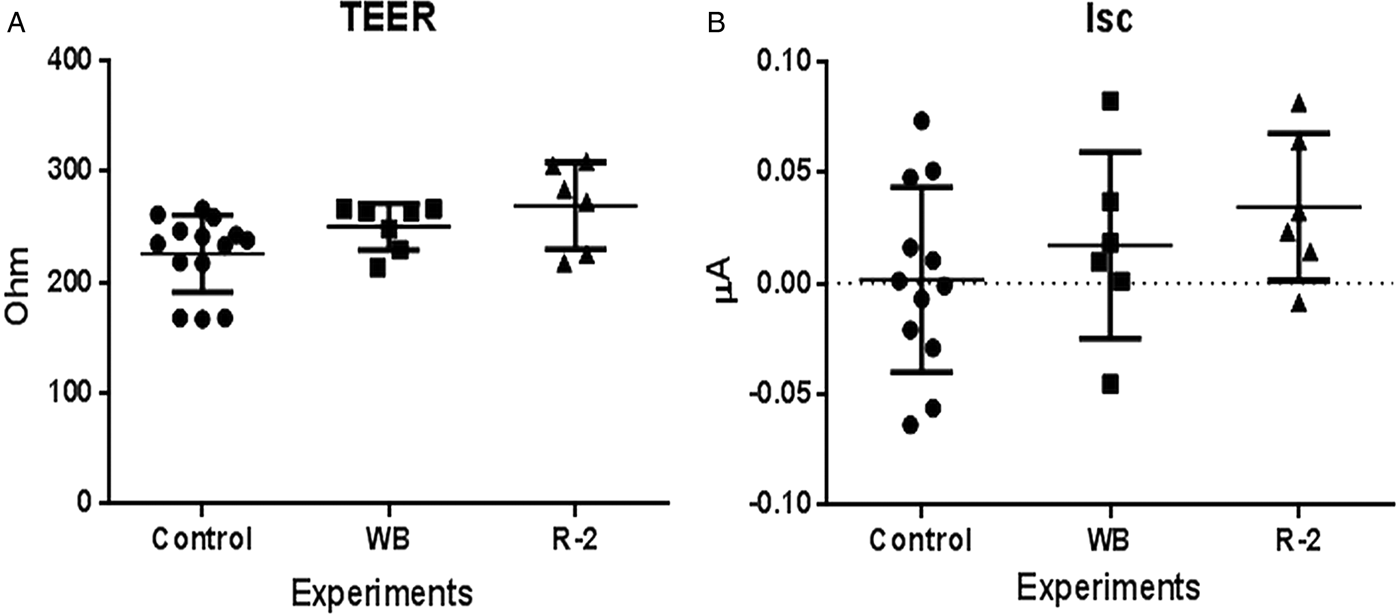
Both Giardia strains resulted in changes in Isc and TEER 1 in the enterocytes. The differences in TEER between control cells and cells exposed to WB and R-2 trophozoite sonicates were 4·4 and 12%, respectively. Exposure of the cells to trophozoite sonicates increased Isc. However, while the increase was only 57% when the cells had been exposed to the WB sonicate, exposure to the R-2 sonicate resulted in a 224% increase. When comparing controls with exposed groups, or R-2 with WB exposed, none of these parameters were statistically significant (Fig. 2).
Comparison of transepithelial electrical resistance (TEER, A) and short circuit current (Isc, B) in the different experimental groups. Both Giardia isolates induced increased TEER and Isc in Caco-2 cells

Transcellular transport
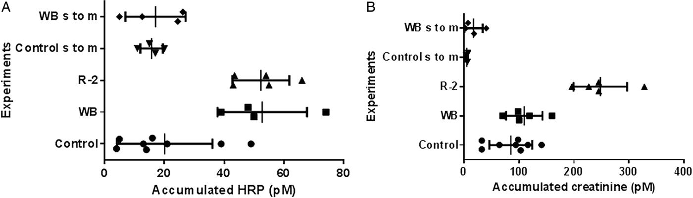
Exposure of the enterocytes to sonicated Giardia trophozoites significantly increased mucosal to serosal transcellular transport of HRP (P = 0·0054), but with no apparent difference between the 2 isolates tested (Fig. 3A). However, transcellular transport in the opposite direction (serosal to mucosal) was only investigated for the WB strain, and no difference from transport in the control cells was observed.
Accumulated HRP (A) and creatinine (B) on the serosal side of the cells after 90 min incubation in the Ussing chamber. Serosal to mucosal (s to m) transport was tested for control and WB-sonicate exposed cells).

Paracellular transport
Exposure of the enterocytes to sonicates of Giardia trophozoites of the WB isolate had no apparent effect on paracellular transport of creatinine in either direction (mucosal to serosal and serosal to mucosal) (Fig. 3). However, cells exposed to trophozoite sonicates of the R-2 strain demonstrated a significant increase in paracellular transport (P = 0·0003), in the mucosal to serosal direction, with more than double the amount of creatinine measured following transportation (Fig. 3B).
DISCUSSION
In this manuscript we describe a user-friendly method for measuring transcellular and paracellular transport across enterocytes in an Ussing chamber set up using 2 probes that can be readily measured by the ELISA method described, and with good reproducibility. We believe that this system could be useful for further investigations, looking at other factors, including microbiome interactions, that may affect the pathophysiology associated with Giardia infection.
These assays, along with electrophysiological measurements, were used to investigate possible effects of exposure to Giardia trophozoite sonicates on transcellular and paracellular transport in an enterocyte cell line. Sonicated Giardia products were selected for these experiments rather than whole live trophozoites in order to avoid the possibility of transport being affected due to physical blocking by the attached trophozoites.
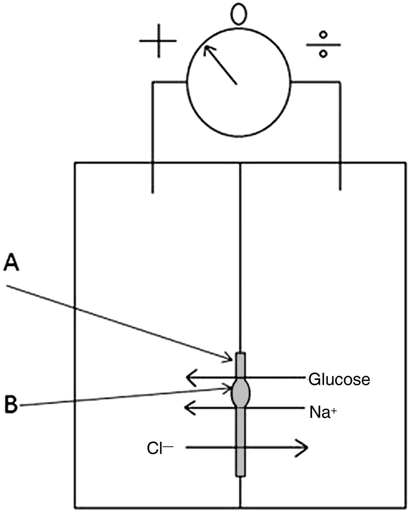
Alterations in intestinal ion secretion during giardiasis have previously been reported in experimental animals and ex vivo in biopsies from human patients (Gorowara et al. Reference Gorowara, Ganguly, Mahajan and Walia1992; Cevallos et al. Reference Cevallos, Carnaby, James and Farthing1995; Troeger et al. Reference Troeger, Epple, Schneider, Wahnschaffe, Ullrich, Burchard, Jelinek, Zeitz, Fromm and Schulzke 2007 ). During the acute phase of disease, mice infected by Giardia showed a net trans-mucosal secretion of both Cl− and Na+ (Gorowara et al. Reference Gorowara, Ganguly, Mahajan and Walia1992). Similarly, in duodenal biopsies from humans suffering chronic giardiasis a net reverse flux of Cl− was shown (Troeger et al. Reference Troeger, Epple, Schneider, Wahnschaffe, Ullrich, Burchard, Jelinek, Zeitz, Fromm and Schulzke2007). This has been proposed as a mechanism causing secretory diarrhoea during giardiasis. Secretory diarrhoea, in which a pathogen, toxins or other external stimuli, triggers chloride ion secretion resulting in osmotic water excretion and diarrhoea, is a theory mostly based on flux experiments using chloride ion isotopes, and models assuming unidirectional passage of chloride ions (Lucas, Reference Lucas2005). However, some evidence suggests that chloride carriers having bidirectional functions, enabling them to transport chloride anions in both directions (Lucas, Reference Lucas2005). Our results show that exposure of cells to Giardia trophozoite sonicates increases Isc in enterocytes in vitro. Although we cannot use these results to determine the specific mechanisms underlying the increase in Isc, alterations of chloride flux may be one of the possible explanations (Fig. 4).
The increased short circuit current induced by exposure to Giardia trophozoite sonicate may be explained by increased chloride ion secretion, and/or increased sodium ion uptake via sodium glucose transporters (SGLT-1). A = layer of epithelial cells and B = SGLT transporter.

This study also shows that Giardia sonicates may increase TEER in vitro. These findings concur with those from in vitro experiments on MDCK performed by Chavez et al.; lab strains of Giardia, Portland-1 and WB, did not alter TEER (1986), while various field strains, isolated from human faecal samples, did increase TEER (1995). Humen et al. (Reference Humen, Perez and Lievin-Le Moal2011) found reduced TEER in epithelial cells stimulated with live trophozoites, and, by pre-treating trophozoites with methyl-b-cyclodextrin, showed that this effect on TEER was dependent on adhesion. Others have compared other Giardia isolates (S2 and NF) with respect to effect on TEER in in vitro grown epithelial cells, and found that that both live and sonicated trophozoites reduced resistance in Caco-2 cells, but without significant difference between the Giardia isolates tested (Teoh et al. Reference Teoh, Kamieniecki, Pang and Buret2000). In another study reduced TEER was found in ex vivo Ussing chamber experiments on duodenal samples from patients suffering from chronic giardiasis (Troeger et al. Reference Troeger, Epple, Schneider, Wahnschaffe, Ullrich, Burchard, Jelinek, Zeitz, Fromm and Schulzke2007). Chronic enteropathies, such as inflammatory bowel syndrome, have been shown to follow in certain cases of giardiasis (Hanevik et al. Reference Hanevik, Dizdar, Langeland and Hausken2009) and such post infectious complications are likely to be multifactorial. Reduced TEER, as observed in chronic giardiasis, could be the result of factors related to bacterial dysbiosis. Several probiotic bacteria have been shown to increase TEER of intestinal epithelial cells (Ramos et al. Reference Ramos, Thorsen, Schwan and Jespersen2013), probably by inducing increased expression of tight and adherence junctions between the cells (Anderson et al. Reference Anderson, Cookson, McNabb, Park, McCann, Kelly and Roy2010). Increased TEER in cells exposed to Giardia products indicate that the bonds between the cells are getting stronger, hence reducing the likelihood of other pathogens or harmful antigens of passing the epithelial barrier. This can provide a possible explanation of why infection with Giardia apparently has protective properties for the host in some situations (Bilenko et al. Reference Bilenko, Levy, Dagan, Deckelbaum, El-On and Fraser2004; Kotloff et al. Reference Kotloff, Nataro, Blackwelder, Nasrin, Farag, Panchalingam, Wu, Sow, Sur, Breiman, Faruque, Zaidi, Saha, Alonso, Tamboura, Sanogo, Onwuchekwa, Manna, Ramamurthy, Kanungo, Ochieng, Omore, Oundo, Hossain, Das, Ahmed, Qureshi, Quadri, Adegbola and Antonio2013; Muhsen et al. Reference Muhsen, Cohen and Levine2014).
Transcellular transport across the intestine during giardiasis has previously been investigated in mice (Mahmood et al. Reference Mahmood, Ganguly, Mahajan and Walia1990) using radiolabelled albumin, and the results suggested that Giardia infection increased macromolecular absorption, but this seemed unrelated to surface-bound receptors. More recently Chen et al. (Reference Chen, Chen, Wu, Lee, Lu, Wu, Ni, Sun, Yu, Buret and Yu2013) investigated post infectious effect of giardiasis in mice. In this study, transepithelial transport of Fluorescein isothiocyanate-dextran and translocation of commensal bacteria was increased. Similarly, in an in vivo study of patients with giardiasis, macromolecular uptake was increased (Troeger et al. Reference Troeger, Epple, Schneider, Wahnschaffe, Ullrich, Burchard, Jelinek, Zeitz, Fromm and Schulzke2007). In accordance with our study, using HRP as an in vitro probe also indicates an increase in transcellular transport in vitro. According to Heyman et al. (Reference Heyman, Crain-Denoyelle, Nath and Desjeux1990), HRP is transported across Caco-2 cell monolayers either in a receptor-mediated fashion or by non-specific transcytosis. Furthermore Heyman et al. (Reference Heyman, Crain-Denoyelle, Nath and Desjeux1990) state that only 10% of this HRP is passaged un-degraded. In their experiments, which involved investigation of mucosal permeability in guinea pigs, undegraded flux of HRP did not seem to be affected by temperature, and thus the authors propose that undegraded HRP passage is due to extracellular leakage. In our experiments only un-degraded HRP was measured, however the flux measured here was higher when compared with the results of Heyman et al. (Reference Heyman, Crain-Denoyelle, Nath and Desjeux1990).
While the results of our study demonstrate that different Giardia isolates can have differing effects on epithelial cells in vitro, it is interesting to speculate whether these findings can be related to the in vivo situation. Similar experiments using sonicates of other diplomonads (e.g. Hexamita or Spironucleus) could have provided further information regarding whether specific effects are genus-specific.
We suggest that these results add further information for helping to explain several phenomena reported from other Giardia studies. Disruption and or reduced expression of tight and adherence junctional proteins may be one of the mechanisms behind increased paracellular transport of creatinine. However, these results contrast with our finding that sonicated isolates resulted in increased TEER. Sonicated Giardia trophozoites have been shown to induce an increase in the apical distribution of sodium glucose transporters 1 (SGLT-1), and this was proposed by the authors to be a possible defence mechanism against apoptosis during giardiasis (Yu et al. Reference Yu, Huang, Kuo, Sayer, Turner and Buret2008). Considering increased intestinal paracellular uptake of creatinine is induced by SGLT-1 glucose transport (Pappenheimer, Reference Pappenheimer1990; Turner et al. Reference Turner, Cohen, Mrsny and Madara2000), this may provide one explanation for the increased creatinine uptake seen in our study. Given that intracellular transport and apical distribution of both SGLT-1 and apical junction proteins have been linked to microtubules (Kipp et al. Reference Kipp, Khoursandi, Scharlau and Kinne 2003 ; Suzuki et al. Reference Suzuki, Matsuzaki, Hagiwara, Aoki, Tajika-Takahashi and Takata2006; Glotfelty et al. Reference Glotfelty, Zahs, Iancu, Shen and Hecht2014), a possible explanation for our somewhat contradictory results regarding TEER and paracellular uptake could be that exposure to Giardia trophozoite sonicates also results in non-specific changes in cellular transport mechanisms. SGLT-1 is only located on the apical side of enterocytes, this provides an explanation as to why serosal to mucosal creatinine flux was lower than in the opposite direction.
The differences in effect between WB and R-2 strains on paracellular transport seen in this study could be explained, in part, by a form of in vitro culture adaptation. Comparisons between a lab-adapted strain and a field strain suggest this is an important factor. The WB strain has been used in experiments since 1979, and may have become attenuated over time. Attenuation is well known effect of axenic culture for other parasites, such as Leishmania (Moreira et al. Reference Moreira, Vitoriano-Souza, Roatt, Vieira, Ker, de Oliveira Cardoso, Giunchetti, Carneiro, de Lana and Reis2012; Magalhaes et al. Reference Magalhaes, Duarte, Mattos, Martins, Lage, Chavez-Fumagalli, Lage, Menezes-Souza, Regis, Manso Alves, Soto, Tavares, Nagen and Coelho2014), Histomonas meleagridis (Hess et al. Reference Hess, Liebhart, Grabensteiner and Singh2008) and Neospora caninum (Bartley et al. Reference Bartley, Wright, Sales, Chianini, Buxton and Innes2006). This should be considered when in vitro studies with culture-attenuated strains, such as WB, produce no apparent effects on the parameters investigated. In such instances it would seem wise not to extrapolate to all isolates, but bear in mind that field isolates may produce differing results.
ACKNOWLEDGEMENTS
We would like to thank the Paul Cos laboratory at the University of Antwerp for advice on how to culture Giardia, and Johan Sörderholm's laboratory at the University of Linköping for directions on the Ussing chamber technique. Andrew Campbell provided important expertise about ELISA, and without him the protocol for creatinine would never have seen the light of day.
FINANCIAL SUPPORT
This study was funded by an internal grant at the Norwegian University of Life Sciences and by Astrid and Birger Torsted's legate.