Aflatoxins (AF) are food contaminants, affecting up to 25 % of the world’s food supply(Reference Gönenç, Yilmaz Sezer and Yilmaz1). A considerable portion of the world’s population, about 4·5 billion individuals, mostly in emerging nations where food systems from harvesting to delivery in the market are not properly controlled, is at risk of chronic AF exposure via consuming contaminated food such as maize and groundnut, which are considered high-risk foods(Reference Smith, Prendergast and Turner2,Reference Wild, Miller and Groopman3) . AF are hazardous secondary metabolites produced by Aspergillus moulds that contaminate foods like maize, rice and legumes(Reference Wild, Miller and Groopman3). There are at least twenty distinct types of AF found(Reference Popescu, Rădulescu and Georgescu4)of which aflatoxin B1 (AFB1), aflatoxin B2 (AFB2), aflatoxin G1 (AFG1) and aflatoxin G2 (AFG2) are the four most known AF, with AFB1 being the most hazardous or toxic and potent(Reference Keskin, Başkaya and Karsli5,Reference Omer, Verhoef and Van’t Veer6) , followed by AFG1, AFB2 and AFG2, respectively. According to the International Agency for Research on Cancer(7), AFB1 is classified as a group 1 carcinogen. The toxicity of AFB1 is often explained by the epoxidation of the 8, 9-double bond and higher efficiency of association with the cyclopentenone ring of the series B as opposed to the six-membered ring of the series G(Reference Ismail, Gonçalves and de Neeff8). The names of AF in the B and G groups are based on whether they are fluorescent in blue or green when exposed to UV light, respectively, whereas the names of AF in the M group are based on whether they are present in milk and milk products(Reference Ismail, Gonçalves and de Neeff8). AFB1 is metabolised in the liver by P450 enzymes in living organisms to form AFB1-exo 8, 9-epoxide. This epoxide reacts with DNA and serum albumin to form guanine adduct (AFB1-N7 guanine adduct) and AFB1-lysine adduct (AFB1-lys), respectively(Reference Essigmann, Croy and Nadzan9,Reference Sabbioni, Skipper and Büchi10) . However, AFB1-guanine adduct has a half-life of 2–3 d and is excreted in the urine, whereas the half-life of AFB1-lysine adduct is 2–3 months in the human serum(Reference Shilong, Muhammad and Yu11). Aflatoxin M1 (AFM1) and aflatoxin M2 (AFM2) are hydroxylated by-products of AFB1 and AFB2 that are excreted by humans and animals exposed to dietary AFB1 and AFB2 in their milk and/or urine. AFM1 is identified in milk 12–24 h after consumption of AF contaminated food and poses a health risk to infants who are breastfed(Reference Galvano, Pietri and Bertuzzi12,Reference Antonius, Wagenaar and Karnekamp13) .
Pregnancy is a crucial physiological state where nutrition plays an important role. During this period, pregnant women are more susceptible to many infections because their immune system is naturally suppressed. Pregnant women are exposed to many environmental threats like pollution, microbial infection and harmful chemical exposure like pesticides, heavy metals and AF. AF exposure is one of the major environmental risks to which pregnant women and developing infants are vulnerable(Reference Smith, Prendergast and Turner2). During this period, the growing fetus might also be affected by the AF-exposed mother.
Prevalence of AF exposure during pregnancy ranged from 6 to 100 %(Reference Smith, Prendergast and Turner2), which suggests that exposure to AF in utero is common where the mother’s diet is contaminated with AF(Reference Groopman, Egner and Schulze14,Reference Obade, Andang’o and Obonyo15) . AF exposure during pregnancy is harmful to the mother and also may have an impact on the health of the fetus directly during crucial periods of development or indirectly through its negative effects on the health of the mother(Reference Smith, Prendergast and Turner2,Reference Obade, Andang’o and Obonyo15) . Therefore, this review is conducted for a better perspective on the AF exposure during pregnancy or infancy and its effects on pregnancy outcomes and infant health. We also calculated the EDI (estimated daily intake) using the empirical formulae based on several reported studies on AF contamination in foods.
Method
This narrative review was conducted by searching relevant literature published in peer-reviewed journals using databases such as PubMed, Google Scholar, Scopus and ScienceDirect. Keywords used included: ‘aflatoxin’, ‘aflatoxin exposure’, ‘pregnancy outcomes’, ‘fetus’, ‘infant’, ‘birth outcomes ’, ‘maternal health ’, ‘infant growth’ and ‘toxicology’. Inclusion criteria: Studies reporting on AF exposure in pregnant women, its transfer to fetus/infant, biomarkers (e.g. AFB1-lysine adducts) and associated health outcomes in fetus, infants or pregnant women. Exclusion criteria: Non-English papers, conference abstracts and studies not directly assessing AF exposure or its effects during pregnancy. Literature was selected from 2000 to 2024, although earlier studies were also included if relevant. Although a formal systematic review protocol was not followed, every effort was made to ensure a comprehensive and representative selection of relevant studies.
Aflatoxin exposure in pregnant women and infants
Maternal aflatoxin exposure
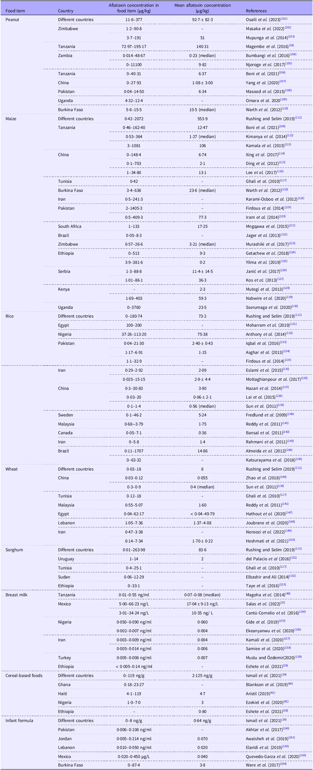
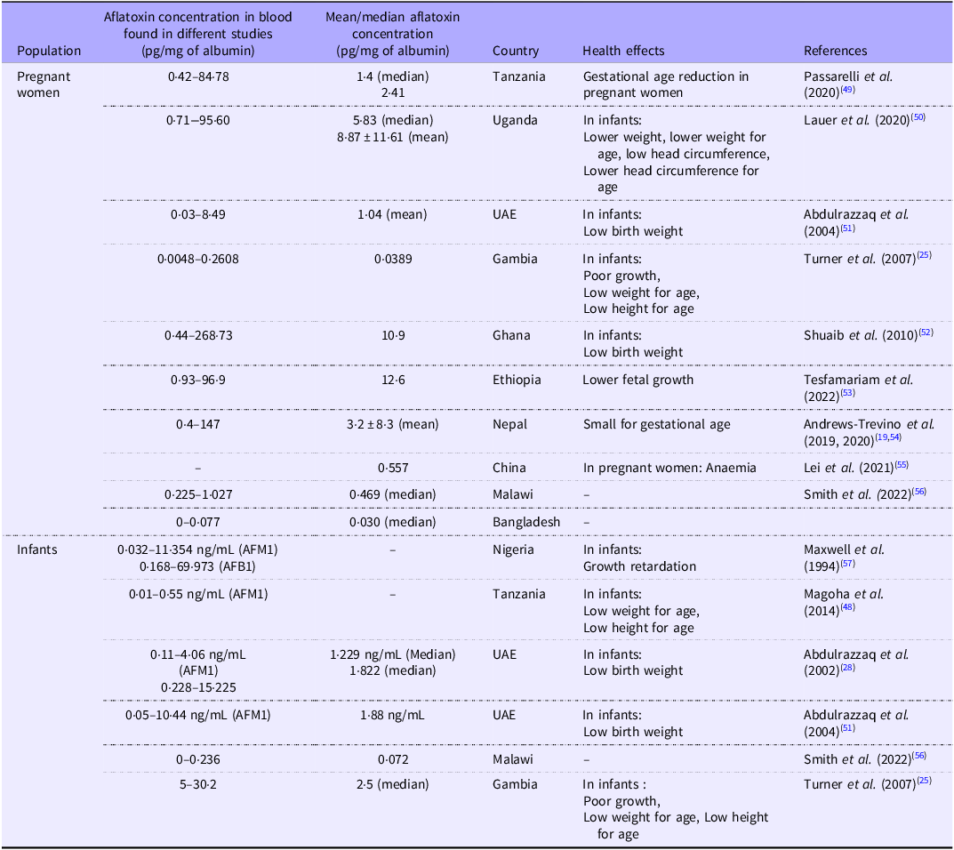
Environment-related factors, including humidity, temperature, availability of nitrogen and plant density, as well as substandard harvesting methods and inadequate storage of grains, all play a significant role in AF accumulation in food(Reference Cotty and Jaime-Garcia16). AF contamination in the food supply is prevalent in a number of developing countries, where the use of maize and peanuts (which are considered high-risk foods) is widespread. Pregnant women are primarily exposed to AF through contaminated foods being consumed(Reference Shuaib, Ehiri and Abdullahi17). The commonly consumed foods by pregnant women, along with AFB1 contamination levels from various countries, are listed in Table 1. Mean AF contamination levels of each food item from different countries were calculated considering the reported contamination levels from different studies in that country (Table 1). The mean AF concentration of peanut was maximum with 140·31 µg/kg being the highest in Africa(Reference Magembe, Mwatawala and Mamiro18). The AFB1-lysine adducts were discovered in 94 % of the maternal serum samples in a longitudinal birth cohort study in Nepal(Reference Andrews-Trevino, Webb and Shively19), and in Uganda, 100 % of the serum samples were detected with AFB1-Lys adduct (AF serum biomarker) in the range 0·71 pg/mg to 95·60 pg/mg albumin, indicating that the population is exposed to dietary AF during pregnancy on a broad basis(Reference Lauer, Duggan and Ausman20). The reported serum AFB1-Lys adduct levels and associated health effects from different countries in pregnant women and infants are listed in Table 2.
Aflatoxin contamination in food items commonly consumed by pregnant women and infants from different countries

Aflatoxin exposure level of pregnant women and infants from different countries

Exposure of pregnant women to AF also depends on the season of the year and stage of the pregnancy(Reference Castelino, Dominguez-Salas and Routledge21). According to a study conducted in Gambia, AF exposure to pregnant women continued throughout their pregnancy, peaking during the dry season. There was some indication that during the dry season, pregnant women who were later in their pregnancies had greater AF-albumin adducts (AF-alb) levels than those who were earlier in pregnancy(Reference Castelino, Dominguez-Salas and Routledge21). The modest increase in AF-alb levels in women during later stages of pregnancy compared with early pregnancy was seen only in the dry season, when overall AF exposure was higher. This suggests that later pregnancy, particularly during the dry season, may be a more vulnerable period for both the mother and fetus due to increased AF accumulation or metabolic changes in pregnancy(Reference Castelino, Dominguez-Salas and Routledge21).
The fetus depends solely on the intrauterine environment during pregnancy, which is controlled by the mother’s health, diet and exposure to chemicals from the environment(Reference Carpenter and Bushkin-Bedient22). The placenta (selectively permeable) works like a barrier for the majority of harmful substances to enter the fetus to avoid the harmful effects; however, it is not impenetrable to or protect against all toxins(Reference Partanen23). Subsequent to being consumed through infected foods, AF are quickly absorbed and dispersed throughout the pregnant woman’s body, where they can reach the placental membrane(Reference Partanen, El-Nezami and Leppänen24) and affect fetal growth and development(Reference Partanen, El-Nezami and Leppänen24). AF and AF-alb were identified in both maternal and cord blood of newborns in a number of different nations(Reference Turner, Collinson and Cheung25–Reference Wild, Rasheed and Jawla27), which confirms AF and/or its metabolites cross the human placenta and the growing fetus is exposed to AF during pregnancy(Reference Abdulrazzaq, Osman and Ibrahim28–Reference De Vries, Maxwell and Hendrickse31). Even if AF was not found in the mother’s blood at the time of delivery, it was found in some cord blood, suggesting that AF may accumulate in the fetus when exposed to these toxins in utero. The metabolism of AF in the fetus may be different from that in adults, and the fetus’s immature liver may be less effective at handling these toxins than a mature organ. The toxin is most likely excreted by the fetus being exposed to transplacental AF into the amniotic fluid, where it is almost certainly reabsorbed, leading to a prolonged exposure cycle(Reference Maxwell, Apeagyei and De32).
Infant aflatoxin exposure
Infant exposure through breast milk
Infants are vulnerable to AF exposure during early life, especially through breastfeeding(Reference Alvito and Pereira-da-Silva33,Reference Gong, Hounsa and Egal34) . AF can pass through breast milk(Reference Alvito and Pereira-da-Silva33,Reference Wu, Jia and Xue35) , which is evident from Ghanian and Nigerian mothers’ breast milk containing both AF, AFM1 and AFB1 in the range of 60·90–299·99 ng/L and 94·50–4123·80 ng/L, respectively(Reference Gürbay, Sabuncuoğlu and Girgin36), impacting both fetal and infant health. Mean AF concentration in breast milk was highest in Mexico with 17·04 ± 9·13 ng/L (Table 1)(Reference Salas, Acosta and Garza37).
Direct exposure of infants through baby foods
As infants grow, they begin weaning after 6 months; that is, they start receiving complementary foods along with breast milk. Infants and children are being exposed to AF through complementary foods such as cereal-based food and infant formula(Reference Alvito and Pereira-da-Silva33,Reference Eshete, Gebremedhin and Alemayehu38–Reference Ezekiel, Abia and Braun42) , as is evident by AF-alb adducts being detected in the serum(Reference Turner, Collinson and Cheung25,Reference Gong, Hounsa and Egal34) and AF in the urine of infants and young children(Reference Polychronaki, Wild and Mykkänen43). As children grow, their exposure to AFB1 increases, probably because their diet starts resembling an adult diet(Reference Andrews-Trevino, Webb and Shively44).
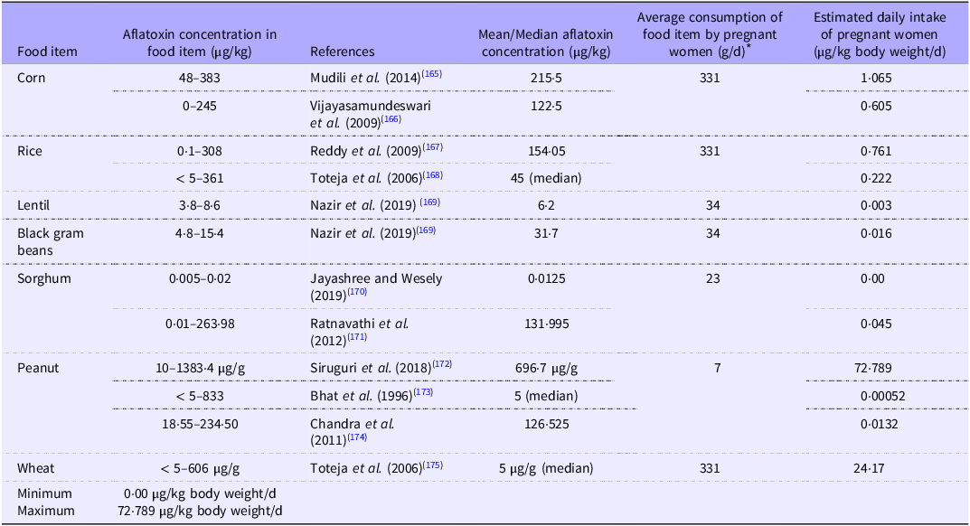
In the Indian context, several studies were done to determine the AF level in various foods. However, no studies were conducted to know the true exposure by determining the serum biomarker in pregnant women and its health effects. Hence, in this review, we made an attempt to calculate the AF EDI (based on available literature and empirical formula) in pregnant women, considering the consumption amount of various food intake data as per the National Nutrition Monitoring Bureau (Table 3).
Estimated dietary exposure of aflatoxin in pregnant women in India

Note: Aflatoxin mean concentration is calculated as the average of the low and high concentrations for those data where the mean/median aflatoxin level in food is not reported.
* Average amount of consumption of each food item by pregnant women (g/d) was taken from the National Nutrition Monitoring Bureau report (NNMB, 2012)(46). National Nutrition Monitoring Bureau (2012) Diet and nutritional status of rural population, prevalence of hypertension and diabetes among adults and infant and young child feeding practices – report of third repeat survey.
Calculation of estimated daily intake
In this review article, we made an attempt to calculate the AF EDI using the empirical formulae based on several reported studies on AF contamination in foods. EDI of AF exposure of the pregnant women is calculated by the following formula:
 $$\small{{\rm{EDI\;}}\left( {{\rm{\mu g}}/{\rm{kgbw}}/{\rm{d}}} \right) = {{{{\rm{Daily\;intake\;food\;}} \times {\rm{\;mean\;concentration\;of\;AF}}}} \over {{{\rm{Average\;body\;weight\;}}\left( {{\rm{kg}}} \right)}}}}$$
where estimated daily intake = estimated amount of AFB1, which can be ingested daily (µg/kg body weight/d), and contamination level of AF = total AF content in each food item (µg/kg).
The average body weight of pregnant women is taken as 67 kg (reference body weight of an adult woman (55 kg) + suggested increase in gestational body weight of adult women during pregnancy (12 kg)(45).
The average amount of consumption of each food item by pregnant women (g/d) was taken from the National Nutrition Monitoring Bureau(46).
Mean AF concentration was calculated by taking the minimum and maximum concentrations of AF in each food item and divided by 2.
The AF EDI based on reported studies on AF contamination in foods was calculated and represented in Table 3.
AF contamination of commonly consumed foods in India was in the range of 0·005 µg/kg −1383·4 µg/g (Table 3). Regulatory limits of total AF contamination in foods in India are 15 ppb according to Food Safety and Standards (contaminants, toxins and residues)(47), and maize, rice, sorghum, peanut and wheat exceeded the limit of 15 ppb according to available studies included in the review article. Calculated EDI of AF contaminated foods by pregnant women were in the range of 0·00–72·789 µg/kg body weight/d with sorghum having the lowest and peanut having the highest EDI (Table 3). Several countries (Tanzania, Uganda, UAE, Gambia, Ghana, Ethiopia, Nepal, China, Malawi, Bangladesh and Nigeria) assessed the health effects of AF exposure in pregnant women and pregnancy outcomes(Reference Turner, Collinson and Cheung25,Reference Abdulrazzaq, Osman and Ibrahim28,Reference Magoha, Kimanya and De Meulenaer48–Reference Maxwell, Familusi and Sodeinde57) (Table 2). However, in India, no such studies were conducted to assess the health effects in pregnant women due to AF exposure. Hence, in India, further studies are warranted to elucidate the AF exposure and pregnancy outcomes.
Health effects in pregnant women and infants due to aflatoxin exposure
Health effects in pregnant women due to aflatoxin exposure
A decreased number of live births were observed on AF exposure of the pregnant rats though it was not significant(Reference Shuaib, Jolly and Ehiri52,Reference Kihara58) , whereas in other studies, there was a significant increase in pregnancy losses on AF exposure of the pregnant rabbits and bovines(Reference Smith, Prendergast and Turner2,Reference Ray, Abbitt and Cotter59,Reference Wangikar, Dwivedi and Sinha60) . Gestational weight gain was low in mothers with higher AF-lys levels, which again is a risk factor for low birth weight of infants(Reference Lauer, Natamba and Ghosh50), whereas Kihara(Reference Kihara58) did not find any effect on gestational period or mother’s weight during pregnancy.
AF exposure during pregnancy also increases the chance of premature delivery(Reference Smith, Prendergast and Turner2,Reference Shuaib, Jolly and Ehiri52,Reference Wang, Tan and Wang61,Reference Kościelecka, Kuć and Kubik-Machura62) . Exposure to AF increases the pro-inflammatory cytokines in the mother and also the fetus, causing cervical ripening, rupture of the amniotic membranes and preterm contractions(Reference Kościelecka, Kuć and Kubik-Machura62). Consumption of AFB1 also increases plasma concentrations of Corticotropin-releasing hormone (CRH) and oestrogen and the expression of Cyclooxygenase-2 (COX-2) in the placenta while lowering the expression of 15-hpgd and Bcl-2, which might be the reason for premature delivery(Reference Wang, Tan and Wang61). The possible effects of AF on the gestational period may perhaps have major implications for public health for people with higher exposure levels since premature births can signal an impairment of ideal prenatal health and instigate a chain reaction of inadequate child growth and development(Reference Passarelli, Bromage and Darling49).
Maternal anaemia
Studies have shown that AF exposure in pregnant women causes anaemia in them(Reference Shuaib, Jolly and Ehiri52,Reference Kościelecka, Kuć and Kubik-Machura62) . It has been found that pregnant women with AF exposure (those having AFB1-alb adduct in their blood) are susceptible to the risk of anaemia, particularly microcytic hypochromic anaemia(Reference Lei, Liu and Ye55). Experimental studies have demonstrated that AF causes microcytic hypochromic anaemia in a variety of animal species by promoting the haemolysis of erythrocytes, inhibiting haematopoiesis, impairing iron absorption and altering Hb levels(Reference Andretta, Kipper and Lehnen63–Reference Kumar and Balachandran65). AF may also worsen iron bioavailability and absorption, as well as hinder erythropoiesis, which are all thought to contribute to anaemia(Reference Smith, Prendergast and Turner2). Prolonged exposure of the mother can also put infants at risk as AF can pass through the placenta(Reference Lei, Liu and Ye55). Further anaemia can trigger a number of mechanisms, including the activation of pro-inflammatory cytokines and a decrease in insulin-like growth hormones, which have detrimental effects and can potentially result in fetal death(Reference Smith, Prendergast and Turner2). However, the dosage used in animal studies was very high, which might not be relevant to humans(Reference Smith, Prendergast and Turner2). Therefore, further clinical trial is needed in the future.
Effects of aflatoxin exposure on fetal growth and intrauterine life
De Vries et al. (Reference De Vries, Maxwell and Hendrickse31), Lamplugh et al. (Reference Lamplugh, Hendrickse and Apeagyei66) and Ibeh and Saxena(Reference Ibeh and Saxena67) have reported that AF exposure during pregnancy can lead to stillbirth, though the mechanism was not studied. AF exposure during pregnancy, especially chronic maternal AF exposure, may impair fetal growth over time(Reference Smith, Prendergast and Turner2,Reference Tesfamariam, Gebreyesus and Lachat53) . AF exposure during pregnancy can have a negative effect on fetal growth during intrauterine life, leading to low birth weight infants(Reference Kyei, Boakye and Gabrysch68). Some studies have reported the negative effect of AF exposure on birth weight(Reference De Vries, Maxwell and Hendrickse31,Reference Abdulrazzaq, Osman and Yousif51,Reference Maxwell, Familusi and Sodeinde57,Reference Supriya, Girish and Reddy69,Reference Supriya, Akhila and Pratap Reddy70) , while Matchado et al. (Reference Matchado, Smith and Schulze71) found a positive association between AF exposure of the mother and the birth weight of the fetus, and others have found no association(Reference Passarelli, Bromage and Darling49,Reference Maxwell, Familusi and Sodeinde57) . This difference in results might be due to a different dietary pattern or the population selected, which was from an urban area with relatively low levels of AF exposure compared with other studies(Reference Passarelli, Bromage and Darling49). Additionally, some animal studies have shown that AF exposure during pregnancy reduces fetal growth, leading to low birth weight of infants(Reference Kihara58,Reference Wangikar, Dwivedi and Sinha60,Reference Butler72–Reference Rotimi, Onuzulu and Dewald79) . AFB1 exposure during pregnancy impairs nutrition and protein transport, which hinders the development of the fetus normally(Reference Ivanovics, Gazsi and Reining80,Reference Nguyen, Li and Eguchi81) . Prenatal AFB1 exposure can alter weight gain and birth weight of the animal, even after the exposure has been discontinued(Reference Rotimi, Onuzulu and Dewald79). Some animal studies have found that AF exposure during pregnancy can also affect bone development of the fetus negatively, causing ossification failures, changes in bone size and shape(Reference El-Nahla, Imam and Moussa78,Reference Abdulrazzaq, Padmanabhan and Bastaki82–Reference da Silva, de Oliveira and Ramalho84) . However, the actual effect on human fetal development is still unclear. Hence, more research is needed in the future to study the effect of AF exposure on fetal growth.
Effect of aflatoxin exposure during pregnancy on infant growth and development
There are some evidences linking maternal AF exposure during pregnancy to undesirable birth outcomes. Earlier studies have shown that AF exposure during pregnancy or fetal life(Reference Andrews-Trevino, Webb and Shively19,Reference Lauer, Duggan and Ausman20,Reference Shuaib, Jolly and Ehiri52) can impair growth in postnatal life(Reference Turner, Collinson and Cheung25,Reference Gong, Hounsa and Egal34) . An Randomized controlled trial (RCT) study from Kenya also found an age-dependent association of AFB1 exposure and negative linear growth, where AF free maize increased the growth at midline but had no effect at endline(Reference Hoffmann, Jones and Leroy85). Few studies have reported that AF exposure is negatively associated with linear growth of infants(Reference Magoha, Kimanya and De Meulenaer48,Reference Natamba86–Reference Watson, Moore and Darboe88) . A longitudinal study in Nepal followed up women from pregnancy till 2 years of age of infants and found that both pregnant mothers and infants are chronically exposed to AF, which is associated with various negative growth outcomes of the infants such as impaired linear and long bone growth and most probably leading to stunting(Reference Andrews-Trevino, Webb and Shively44). Hence, proving that AF exposure during pregnancy can have negative effects on the growth of infants. On the other hand, some studies(Reference Shirima, Kimanya and Routledge89–Reference Chen, Mitchell and Gratz91) found no association between AF exposure and infant growth or body weight. The inconsistency in results might be due to differences in study design, sample size, duration or methods of experiment.
Despite the fact that earlier investigations showed that AFB1 can pass the blood-brain barrier(Reference Qureshi, Hamid and Ali92), the first study on humans to look at the relationship between AFB1 exposure of the pregnant mother and head circumference of the infant at birth was carried out by Lauer et al. (Reference Lauer, Duggan and Ausman20). They found that AF exposure is associated with smaller head circumference in infants(Reference Lauer, Duggan and Ausman20). The same was reported in a prospective cohort study by Matchado et al. (Reference Matchado, Smith and Schulze71), indicating reduced brain size that lasts beyond 2 years of age.
AF exposure during pregnancy can also affect the behaviour of offspring, which is shown by delay of early response development, impaired locomotor coordination and impaired learning ability(Reference Kihara58,Reference Supriya, Akhila and Pratap Reddy70) . The mechanism behind behavioural change is not clear, but the toxic effect of AF on RNA inhibition, DNA methylation, chromosomal aberrations and transplacental carcinogenesis might be the reason(Reference Kihara58,Reference Rotimi, Onuzulu and Dewald79,Reference Hernandez-Vargas, Castelino and Silver93) . However, more research is needed to know the exact mechanism of action and confirm the findings on humans.
Aflatoxin exposure during pregnancy and immune suppression
There are only a few studies that have been done on AF exposure during pregnancy to see the effect on the immune function of the children. AF exposure during pregnancy causes DNA methylation in white blood cells of infants, changing the cell types involved in the immune system and thus suppressing the immune system(Reference Hernandez-Vargas, Castelino and Silver93). Maternal exposure to AF causes an increase in the number of lymphocytes, decreases cytokine production by intestinal type 2 innate lymphoid cells (ILC2), reduces microbial-related epithelial gene expression and changes gut microbiota of the fetus or infant, thereby suppressing their immune system(Reference Bastos-Amador, Duarte and Torres94). Immune suppression can further make the infant more prone to many infectious diseases(Reference Gong, Watson and Routledge95). However, more research is needed to establish a causal relationship between prenatal AF exposure and immunological suppression in children.
Effect of aflatoxin during pregnancy on the organ system and its dysfunction
Animal studies have shown that visceral changes in the embryo or fetus are also observed upon AF exposure of the mother, such as a decrease in size and weight of organs like the liver, kidneys and heart(Reference El-Nahla, Imam and Moussa78,Reference Abdulrazzaq, Padmanabhan and Bastaki82) . Degeneration and congestion of the heart, liver and kidney are also observed in fetuses exposed to AF during pregnancy(Reference El-Nahla, Imam and Moussa78,Reference da Silva, de Oliveira and Ramalho84,Reference Fetaih, Dessouki and Tahan96) .
AF exposure during pregnancy is also associated with neonatal jaundice or jaundice in infants(Reference Shuaib, Ehiri and Abdullahi17,Reference Kyei, Boakye and Gabrysch68) . In two studies(Reference Abulu, Uriah and Aigbefo Hs97,Reference Sodeinde, Chan and Maxwell98) , compared with non-jaundiced newborns, neonates exposed to maternal AF during pregnancy had a more than 2-fold greater risk of developing neonatal jaundice; however, two other investigations failed to detect this association(Reference Abdulrazzaq, Osman and Yousif51,Reference Ahmed, Hendrickse and Maxwell99) . Despite small sample sizes, there was some evidence of a negative relationship between exposure to AF and the onset of neonatal jaundice, with two out of four studies indicating a distinct increase, while the other two showed trends in this way. The apparent correlation between fetal exposure to AF and neonatal jaundice may be partially explained by the toxic effects of AF on the newborn’s liver and enhanced haemolysis of fetal Hb(Reference Smith, Prendergast and Turner2).
Animal studies have shown that AF exposure during pregnancy at different doses significantly decreased cholesterol and increased TAG levels in infants, though this increased TAG was not associated with body weight(Reference Rotimi, Onuzulu and Dewald79). Since the cholesterol level is decreased, levels of testosterone, progesterone and luteinising hormone, which are steroid hormones and need the presence of cholesterol for biosynthesis, are also decreased on prenatal exposure to AF(Reference Supriya, Girish and Reddy69,Reference Rotimi, Onuzulu and Dewald79,Reference Supriya and Reddy100) . The decrease in testosterone might also be due to the binding of AF to steroidogenic acute regulatory (StAR) protein, the protein involved in the transport of cholesterol from the outer to the inner mitochondrial membrane for testosterone synthesis. Prenatal AFB1 exposure also reduced thyroxin at high exposure doses and prolactin at low exposure doses. All these altered hormonal conditions of the prenatal AF-exposed rats indicate that AF can act as an endocrine disruptor and it might increase the possibility of developing non-communicable diseases in later life(Reference Rotimi, Onuzulu and Dewald79).
Conclusion
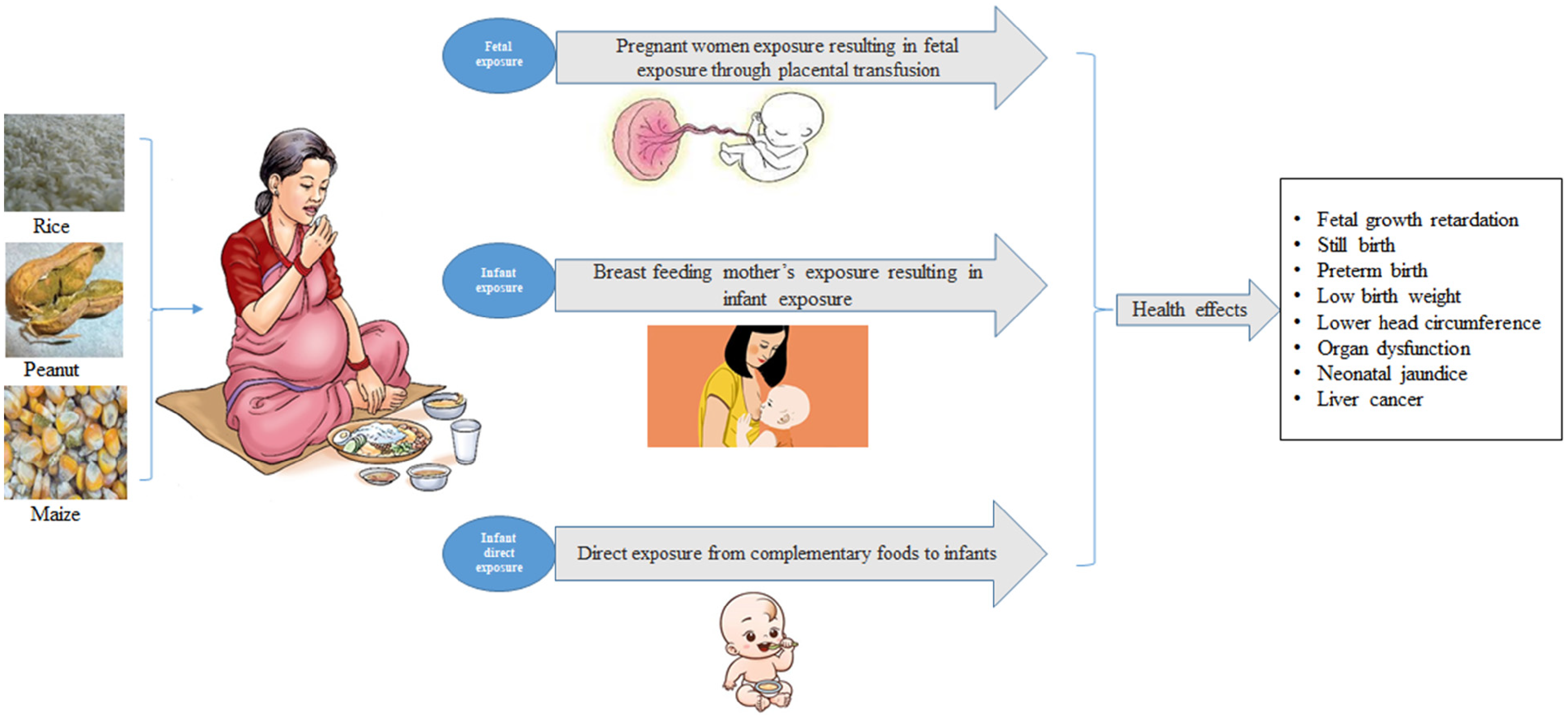
AF exposure during pregnancy or infancy has been associated with various adverse outcomes, with infants ultimately experiencing the most significant health effects due to both prenatal (via placenta) and postnatal (via breast milk and foods) exposures. It affects both the mother and the child in the long term. It may result in maternal anaemia, premature birth and loss of pregnancy. AF exposure in mothers has been shown to cause deleterious effects on fetal development, which ultimately impacts infant growth and health, such as low birth weight, neonatal jaundice and organ dysfunction (Figure 1). It may also have an adverse effect on the endocrine system and immune system. This review summarised the AF exposure of pregnant women by consumption of AF contaminated foods globally and its adverse health effects on the infant and overall health. The calculated EDI of AF by pregnant women showed greater exposure to AF through commonly consumed foods. However, extensive clinical studies on maternal AF exposure are lacking in India. Therefore, more clinical studies are warranted on maternal AF exposure to elucidate the birth outcomes and associated infant health outcomes. The outcome of the research will bring a better understanding of the maternal AF exposure and health outcomes, which might recommend the government bodies to develop AF management strategies such as mitigating AF contamination in food commodities, conducting awareness programmes and educating people to minimise AF exposure for improving public health.
Aflatoxin exposure during pregnancy or infancy and its effect on child health.

Acknowledgements
The authors acknowledge the support and guidance of the Director, ICMR-National Institute of Nutrition, Hyderabad, India.
Not applicable since this is a review article.
S. G. contributed to the original draft writing and revising, V. V. provided valuable inputs and contributed to editing and revising the manuscript, S. K. and Y. S. contributed to the literature search and reviewing and S. N. S. provided necessary suggestions.
The authors declare no conflict of interest associated with this manuscript.