Human adenoviruses (HAdVs) are nonenveloped, double-stranded DNA viruses, highly resistant to inactivation that can cause a wide spectrum of clinical manifestations, from mild to life-threatening conditions. Respiratory, gastrointestinal and ocular infections are among the most frequent presentations. In immunocompromised patients, HAdVs can also cause hemorrhagic cystitis, hepatitis, meningitis, and disseminated disease.Reference Echavarría1 HAdVs are classified into 7 species (A–G) and are traditionally typed into serotypes (1–52) by neutralization assays. Recently, typing by sequencing analysis has become more broadly used; therefore, strains are now classified into genotypes. To date, >100 genotypes have been described by complete genome-sequencing analysis.Reference Harrach, Tarján and Benkő2,Reference Marcone, Culasso and Campos3
Ocular infections include conjunctivitis, epidemic keratoconjunctivitis (EKC), pharyngoconjunctival fever (PCF), and follicular conjunctivitis. HAdV serotype 8 (HAdV-8) is one of the main causes of EKC, along with HAdV-19 (now reclassified as HAdV-64)Reference Zhou, Robinson and Rajaiya4 and HAdV-37. In addition, HAdV genotypes 53, 54, and 56 have also been associated with EKC with lower frequency.Reference Robinson, Singha and Henquellb5 HAdVs are transmitted through direct contact or droplets that can reach the conjunctiva, nose, or throat. HAdV can also be transmitted by contaminated fomites or by the fecal–oral route. Some serotypes (in particular HAdV-8 and -19) are highly resistant and can persist for up to 3–4 weeks in the environment and fomites.Reference Gordon, Gordon and Romanowski6
Hospital outbreaks of HAdV conjunctivitis can occur by direct transmission through infected secretions from other patients, healthcare workers (HCWs), or visitors.Reference Faden, Wynn and Campagna7 Contaminated handheld ophthalmic equipment used for retinopathy of prematurity (ROP) screening can also be a source of infection.Reference Sammons, Graf and Townsend8 In this study, we report a nosocomial conjunctivitis outbreak in a neonatology unit. Epidemiology, demographic, clinical characteristics, viral typing, associated economic loss, and outbreak containment are described.
Methods
A nosocomial conjunctivitis outbreak occurred from January 30 to April 28, 2018, in the neonatology unit at Centro de Educación Médica e Investigaciones Clínicas (CEMIC) University Hospital in Buenos Aires, Argentina. An observational prospective study was begun to report the outbreak and to determine the associated economic loss. CEMIC is a private tertiary-care healthcare institution with inpatients and outpatients including newborns, children, and adults. CEMIC has 125 hospital beds including 18 rooming-in beds in the maternity ward, 17 beds in the neonatology unit, and 14 beds in the pediatric unit for both the general ward and the pediatric intensive care unit (PICU). On average, 1,300 healthy deliveries are performed and 230 critically ill newborns are admitted annually. The neonatology unit includes both the general nursery and the neonatology intensive care unit (NICU). Patients are distributed along 4 open bay units with up to 4–5 beds per unit. Premature or critically ill newborns are admitted to the NICU, and healthy newborns are examined daily in the general nursery but otherwise remain in rooming-in beds with their mothers. This study complied with the principles outlined by the Declaration of Helsinki, and it was evaluated and approved by the CEMIC ethics committee (qualified by the Department of Health and Human Services, HHS no. IRB00001745- IORG 001315).
Population
During the outbreak period, all neonates admitted at the NICU (n = 52) either with or without ocular symptoms were included in the study. Ocular swabbing and HAdV PCR were performed in 11 of 14 symptomatic neonates and in 2 of 38 asymptomatic neonates. In total, 59 NICU-related HCWs were enrolled and tested for HAdV between March 22 and March 28. Of 59 HCWs, 7 were symptomatic and 52 were asymptomatic. In total, 101 conjunctival swabs from 13 neonates and 59 HCWs were tested for HAdV using real-time polymerase chain reaction (PCR). A questionnaire form was specifically designed to collect data on newborn demographic and clinical characteristics, ophthalmologic controls, and treatment. A different questionnaire form was designed to collect data on HCWs.
Definitions
Symptomatic patients were defined as hospitalized neonates with conjunctivitis symptoms: lacrimation, purulent discharge and/or lachrymal swelling in one or both eyes. Asymptomatic patients were defined as hospitalized neonates without ocular symptoms. Infected patients were defined as symptomatic patients with a positive HAdV PCR. Days with conjunctivitis symptoms refers to the period of time (in days) from symptoms onset until ophthalmologic resolution or hospital discharge. Patients were not followed after hospital discharge. We defined infectious positivity range (by culture isolation) as the time (in days) from symptom onset until last available ocular positive sample for HAdV by culture. And we defined PCR positivity range as the time (in days) from symptom onset until last available ocular positive sample for HAdV by real-time PCR.
Virological detection
For HAdV detection, a commercial real-time PCR (LightMix Kit human adenovirus, Tib Molbiol GmbH, Berlin, Germany) that amplifies a 129-bp fragment of the hexon gene was performed in a Light Cycler 2.0 instrument (Roche, Mannheim, Germany). Viral DNA was extracted from conjunctival swabs using the Magna Pure System Compact (Roche, Basel, Switzerland) according to the manufacturer’s instructions.
Cell culture and viral isolation
To determine viral replication throughout time, follow-up ocular swabs were cultured. Specifically, ocular swabs from patients with at least 2 HAdV-positive PCR results were inoculated in A549 cells. Samples were maintained in culture for up to 4 weeks or until adenovirus-like cytopathic effect (CPE) was observed. In addition, 2 blind passages were performed for all samples. CPE was confirmed by indirect immunofluorescence (IFI) or PCR.
PCR amplification and sequencing HAdV typing was done by sequencing the hypervariable region (HVR) 1–6 from the loop 1 of the hexon gene (900 nucleotides).Reference Lu and Erdman9 PCR products were purified and sequenced by Sanger dideoxy method (Macrogen, Korea).
Phylogenetic analysis
Nucleotide sequences were visually inspected with BioEdit and aligned in aminoacids with ClustalW. The phylogenetic analysis was performed using the maximum likelihood method (PhyML 3.0), and the topology support of the tree was estimated with bootstrap using 1,000 replicates. Bootstrap values of ≥70% were considered statistically significant for the grouping. All strains that were sequenced were identical; the strain was submitted to GenBank under accession no. MT024787.
Economic analysis
Economic costs were determined by analyzing data from billing records. Direct costs included extra costs and productivity loss. Extra costs were calculated from extra personnel professional salaries. Productivity loss was calculated as profit loss from the neonatology unit partial closure and closure of the high-risk pregnancy unit of the gynecology and obstetrics department from March 16 to April 4, 2018.
Statistical analysis
Data were expressed as the mean ±SD or median and interquartile range for continuous variables and as frequencies for categorical variables. Stata version 13.0 software (StataCorp, College Station, TX) was used for all data management and analysis. The differences between the groups were assessed using the Wilcoxon rank-sum test for continuous variables and χ2 test for categorical variables. P < .05 was considered statistically significant.
Results
On February 27, 2018, 1 newborn (patient 4) and 1 HCW presented with conjunctivitis symptoms within the same week. This observation prompted an outbreak alert on March 6 by the neonatology authorities and the infectious disease control team.
When retrospectively analyzed, it was noted that, at the end of January, the father of patient 1 had arrived at the NICU with conjunctivitis symptoms. He was asked not to enter the NICU to visit his premature newborn daughter, but he claimed to be under treatment and to be noncontagious. However, patient 1 developed conjunctivitis 1 week later. At the time, HAdV was not suspected and only bacterial cultures were performed. In addition, the mother of patient 1 developed conjunctivitis 1 week later.
From January 30 to April 28, 2018, 52 neonates were hospitalized in the NICU. Among them, 14 (21%) had ocular symptoms diagnosed as conjunctivitis, and the remaining 38 never developed ocular symptoms. The initial 3 patients with conjunctivitis were only tested for bacterial infections in their ocular swabs. HAdV was included in the differential diagnosis after 1 of these patients tested positive for HAdV in a respiratory sample. Thus, these initial 3 patients with conjunctivitis were included in the study as infected patients even though HAdV PCR was not performed. The remaining 11 symptomatic neonates were HAdV positive by PCR. Of 38 asymptomatic neonates, 2 were tested by PCR and were HAdV negative.
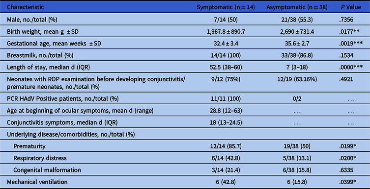
Demographic and clinical characteristics for symptomatic and asymptomatic neonates were compared (Table 1). Most symptomatic neonates (85.7%) were premature, and only half of the asymptomatic neonates were premature (P = .019). Gestational age, birth weight, and length of stay were significantly different between symptomatic and asymptomatic neonates.
Demographic and Clinical Characteristics of Symptomatic and Asymptomatic Neonates During the HAdV Outbreak (n = 52)

Note. SD, standard deviation; IQR, interquartile range; ROP, retinopathy of prematurity.
*P ≤ .05; **P ≤ .01; ***P ≤ .001.
The percentage of premature neonates with ROP examinations before developing ocular symptoms was not significantly different between neonates with or without conjunctivitis (Table 1). The mean age at discharge was 47.6 days old for symptomatic neonates and 16.3 days old for asymptomatic neonates.
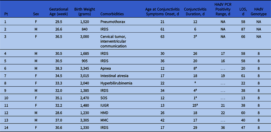
The youngest neonate who developed HAdV conjunctivitis was 12 days old. All cases developed bilateral conjunctivitis with pseudomembranes, none presented keratitis or fever. All patients received treatment with topical corticosteroids and ophthalmic tobramycin drops. Clinical characteristics and HAdV typing of the 14 neonates with conjunctivitis are described in Table 2.
Clinical Characteristics of Neonates With Conjunctivitis (n=14)
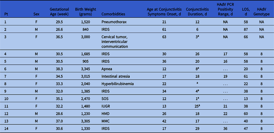
Note. aPatients were discharged from the hospital before ocular symptoms resolution. Pt, patient; NA, not available; IRDS, infant respiratory distress syndrome; IUGR, intrauterine growth restriction; HMD, hyaline membrane disease; MMC, myelomeningocele; SOS, suspicion of sepsis; LOS, length of stay.
Some patients were discharged from the NICU when their underlying condition or prematurity permitted even if they remained with conjunctivitis. Conjunctivitis symptoms duration, calculated only with patients who resolved their conjunctivitis before discharge, ranged from 6 to 32 days, with a median duration of 18 days (Table 2).
In total, 11 neonates had at least 1 conjunctival sample for HAdV PCR, 6 neonates had 2 conjunctival samples and 3 neonates had 3 conjunctival samples. All second samples from aforementioned 6 patients were PCR positive and corresponded to 12, 14, 16, 21, 22 and 25 days after symptom onset. The third samples from aforementioned 3 patients revealed that 2 were HAdV negative and 1 was HAdV PCR positive, corresponding to a sample obtained 32 days after symptom onset. Furthermore, this last neonate was HAdV PCR positive on day 4 (4 days before developing conjunctivitis). In addition, another neonate was HAdV PCR positive while asymptomatic (8 days before symptoms onset). These patients were swabbed while asymptomatic due to their close proximity with positive patients.
The PCR positivity range had a median period of 18.5 days. Cultures were performed on samples after a second PCR was positive. HAdV cultures performed in these patients (n = 6) were also positive. All blind passages showed a slow CPE. The median infectious positivity range was 18.5 days.
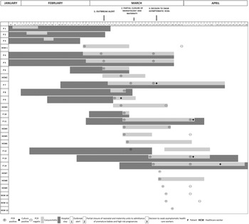
Outbreak progression in neonates and HCWs with positive HAdV PCR was studied throughout time. Specifically, length of stay, length of conjunctivitis symptoms, date and PCR result, and culture are shown in relation to infection control measures (Fig. 1).
Outbreak timeline in neonates with conjunctivitis and HCW with a positive HAdV PCR in relation to infection control measures, January–April 2018.

All symptomatic HCWs were HAdV positive by PCR. Of 52 asymptomatic HCWs, 5 (9.6%) were also HAdV positive by PCR. After March 29, there were no new cases, neither neonates nor HCWs. The last patient, who became symptomatic on March 22, was discharged on April 28, and this was considered the end of the outbreak.
Infectious control interventions
The infection control committee issued an outbreak alert on March 6. Partial closure of the neonatology unit and redirection of high-risk pregnancies were performed 10 days later, on March 16. After the outbreak alert, contact precautions, cohorting of patients and changes in room allocation were implemented. Disposable gowns and gloves were worn by HCWs and parents for any contact with infected patients. Cleaning of high-touch surfaces (eg, counters, desktops, telephones, door knobs, and computer keyboards) was intensified to 3 times per day with a virucidal quaternary-based disinfectant. All patients had exclusive stethoscopes and thermometers, and shared equipment such as scales was disinfected after each use. Hand hygiene practices were reinforced. All HCWs with positive HAdV PCR results (symptomatic or asymptomatic) were furloughed for at least 15 days.
After the outbreak alert, high-risk pregnancies were not admitted to CEMIC and were redirected to other hospitals. In addition, the NICU was divided into 2 separate areas for symptomatic and asymptomatic neonates. The general nursery was transferred to a different room, and no contact between staff from the NICU and the general nursery was allowed. Extra personnel were hired to cover for additional areas and licensed personnel.
Despite strict containment measures, 2 additional cases appeared in the NICU 17 days after the outbreak alert, a week after partial closure. Therefore, additional sources of infection, including asymptomatic carriage by HCWs, were evaluated.
Typing and phylogenetic analysis
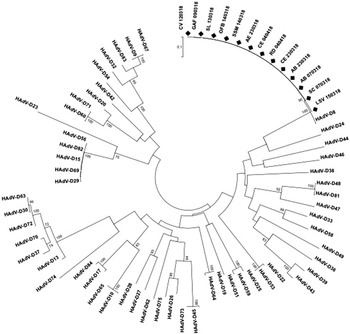
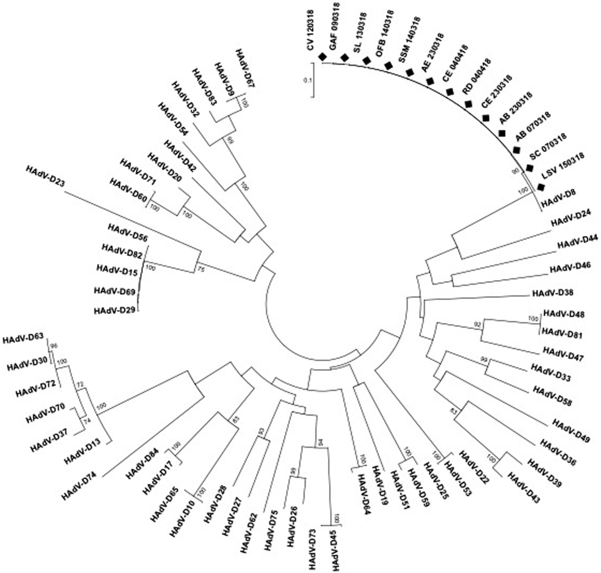
All HAdV-positive samples were successfully amplified for sequencing by PCR targeting the partial hexon gene. All sequences were identical and clustered together with the HAdV-D8 reference strain, with a high bootstrap support (Fig. 2). Phylogenetic analysis by maximum likelihood performed with strains from other countries revealed that strains from this outbreak clustered together with strains reported since 2001 from China, Denmark, Japan, Tunisia, and the United State (data not shown).
Phylogenetic analysis of HAdV strains from the outbreak and references. A maximum likelihood tree was constructed based on hypervariable regions (HVR) 1–6 of the hexon gene (900 bp), using the GTR+I+Γ model with 1,000 pseudoreplicates. Our studied strains are indicated with a diamond shape.
Economic burden
As an outbreak result, the NICU was closed to new admissions. Therefore, the number of occupied beds was significantly lower. The high-risk pregnancy unit returned to its normal activity on April 30. The NICU median percentage of occupied beds during the outbreak was 43.1%, significantly lower than same period a year before, in 2017 (59.4%; P < .001). Based on decreased occupancy and median daily charge per patient, lost productivity was US$175,000. Direct costs of preventive measures (ie, implementing a new examination room) and salaries of extra personnel cost US$30,350. In total, this outbreak cost the hospital US$205,350.
Discussion
Epidemic conjunctivitis outbreaks in NICUs have been broadly reported, and HAdV-D8 is one of the most frequent genotypes.Reference Sammons, Graf and Townsend8,Reference Calkavur, Olukman and Ozturk10-Reference Ersoy, Otlu and Türkçüoⓖlu12 Due to its unusual resistance, this virus is harder to inactivate and control.Reference Gordon, Gordon and Romanowski6 Even though the associated costs and economic impact of these outbreaks are significant, data are very limited.
We report a conjunctivitis outbreak in the NICU associated with HAdV genotype 8. This outbreak affected 14 neonates and 12 HCWs and led to a partial unit closure and redirection of high-risk pregnancies to other institutions. A possible external source was a visitor with conjunctivitis (father of patient 1) who refused to step out of the NICU, claiming to have his ocular disease under control. Prohibition of parental visits is an extremely delicate topic, especially when dealing with neonates. Viral spreading is easier in closed settings such as NICUs. Furthermore, premature patients require periodical eye examinations for ROP screening. This procedure may represent another source of transmission because lenses and ophthalmoscopes are usually owned by the healthcare provider, cleaning procedures are not standardized, and ophthalmologic solutions can also become contaminated.Reference Sammons, Graf and Townsend8 In our study, the number of premature neonates with ROP examinations was similar in patients who developed conjunctivitis and those who did not, suggesting that this may not have been the source of transmission.
HAdV conjunctivitis outbreaks are also hard to control due to possible carriage by asymptomatic individuals. Kaneko et alReference Kaneko, Maruko and Iida13 detected HAdV in conjunctiva from asymptomatic inpatients during a nosocomial infection. They suggest that mild or unapparent infection could be common during conjunctivitis outbreaks and might prolong them.Reference Kaneko, Maruko and Iida13 Based on this study, the infection control committee decided on March 23 to screen all asymptomatic HCWs to look for other possible sources of transmission. As a result, 5 asymptomatic HCWs were HAdV positive by PCR.
Typing of HAdV was important to determine whether all neonates were infected with the same genotype. Partial hexon sequencing is a rapid and accurate assay for typing. Genotyping using this genome area showed a high agreement compared to typing by restriction enzyme analysis.Reference Marcone, Culasso and Reyes14 Phylogenetic analysis proved all tested neonates had HAdV genotype 8 (species D). This genotype is especially resistant to environmental conditions and can be recovered for up to 35 days from metal surfaces and for 49 days from plastic surfaces.Reference Gordon, Gordon and Romanowski6 HAdV-D8 is one of the major agents of epidemic keratoconjunctivitis.Reference Adhikary and Banik15 Our strain was identical to that reported in studies from China, Denmark, Japan, Tunisia, and the United States since 2001.Reference Fedaoui, Ben and Ben16-Reference Lamson, Kajon and Shudt19 Lamson et alReference Lamson, Kajon and Shudt19 described outbreaks in NICUs in 3 New York hospitals in 2012. All of these were associated with genotype D8 and were detected not only in infants but also in HCWs.Reference Lamson, Kajon and Shudt19 Ersoy et alReference Ersoy, Otlu and Türkçüoⓖlu12 described an outbreak in NICUs associated with HAdV-D8 that lasted 41 days. Infection control interventions permitted the control of our outbreak, which lasted 56 days from the first until the last patient’s onset of symptoms.
Conjunctivitis symptoms associated with HAdV usually last 7–21 days.Reference Jhanji, Chan and Li20 Our infected neonates had a median duration of symptoms of 18 days, and 1 premature neonate had conjunctivitis symptoms for 32 days. PCR positivity range calculated with patients’ second samples had a median duration of 18 days. Because all second positive PCR assays were also culture positive, both PCR positivity range and infectious positivity range had the same median. Therefore, in our study, neonates remained infectious longer than the usually reported 10–14 days.Reference Jhanji, Chan and Li20
The viral incubation period is 2–12 days.Reference Meyer-Rüsenberg, Loderstädt and Richard21 We were able to detect the virus by PCR prior to symptoms onset because 2 neonates were HAdV positive by PCR while they were still asymptomatic. Thus, we demonstrated that the incubation period can be at least 8 days.
Economic impact
Outbreaks have a significant economic impact in institutions and hospitals. Very few publications have reported the real losses and costs associated with nosocomial conjunctivitis outbreaks. Even though these outbreaks are frequent in NICUs, no reports have included their economic burden. Piednoir et alReference Piednoir, Bureau-Chalot and Merle22 reported a cost of US$29,527 during an adenoviral conjunctivitis outbreak among 41 adults in a long-term care institution. In our study, we determined the associated cost of this NICU outbreak by adding the direct costs and calculating the lost productivity. The cost reached a significant amount of US$205.350, representing a serious economic impact. This amount may be even higher since other direct or indirect outbreak-associated costs were not evaluated. We suggest that preventive measures such as education, strict infection control strategies, and active surveillance are cost-effective ones.
Laboratory diagnosis confirmed the cause of this outbreak as HAdV-D8. The immediate adoption and reinforcement of rigorous infection control measures helped to limit nosocomial spread. This outbreak represented a serious institutional problem, causing remarkable morbidity, significant costs and absenteeism. Rapid and sensitive HAdV diagnosis by PCR was a key factor in limiting the outbreak. Reinforcement of established infection control measures and implementation of new strategies to avoid nosocomial transmission should be constantly revised and supervised. Strict infection control measures are key factors to avoid transmission of preventable diseases and are cost-effective when analyzing the significant economic loss and patient harm associated with an outbreak.
Acknowledgments
We are deeply grateful to Francisco Desmery for his significant contribution in filling forms. We especially thank Carmen Ricarte for performing PCR assays and viral cultures. Sequencing analysis was supported by Altona Diagnostics Argentina SRL. We also thank staff members from Virology Laboratory, Natalia Rios, Vanesa Romano and Cristina Videla. We acknowledge all medical staff and nurses from the neonatology unit. We appreciate the collaboration from Andrea Perrot and Javier Chocho for billing information.
Financial support
This study was supported by the Virology Unit funds (UNVIR-CONICET).
Conflicts of interest
All authors report no conflicts of interest relevant to this article.