Introduction
The red fox (Vulpes vulpes) is the most widespread European wild canid and is considered as the main wild reservoir for many parasites, including extraintestinal nematodes which represent a wide group of helminths transmittable to other animals and occasionally to humans. The most significant species are Angiostrongylus vasorum, Crenosoma vulpis, Dirofilaria immitis, Filaroides spp., Eucoleus aerophilus (syn. Capillaria aerophila) and Eucoleus boehmi (syn. Capillaria boehmi) which inhabit the cardiorespiratory system, Dirofilaria repens and Acanthocheilonema spp. in subcutaneous tissues, Spirocerca lupi in the stomach, Pearsonema plica in the urinary tract and Trichinella spp. in striated muscles.
Over the past decade an increasing number of studies concerning extraintestinal nematodes of foxes have been conducted in several European countries (table 1). Other studies have been carried out worldwide (Jeffery et al., Reference Jeffery, Lankester, McGrath and Whitney2004; Nevárez et al., Reference Nevárez, López, Conboy, Ireland and Sims2005; Jefferies et al., Reference Jefferies, Shaw, Viney and Morgan2009). This interest could be correlated to the growing awareness of the diffusion of these parasites in pets (Conboy, Reference Conboy2009; Callegari et al., Reference Callegari, Kramer, Cantoni, Di Lecce, Dodi and Grandi2010; Clark et al., Reference Clark, Lopez, Levine, Cooper, Craig, Voges, Johnson and Porter2013; Schulz et al., Reference Schulz, Seybold, Sauter-Louis and Hartmann2013).
The prevalence (%) of extraintestinal nematodes in red foxes in Europe from 2003 to 2013: D, Germany; DK, Denmark; ES, Spain; GB, Great Britain; H, Hungary; IT, Italy; LTU, Lithuania; NOR, Norway; PT, Portugal; S, Serbia; 0, not infected; –, not recorded.

1, Manfredi et al. (Reference Manfredi, Giacometti, Fraquelli and Piccolo2003); 2, Ferroglio et al. (Reference Ferroglio, Ragagli and Trisciuoglio2009); 3, Magi et al. (Reference Magi, Macchioni, Dell'Omodarme, Prati, Calderini, Gabrielli, Iori and Cancrini2009); 4, Ferrantelli et al. (Reference Ferrantelli, Riili, Vicari, Percipalle, Chetta, Monteverde, Gaglio, Giardina, Usai and Poglayen2010); 5, Segovia et al. (Reference Segovia, Torres and Miquel2004); 6, Mañas et al. (Reference Mañas, Ferrer, Castellà and Loppez-Martin2005); 7, Martinez-Carrasco et al. (Reference Martinez-Carrasco, Ruiz de Ybanezj, Sagariminaga, Garijo, Moreno, Acosta, Hernandez and Alonso2007); 8, Gerrikagoitia et al. (Reference Gerrikagoitia, Barral and Juste2010); 9, Sréter et al. (Reference Sréter, Szell, Marucci, Pozio and Vargac2003); 10, Tønsberg et al. (Reference Tønsberg, Saeed and Koch2004); 11 Saeed et al. (Reference Saeed, Maddox-Hyttel, Monrad and Kapel2006) [aonly from the endemic area of New Zealand with 436 foxes examined]; 12, Eira et al. (Reference Eira, Viganda, Torres and Miquel2006); 13, Davidson et al. (Reference Davidson, Gjerde, Vikoren, Lillehaug and Handeland2006); 14, Morgan et al. (Reference Morgan, Tomlinson, Hunter, Nichols, Roberts, Fox and Taylor2008); 15, Bružinskaitė-Schmidhalter et al. (Reference Bružinskaitė-Schmidhalter, Šarkūnas, Malakauska, Mathis, Torgerson and Deplazes2011); 16, Bork-Mimm & Rinder (Reference Bork-Mimm and Rinder2011); 17, Lalošević et al. (Reference Lalošević, Lalošević, Capo, Simin, Galfi and Traversa2013) [bnine of ten foxes infected with E. boehmi]. The search for Filaroides spp. in references 1, 3 and 15 was negative; 0.7% were infected with Dirofilaria repens and 9.3% with Acanthocheilonema reconditum (Magi et al., Reference Magi, Macchioni, Dell'Omodarme, Prati, Calderini, Gabrielli, Iori and Cancrini2009), and 2.3% with A. dracunculoides (Bružinskaitė-Schmidhalter et al., Reference Bružinskaitė-Schmidhalter, Šarkūnas, Malakauska, Mathis, Torgerson and Deplazes2011). The search for Aonchotheca putorii was negative in 7 and 15.
The aim of the present work was to investigate the extraintestinal parasitic fauna of foxes from north-west Italy (Liguria and Piedmont), an area previously little studied, in order to increase knowledge on the diffusion of these parasites in Italy.
Materials and methods
Collection and examination of foxes
From 2009 to 2012, 165 foxes (98 males and 67 females) from north-west Italy (the province of Imperia in Liguria and Cuneo in Piedmont) were examined. The foxes had been culled according to Italian law (157/92) and collected by the Istituto Zooprofilattico Sperimentale of Piedmont, Liguria and Aosta Valley (Imperia section). Individual data on the area of origin, sex, weight and age were recorded for each fox. The age was estimated from body size, dentition and extent of tooth wear (Harris, Reference Harris1978). Foxes were classified as juvenile ( ≤ 1 year of age, 37 foxes) or adult (>1 year of age, 128 foxes).
The cardiorespiratory system was examined as follows. The trachea was observed under a stereomicroscope; pulmonary tissue smears were taken to search for eggs, microfilariae and larvae; heart and lungs were cut and examined. All the above-mentioned dissected organs were washed with tap water in a conical beaker to collect parasites. Only two foxes were sent to our laboratory with nasal cavities, which were inspected.
The stomachs of 81 foxes were examined. Gastric walls were observed macroscopically, gastric contents were submitted to sedimentation and examined under a stereomicroscope. Kidneys of all the foxes were opened longitudinally and inspected. The urinary bladders of 74 foxes were opened, washed in a conical beaker and the sediment was examined by stereomicroscopy. The sediment was then centrifuged at 700 × g for 5 min to collect eggs. Livers of 78 foxes were examined. The results are reported in Macchioni et al. (Reference Macchioni, Chelucci, Guardone, Mignone, Prati and Magi2013). Muscle samples (5 g) of all 165 foxes, taken from the tibialis anterior muscle, were subjected to in vitro digestion for the detection of larvae of Trichinella spp., according to Reg. EC No. 2075/2005. Rectal faecal samples of all foxes were subjected to coprological examination with flotation in centrifuge in a solution of zinc sulphate (specific gravity 1.350).
Specific identification of parasites was based on the observation by optical microscopy of the morphology of eggs, larvae or adult worms, according to taxonomical keys (Levine, Reference Levine1968; Campbell, Reference Campbell1991). All adult parasites found were isolated, counted, separated by gender, and stored in 70% alcohol.
Data analysis
Prevalences with a 95% confidence interval (CI), abundance, mean intensity and range were calculated (Thul et al., Reference Thul, Forrester and Abercrombie1985; Bush et al., Reference Bush, Lafferty, Lotz and Shostak1997). Multiple infections were also described. The negative binomial distribution was fitted to investigate the ecological equilibrium among hosts and the most prevalent parasites, as an indication of overdispersion (Bliss & Fisher, Reference Bliss and Fisher1953). Results of the coprological tests were compared to necropsy (gold standard). Pearson's chi squared test and Fisher's exact test were carried out. Multivariate logistic regression was carried out, taking positivity for parasites as the dependent binary variable and gender and age as covariates. The significance of tests was reached for P values less than 0.05.
Results and discussion
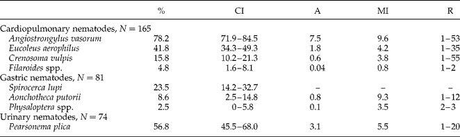
The present epidemiological research shows that the red fox in the study area hosts a wide variety of extraintestinal nematodes (table 2). Nine nematode species (A. vasorum, E. aerophilus, E. boehmi, C. vulpis, Filaroides spp., S. lupi, Aonchotheca putorii, Physaloptera spp. and P. plica) were found. The most prevalent parasite was A. vasorum (78.2%) in the heart and lung arteries, followed by P. plica (56.8%) in the urinary bladder. No fox was found to be completely negative for parasites.
The prevalence (%), abundance (A), mean intensity (MI) and range (R) of cardiopulmonary, gastric and urinary nematodes identified in the present study; N=number of foxes examined; CI=95% confidence intervals; –, not recorded.

For the most prevalent parasitic species a negative binomial distribution fitted the data, indicating an ecological equilibrium between hosts and parasites (all P values of the goodness-of-fit chi square tests were larger than 0.05). The parameters of the fitted distributions were: A. vasorum, k= 0.545, P= 0.068; E. aerophilus, k= 0.214, P= 0.108; P. plica, k= 0.485, P= 0.133.
No significant difference in the prevalence of parasites was observed between genders and age classes, except for P. plica (P value = 0.0001). For this parasite, juvenile animals (93.3%) were more infected than adults (47.5%). These results were confirmed by logistic regression with positivity for parasite as the dependent variable and gender and age as covariates.
Angiostrongylus vasorum (prevalence 78.2%), E. aerophilus (41.8%), C. vulpis (15.8%) and Filaroides spp. (4.8%) were found in the cardiopulmonary system. Altogether 67 multiple infections were recorded: two cases of quadruple infection with A. vasorum, E. aerophilus, C. vulpis and Filaroides spp.; 13 cases of triple infection and 52 cases of double infection, 38 of which included infections with A. vasorum and E. aerophilus. Co-infections with A. vasorum and E. aerophilus were the most frequent; however, the correlation was not statistically significant.
The results of the copromicroscopy concerning extraintestinal parasites were as follows: E. aerophilus prevalence 25.5% (95% CI: 18.8–32.1%), A. putorii 6.7%, S. lupi 4.2% (1.2–7.3%), Physaloptera spp. 1.2% (0–2.9%). Two foxes were found to be positive for Physaloptera spp. with the coprological test, but it was not possible to examine their stomachs. Two other foxes were positive at necropsy, but negative after the coprological examination.
The results of the coprological test concerning extraintestinal species were compared with necropsy considered as a gold standard and the sensitivity (S) and specificity (S′) were calculated for E. aerophilus, S = 0.49, S′ = 0.92; S. lupi, S = 0.26, S′ = 0.97; A. putorii, S = 0.33, S′ = 0.97; and Physaloptera spp., S = 0, S′ = 1. A low sensitivity of the coprological test is expected and was observed for E. aerophilus in foxes by Lalošević et al. (Reference Lalošević, Lalošević, Capo, Simin, Galfi and Traversa2013) and for S. lupi in dogs by Mylonakis et al. (Reference Mylonakis, Koutinas, Liapi, Saridomichelakis and Rallis2001).
The coprological test also revealed the presence of intestinal parasites: Ancylostomatidae (prevalence 48.5%), Toxocara canis (18.2%), Molineus spp. (18.2%), Toxascaris leonina (14.5%), Coccidia (10.3%), Trichuris vulpis (4.8%), Mesocestoides spp. (3.6%) and Hymenolepis diminuta (3.0%).
Angiostrongylus vasorum was found to be the most frequent cardiorespiratory parasite. The prevalence recorded in the present study is among the highest in Europe (table 1) and worldwide (56% in Canada, Jeffery et al., Reference Jeffery, Lankester, McGrath and Whitney2004). The presence of A. vasorum in our study area matches the predicted distribution described in the model by Morgan et al. (Reference Morgan, Jefferies, Krajewski, Ward and Shaw2009) based on climatic conditions. This species has been reported as a common parasite in foxes, and less frequently in dogs, in Europe, Africa, and North and South America (Morgan et al., Reference Morgan, Shaw, Brennan, De Waal, Jones and Mulcahy2005). However, over the past two decades, the number of reports in dogs has increased (Conboy, Reference Conboy2011).
The prevalence of E. aerophilus found in the present work (41.8%) is the highest reported in Italy and is comparable with other studies available (table 1). Most surveys reported prevalences higher than 30%, although values vary from 5% (Eira et al., Reference Eira, Viganda, Torres and Miquel2006, Portugal; Martinez-Carrasco et al., 2007, Spain) to 97.1% (Bružinskaitė-Schmidhalter et al., Reference Bružinskaitė-Schmidhalter, Šarkūnas, Malakauska, Mathis, Torgerson and Deplazes2011, Lithuania)) (table 1). Eucoleus aerophilus may be responsible for rare zoonotic infections, and 12 cases have been reported in the literature: 8 in Russia, 1 in Morocco, 1 in Iran (Aftandelians et al., Reference Aftandelians, Raafat, Taffazoli and Beaver1977), 1 in France and 1 in Serbia (Lalošević et al., Reference Lalošević, Lalošević, Klem, Stanojev-Jovanović and Pozio2008).
The prevalence of C. vulpis observed in the present investigation is consistent with values found in other surveys (table 1). Higher values (78.4%) have been found in foxes in Canada by faecal examination (Nevárez et al., 2005).
Lung nematodes of the genus Filaroides were detected in eight foxes (4.8%). These parasites are quite rare in canids. In Europe, to the best of our knowledge, they have only been found in dogs (Caro-Vadillo et al., 2005).
One of the two foxes whose nasal cavities we were able to examine was positive for E. boehmi and three adult worms were collected. Eucoleus boehmi is similar to E. aerophilus, but lives in the nasal and paranasal cavities of canids instead of in the trachea. Eggs of E. boehmi measure 54–60 × 30–35 μm, contain a multicellular embryo that frequently does not entirely fill the egg, have a surface marked by delicate pits, and are bipolar plugged. The eggs of E. aerophilus are bigger, 60–72 × 26–34 μm, have a net-like outer layer, polar plugs, and are entirely filled by one- or two-cell embryos (Campbell, Reference Campbell1991). Eucoleus boehmi was identified for the first time in foxes from Moravia and Austria (Supperer, Reference Supperer1953) and was then found in red foxes and wolves in Poland (Zarnowsky & Patyk, Reference Zarnowsky and Patyk1960), in foxes in Austria (Hinaidy, Reference Hinaidy1971), Germany (Schöffel et al., Reference Schöffel, Schein, Wittstadt and Hentsche1991), Hungary, Norway and Serbia (table 1). To the best of our knowledge, this is the first published report of E. boehmi in foxes in Italy.
Filarial infections were not found in any animal in the present work. Wild canids are infected by D. immitis in areas where this parasite is endemic (Gortázar et al., Reference Gortázar, Castillo, Lucientes, Blanco, Arriolabengoa and Calvete1994). This confirms that foxes are only epiphenomena in the epidemiology of D. immitis (Stancampiano et al., Reference Stancampiano, Capelli, Schiavon, Mutinelli and Bozzolan1998).
Spirocerca lupi (23.5%), A. putorii (8.6%) and Physaloptera spp. (2.5%) were found in the stomach (table 2). Spirocerca lupi lives in the oesophagus and stomach of wild canids (foxes, wolves, jackals) and domestic dogs in the Middle East, China, India, Japan, South America and southern USA (Pence & Stone, Reference Pence and Stone1978; Ferrantelli et al., Reference Ferrantelli, Riili, Vicari, Percipalle, Chetta, Monteverde, Gaglio, Giardina, Usai and Poglayen2010). Reports of S. lupi in European red foxes are limited to the Iberian peninsula (Gortázar et al., Reference Gortázar, Villafuerte, Lucientes and Fernandez-de-Luco1998; Segovia et al., Reference Segovia, Torres and Miquel2004; Eira et al., Reference Eira, Viganda, Torres and Miquel2006) and Belarus (Shimalov & Shimalov, Reference Shimalov and Shimalov2002). In Italy, this parasite has been found in foxes in Sardinia (Leoni et al., Reference Leoni, Masala and Garippa1985; Polidori et al., Reference Polidori, Mura, Moretti, Balzano and Principato1985) and in Sicily (Ferrantelli et al., Reference Ferrantelli, Riili, Vicari, Percipalle, Chetta, Monteverde, Gaglio, Giardina, Usai and Poglayen2010) (table 1).
Reports of Physaloptera spp. and A. putorii in foxes are rare (table 1): P. sibirica was found for the first time in Italy in foxes and badgers from Aosta Valley and Piedmont (Ferroglio et al., Reference Ferroglio, Ragagli and Trisciuoglio2009), with a similar prevalence in foxes (2.63%) to the value found in the present study. This gastric parasite had previously only been reported in Spain (Miquel et al., Reference Miquel, Segovia, Feliu and Torres1996; Rodriguez & Carbonell, Reference Rodriguez and Carbonell1998; Segovia et al., Reference Segovia, Torres and Miquel2004) and in Belarus (Shimalov & Shimalov, Reference Shimalov and Shimalov2002). Aonchotheca putorii is more frequently found in mustelids (Sato et al., Reference Sato, Inaba, Ihama and Kamiya1999).
Pearsonema plica was found in the urinary bladder of 42 foxes (56.8%), while kidneys were negative. Eggs of this Trichuridae are shed in the urine and are characterized by a lemon shape, similar to the eggs of E. aerophilus and E. boehmi (Campbell, Reference Campbell1991). Pearsonema plica was found in Italy in foxes more than 20 years ago (Iori et al., Reference Iori, Costantini and Cancrini1990) and more recently in pets (Callegari et al., Reference Callegari, Kramer, Cantoni, Di Lecce, Dodi and Grandi2010; Rossi et al., Reference Rossi, Messina, Ariti, Riggio and Perrucci2011). The prevalence of this parasite in the present study is in agreement with other epidemiological studies (table 1).
Although all the foxes examined were negative for Trichinella spp., we believe that monitoring should continue, since this host is one of the main reservoirs of many Trichinellaspecies (Pozio, Reference Pozio2007; Chmurzyńska et al., Reference Chmurzyńska, Różycki, Bilska-Zajęc, Nöckler, Mayer-Scholl, Pozio, Cencek and Karamon2013). Foxes that are positive for Trichinella spp. have been found in Liguria and Piedmont (Rossi et al., Reference Rossi, Pozio, Mignone, Ercolini and Dini1992) and a human case was reported in Cuneo (Romano et al., Reference Romano, Motta, Melino, Negro, Gavotto, Decasteli, Careddu, Bianchi, Bianchi and Pozio2011).
The present study confirms that red foxes represent an important wild reservoir of extraintestinal parasites that can be transmitted to domestic animals, particularly to dogs (Bolt et al., Reference Bolt, Monrad, Henriksen, Dietz, Koch, Bindseil and Jensen1991). Furthermore, the presence of E. aerophilus is important, because this parasite can be responsible for zoonotic infections (Lalošević et al., Reference Lalošević, Lalošević, Klem, Stanojev-Jovanović and Pozio2008). Extraintestinal nematodes have been reported increasingly in pets worldwide and are considered as emerging parasites (Rinaldi et al., Reference Rinaldi, Calabria, Carbone, Carrella and Cringoli2007; Conboy, Reference Conboy2009; Bork-Mimm & Rinder, Reference Bork-Mimm and Rinder2011; Clark et al., Reference Clark, Lopez, Levine, Cooper, Craig, Voges, Johnson and Porter2013), due to the greater focus of parasitologists on these species. The apparent spread to domestic animals could be associated with the increase in the fox population (Deplazes et al., Reference Deplazes, Hegglin, Gloor and Romig2004). Epidemiological research on foxes could therefore provide useful information on these parasites.
Financial support
This research received no specific grant from any funding agency, commercial or not-for-profit sectors.
Conflict of interest
None.