Minimally invasive surgical approaches to repair congenital heart defects have been introduced for over a decade, and initially strived to improve cosmetic appearance after the closure of an atrial septal defect. Reference Mihalyi, Kiraly, Prodan, Bodor, Tamas and Hartyanszky1–Reference Heinisch, Bartkevics and Beck27 Owing to a steep surgical learning curve, concerns with regards to safety and technique complexity were understandably expressed, and have hampered more widespread acceptance in the surgical community, since all minimally invasive techniques potentially require alteration of conventional cannulation sites for cardiopulmonary bypass, with reduced surgical access and control of certain cardiac structures. Reference Mavroudis, Backer, Stewart and Heraty28 However, increasingly, requests by parents, peer pressure, and competition with catheter-based percutaneous interventional techniques have played a role towards developing and expanding minimally invasive surgical approaches, Reference Dodge-Khatami and Salazar15 thereby broadening the indications to safely repair a wider range of congenital heart defects, including partial anomalous pulmonary venous connection, partial and complete atrio-ventricular septal defect with a cleft in the left-sided atrio-ventricular valve, cor triatriatum, scimitar syndrome, ventricular septal defect, subaortic fibromuscular ridge, and tetralogy of Fallot. Reference Mihalyi, Kiraly, Prodan, Bodor, Tamas and Hartyanszky1–Reference Heinisch, Bartkevics and Beck27 Together with enhanced anaesthesia pain management protocols, various surgical incisions such as a right horizontal or vertical axillary thoracotomy, and the anterolateral thoracotomy have been compared to the conventional median sternotomy, Reference Mihalyi, Kiraly, Prodan, Bodor, Tamas and Hartyanszky1–Reference Heinisch, Bartkevics and Beck27 and aim not only to improve cosmetics, but merit consideration to reduce post-operative pain and discomfort, achieve faster post-operative functional recovery, and perhaps decrease hospital costs. While the duration of ICU and hospital stay, as well as chest drain output and blood transfusions, have extensively been studied after minimally invasive techniques, Reference Schreiber, Bleiziffer and Kostolny3,Reference Mishaly, Ghosh and Preisman4,Reference Palma, Giordano and Russolillo6–Reference Hu, Tan, Chen, Ding and Xu10,Reference Yaliniz, Topcuoglu and Gocen12–Reference An, Zhang, Zheng, Wang, Wu and Xing14,Reference An, Zhang, Zheng, Wang and Ma17,Reference Heinisch, Wildbolz and Beck19,Reference Lee, Weiss, Williams, Kiblawi, Dong and Nguyen21,Reference Wang, Ye, Ge and Wang23–Reference Heinisch, Bartkevics and Beck27 to the best of our knowledge, only one group evaluated pain and comfort of patients in the immediate post-operative period, and subsequent quality of life. Reference Kara and Caner29 In our single-centre study, we sought to evaluate the quality-of-life metrics by comparing paediatric congenital heart repairs through a mini right axillary thoracotomy, with children undergoing repair of similar defects through a median sternotomy.
Materials and methods
Patient population
The Institutional Review Board approval was obtained before all data collection (#HSC-MS-19-1082, approved 20 February, 2020). Between July, 2018 and June, 2020, all consecutive patients under 18 years of age with the principal first diagnoses of peri-membranous, muscular or inlet VSD (n = 56), atrial septal defect (n = 20), partial anomalous pulmonary venous connection (n = 6), scimitar syndrome (n = 2), double-chambered right ventricle (n = 2), and partial atrio-ventricular septal defect (n = 1), who underwent surgical repair via a minimally invasive right axillary thoracotomy (n = 54) or median sternotomy (n = 33), were included in this retrospective study. The patients were assigned according to the surgeon and referring cardiologist preference after discussion with the patient’s parents/guardian, and offered either of the two approaches. Groups were comparable with regards to the distribution of types of diagnoses/repairs amongst the two approaches.
Surgical approach
Our surgical approach for the minimally invasive right axillary thoracotomy includes a short muscle-sparing right mid-axillary incision underneath the right arm close to the armpit, while leaving the right groin vessels free and prepped without any anaesthesia lines in case peripheral cannulation becomes necessary for cardiopulmonary bypass. Reference Prêtre, Kadner, Dave, Dodge-Khatami, Bettex and Berger2,Reference Dave, Comber, Solinger, Bettex, Dodge-Khatami and Prêtre5,Reference Dodge-Khatami and Salazar15 In the right axillary group, intracardiac repair was performed using either induced ventricular fibrillation or aortic cross-clamping with cardioplegia; cross-clamping with cardioplegia was used in all median sternotomy patients. Peripheral femoral vessel cannulation (venous, n = 6 and arterial, n = 2) was required in six patients in the right axillary thoracotomy group, and in none for the sternotomy group. All patients in both groups were treated with the same protocols of general anaesthesia and peri-operative management, with identical intention to treat with regards to blood transfusion, weaning from mechanical ventilation and tracheal extubation, removal of chest drains, transfer from the ICU and hospital discharge.
Data collection
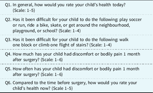
In addition to the usual demographic details, the following data and outcomes were analysed and compared between the two groups: duration of induced ventricular fibrillation/aortic cross-clamping, duration of cardiopulmonary bypass, length of tracheal intubation, duration of chest drains, requirement for blood transfusion, duration of ICU and hospital stay. To evaluate post-operative quality of life, a questionnaire based on the Child Health and Infant/Toddler Health Questionnaires focused on the patient’s global health, physical activity, and pain/discomfort was administered to the parents/guardians after consent/assent was obtained. Questions included in the quality-of-life metrics questionnaire are reported in Table 1, while the scoring system used to evaluate and quantitate the answers received is reported in Table 2. All responses were obtained within the first two 2 years after each patient’s surgery.
Questionnaire with quality-of-life metrics

Scoring system for the quality-of-life metrics

Statistical analysis
Data are described as numbers with frequencies and percentages, medians with interquartile ranges, and means with standard deviation. Two-tailed p-values of less than 0.05 were considered statistically significant.
Results
Demographic and clinical data
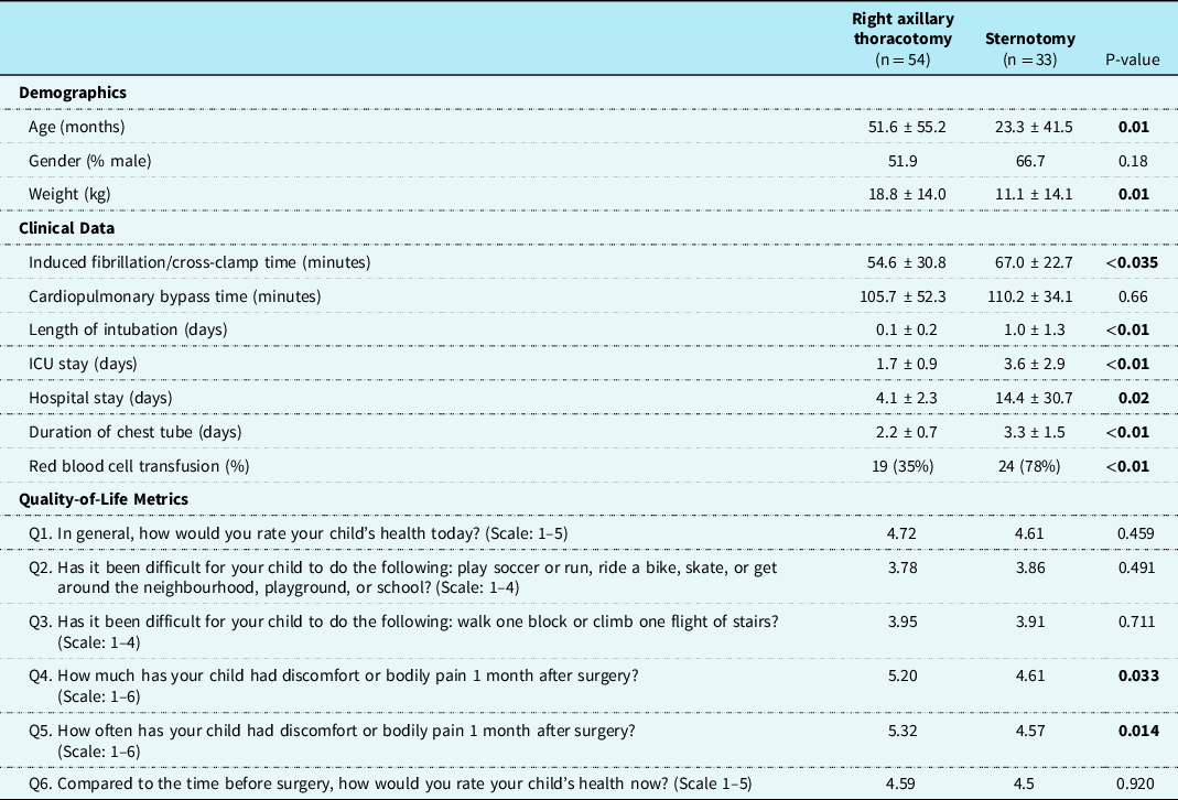
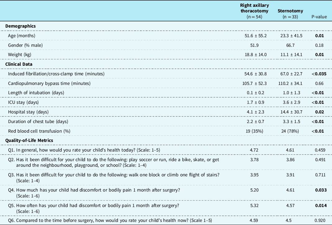
A total of 87 patients responded to the QOL questionnaire (n = 54 in the right axillary thoracotomy group and n = 33 in the median sternotomy group). Patients who received a right axillary thoracotomy were significantly older (51.6 months ± 55.2 versus 23.3 months ± 41.5; p = 0.01) and weighed more (18.8 versus 11.1 kg; p = 0.01). Length of intubation (p < 0.01), ICU length of stay (p < 0.01), hospital length of stay (p = 0.02), chest tube duration (p < 0.01), and need for red blood cell transfusion (p < 0.01) were all statistically significantly lower in the right axillary thoracotomy group (Table 3). Induced ventricular fibrillation/cross-clamp time was slightly shorter in the right axillary group compared to the median sternotomy group (54.6 ± 30.8 minutes versus 67.9 ± 22.7 minutes, respectively; p < 0.035). There was no statistically significant difference between the cardiopulmonary bypass times and between the two groups (105.7 ± 52.3 versus 110.2 ± 34.1 minutes; p = 0.66). In the right axillary group, there were two unplanned reoperations during the same hospital stay for residual findings: both were for baffle leaks after low inferior sinus venosus atrial septal defect repairs at the inferior caval vein-right atrial/left atrial junction, one leading to streaming of desaturated blood and mild cyanosis, and the other to residual left-to-right shunting deemed unsatisfactory. Both patients underwent baffle revisions, performed through the same mini right axillary incision, without complications.
Comparative results between both approaches
Quality-of-life metrics
The perceived perception by parents was a significantly less degree and frequency of their child’s post-operative pain at 1 month in those who underwent a minimally invasive right axillary thoracotomy approach (p = 0.033 and p = 0.014, respectively), compared to after repair through a sternotomy. There were no statistically significant differences noted in the patient’s global health or physical activity limitations at last follow-up after hospital discharge between the two groups.
Discussion
As the substantial reduction in mortality and even morbidity for the treatment of certain congenital heart defects has become the expected norm, patient parents, the media, and society at large have challenged the surgical community to become “minimally invasive”, avoiding a median sternotomy for surgical repairs not amenable to catheter intervention, without compromising the quality of repair. To offer valid alternatives to less invasive transcatheter interventional techniques, various types of surgical incisions have been introduced to avoid a median sternotomy including the anterior, anterolateral (or sub-mammary), posterolateral, and muscle-sparing right axillary thoracotomy. Reference Mihalyi, Kiraly, Prodan, Bodor, Tamas and Hartyanszky1–Reference Heinisch, Bartkevics and Beck27 With the anterolateral or sub-mammary approach, rib deformation and atrophy of severed pectoral muscles, and asymmetrical breast development in females have been reported, resulting in suboptimal cosmetic results. Reference Schreiber, Bleiziffer and Kostolny3,Reference Mishaly, Ghosh and Preisman4,Reference Palma, Giordano and Russolillo6,Reference Ding, Wang and Dong7,Reference Hu, Tan, Chen, Ding and Xu10–Reference Yaliniz, Topcuoglu and Gocen12,Reference Gil-Jaurena, Pérez-Caballero, Pita-Fernández, González-López, Sánchez and De Agustín16,Reference Hong, Chen and Lin20,Reference Liu, Rui, Zhang, Lin, Li and Hua24–Reference Luo, Chen, Yu, Chen and Huang26,Reference Mavroudis, Backer, Stewart and Heraty28 The suggested advantages of the mini right axillary incision include: (a) a muscle-sparing approach with resultant shorter recovery time and return to full capacity of the right arm and shoulder function, as only longitudinal muscle fibres of the serratus anterior are split parallel to the ribs; (b) avoidance of breast tissue, which is especially important in young female infants given the absence of any visible landmark at the time of surgery, thus avoiding future asymmetrical breast growth; (c) a completely hidden scar underneath the resting arm, resulting in a superior cosmetic appearance in comparison with any other thoracic incision. Reference Mihalyi, Kiraly, Prodan, Bodor, Tamas and Hartyanszky1–Reference Dave, Comber, Solinger, Bettex, Dodge-Khatami and Prêtre5,Reference Gil-Jaurena, Castillo and Gonzalez8–Reference Hu, Tan, Chen, Ding and Xu10,Reference An, Zhang, Zheng, Wang, Wu and Xing14–Reference An, Zhang, Zheng, Wang and Ma17,Reference Heinisch, Wildbolz and Beck19–Reference Lee, Weiss, Williams, Kiblawi, Dong and Nguyen21,Reference Wang, Ye, Ge and Wang23–Reference Heinisch, Bartkevics and Beck27
As with introducing any new technique, during the early development of the minimally invasive right axillary approach, our goal was to demonstrate its safety, and reproducibly match standard hospital outcomes or even improve upon them, compared to surgery through a conventional median sternotomy. Several reports from groups who perform variations of our minimally invasive technique have presented similar if not better data related to the length of tracheal intubation, duration of need for chest drains, requirement for blood transfusion, duration of ICU and hospital length of stay, Reference Mihalyi, Kiraly, Prodan, Bodor, Tamas and Hartyanszky1–Reference Dave, Comber, Solinger, Bettex, Dodge-Khatami and Prêtre5,Reference Gil-Jaurena, Castillo and Gonzalez8–Reference Hu, Tan, Chen, Ding and Xu10,Reference An, Zhang, Zheng, Wang, Wu and Xing14–Reference An, Zhang, Zheng, Wang and Ma17,Reference Heinisch, Wildbolz and Beck19–Reference Lee, Weiss, Williams, Kiblawi, Dong and Nguyen21,Reference Wang, Ye, Ge and Wang23–Reference Heinisch, Bartkevics and Beck27 as confirmed in our current study. The two patients requiring reoperations during the same hospital stay merit a discussion regarding optimal pre-operative imaging, considerations with regards to cannulation, and sufficient exposure required for an optimal repair. It has been an integral part of the pre-operative imaging protocol to use low-dose CT scanning for lesions involving anomalous pulmonary vein connections and either very high or very low sinus venosus atrial septal defects. Reference Dodge-Khatami and Adebo30 In patients with anomalous pulmonary venous connections, documenting a high insertion of upper and middle lobe pulmonary veins may allow a planned Warden operation and very high venous cannulation of the innominate vein or even internal jugular vein. With very low sinus venosus defects, pre-operative CT imaging may point to the need for peripheral venous cannulation of the femoral vein, so as to leave the area of repair free from cluttering cannulae. In our two patients, despite adequate pre-operative advanced imaging and satisfactory exposure after peripheral venous cannulation, imperfect technique resulted in low sinus venosus atrial septal baffle leaks requiring revision during the same hospital stay, through the same mini right axillary incision.
With regard to the quality-of-life metrics, we identified only one prior study comparing patients operated on through an anterolateral thoracotomy with those who underwent median sternotomy, which evaluated pain score systems on two occasions in the immediate post-operative period. Reference Kara and Caner29 After initial slightly higher pain scores in patients undergoing surgery through the anterolateral thoracotomy, a general state of well-being, comfortable mobilisation, and greater success in post-operative exercises were observed, starting from the first post-operative day. Additionally, discharge occurred earlier, lower subsequent pain scores were reported, along with improved signs of well-being during the first post-operative week. Reference Kara and Caner29
For our study, an abbreviated questionnaire derived from the Child Health and Infant/Toddler Health Questionnaires system was used, focusing primarily on the patient’s global health, physical activity, and pain/discomfort. The results of the questionnaires (Table 3) didn’t show any statistical difference in perceived quality of life between the two groups at last follow-up when beyond a month. During the early post-operative period up to 1 month after hospital discharge, the amount and duration of discomfort or bodily pain were significantly less in the right axillary thoracotomy group, compared to patients undergoing a conventional median sternotomy (p = 0.033 and p = 0.014, respectively). All data in this study were obtained within 2 years of the patient’s surgical procedure, and two out of six QOL questions (question 4 and question 5) include a specific time frame of 1 month post-operatively, which limits or could influence answers should the time frame be left open and/or asked at an earlier or later date. Although conjecture, it is uncertain whether this time frame after surgery is enough to capture potential differences in a patient’s global health and physical limitations, or highlights psychological benefits pertaining to self-perception with a hidden scar. It is possible that with increased follow-up time after surgery, physical limitations, symptoms, or simply perceived psychological status and body perception could be different between the two incisions, which begs for longer follow-up studies of similar design.
Study limitations
The major limitation of this study is its retrospective design. In addition, the groups included all consecutive patients coming for repairs of the same diagnoses during the study period, and are not propensity matched in terms of patient age and weight, which could potentially impact the QOL scores due to differences between age groups (infant/toddler versus child). Lastly, this study would benefit from longer follow-up than 2 years, perhaps up to decades, as patient’s and parents’ perceptions regarding QOL measures may change throughout a child’s lifetime, well into adolescence and early adulthood.
Conclusion
In conclusion, the minimally invasive right axillary thoracotomy approach for the surgical repair of certain congenital heart lesions was associated with decreased red blood cell transfusion, earlier chest tube removal, decreased time to tracheal extubation, and decreased ICU and hospital length of stay, compared to a similar group of children with regards to diagnoses and repaired defects during the same period, although not matched for age or weight. With regards to quality of life, the parents/guardians perceived a significantly less degree and frequency of post-operative pain in patients who underwent a right axillary thoracotomy until 1 month, but not beyond that. The mini right axillary thoracotomy should be taken into consideration as a safe and reproducible alternative approach for the surgical treatment of the less complex congenital heart defects, and does not add to surgical complexity or post-operative burden to the children. Future studies could focus on longer term differences in the QOL metrics between the right axillary approach and median sternotomy, in age and weight propensity-matched patients.
Acknowledgements
None.
Financial support
This research received no specific grant from any funding agency, commercial, or not-for-profit sectors.
Conflicts of interest
None.
Ethical standards
The research does not involve human and/or animal experimentation.