Introduction
Although microscopic myringoplasty is the most frequently performed procedure for repairing tympanic membrane perforations, anterior perforations pose a difficult and risky reconstructive challenge with a poor surgical outcome, as the anterior canal wall limits visualisation of the tympanic ring.Reference Tarabichi1 In addition, a reduced vascular supply that decreases graft viability, and medialisation of the graft (e.g. because of negative pressure produced by sniffing), lower the success rate compared to posterior perforations.Reference Celik, Samim and Oztuna2
The post-auricular approach is usually the preferred approach, and anterior canalplasty is performed when the anterior canal wall is prominent and the anterior annulus cannot be visualised. This requires considerable surgical skill.Reference Peng and Lalwani3, Reference Gavriel and Eviatar4
An endoscopic transcanal approach bypasses the narrow segment of the ear canal, and provides a wide view, which enables surgeons to look ‘around the corner’, even when a 0-degree endoscope is used. This can decrease the need for the post-auricular approach and canalplasty, without increasing the failure rate.Reference Ayache5, Reference Tarabichi, Nogueira, Marchioni, Presutti, Pothier and Ayache6
Incorporating the endoscope as a tool into the surgical armamentarium in otology has contributed considerably to minimally invasive surgery. The increasing use of endoscopes in ear surgery is justified by their proven advantages over the microscope, including wider viewing angles, and the visualisation of deep recesses and hidden structures beyond the shaft of the surgical instruments.Reference Badr-El-Dine, James, Panetti, Marchioni, Presutti and Nogueira7
This study compares the clinical and audiological results of the endoscopic transcanal push-through technique with the commonly used microscopic underlay technique in myringoplasty performed for anterior tympanic membrane perforations. We aimed to improve the outcome of surgery in patients with anterior tympanic membrane perforations.
Materials and methods
Study design and setting
An open-label randomised clinical trial was performed in the Otolaryngology Department, Suez Canal University Hospital (Ismailia, Egypt) between June 2015 and May 2017. The study protocol was approved by the local ethics committee and written informed consent was obtained from all patients.
Patient eligibility and enrolment
The study included patients of both sexes, aged over 18 years, who attended the ENT out-patient clinic during the study period. All patients had a small anterior tympanic membrane perforation, with a dry ear for at least three months. Patients with an air–bone gap of more than 25 dB were excluded from the study. Patients were also excluded if they had significant co-morbidities, such as a cholesteatoma, a retraction pocket, Eustachian tube dysfunction, active chronic suppurative otitis media or mixed hearing loss.
Study plan
All patients had their complete history taken. Complete ENT examination was performed, including otoendoscopy to assess the external canal (narrow, wide, anterior bulge), the tympanic membrane perforation (site, size, shape, edge) and the middle-ear mucosal status. Audiometric assessment using pure tone audiometry and the necessary pre-operative laboratory investigations were performed for all patients. High-resolution computed tomography of the temporal bone was not routinely carried out on our patients.
Randomisation
Fifty-six patients were divided randomly into two groups: an endoscopic transcanal push-through myringoplasty group (n = 28) and a microscopic underlay myringoplasty group (n = 28). Randomisation was performed prior to surgery as follows. Opaque envelopes were numbered sequentially from 1 to 56. A computer-generated table of random numbers was used for group assignment; if the last digit of the random number was 0–4, assignment was to the endoscopic transcanal push-through myringoplasty group, and if the last digit was 5–9, assignment was to microscopic underlay myringoplasty group.
Surgical technique
Endoscopic transcanal push-through myringoplasty
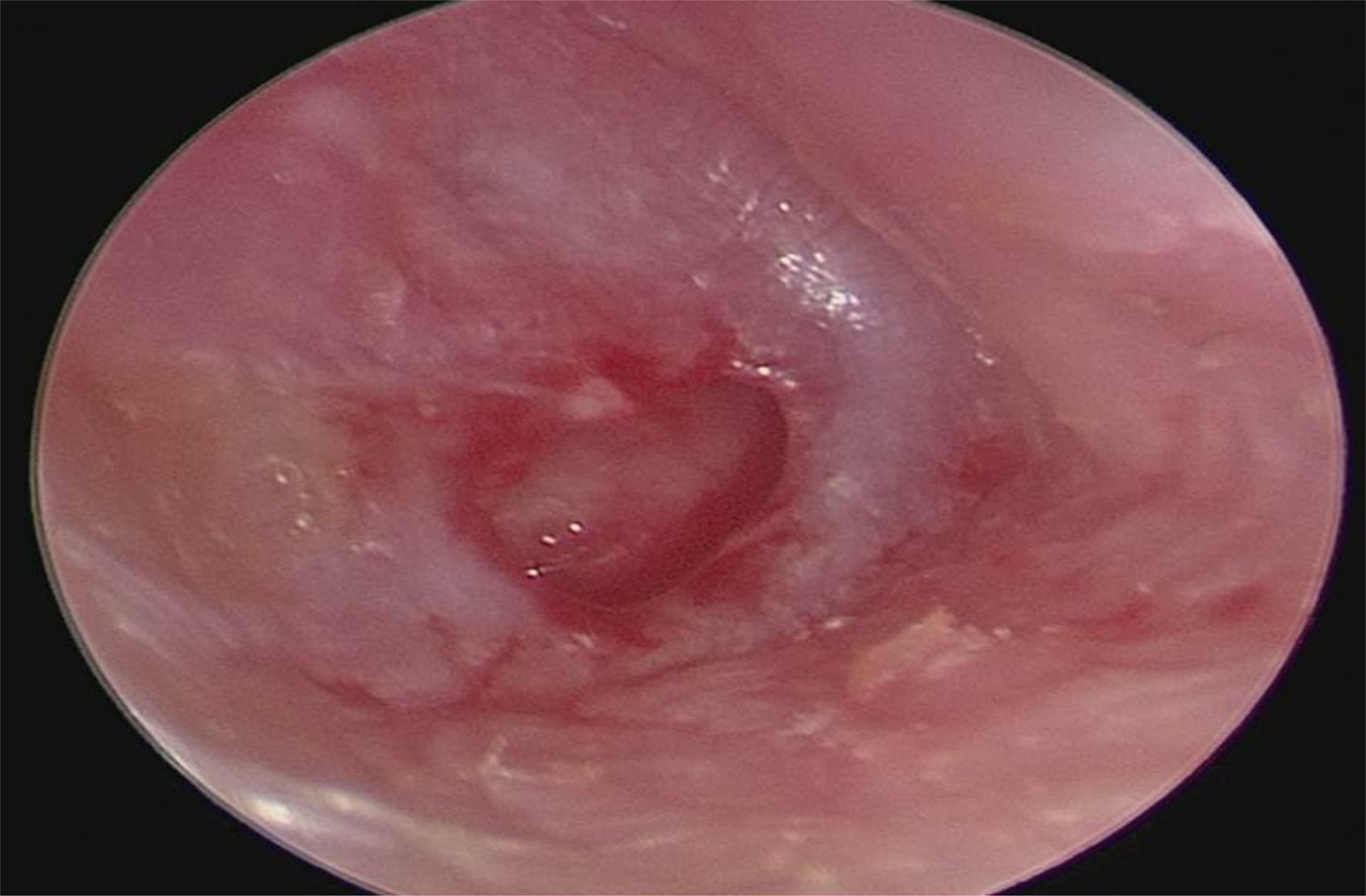
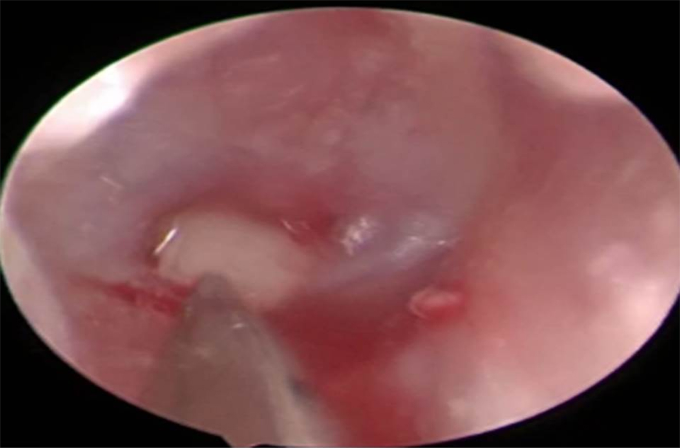
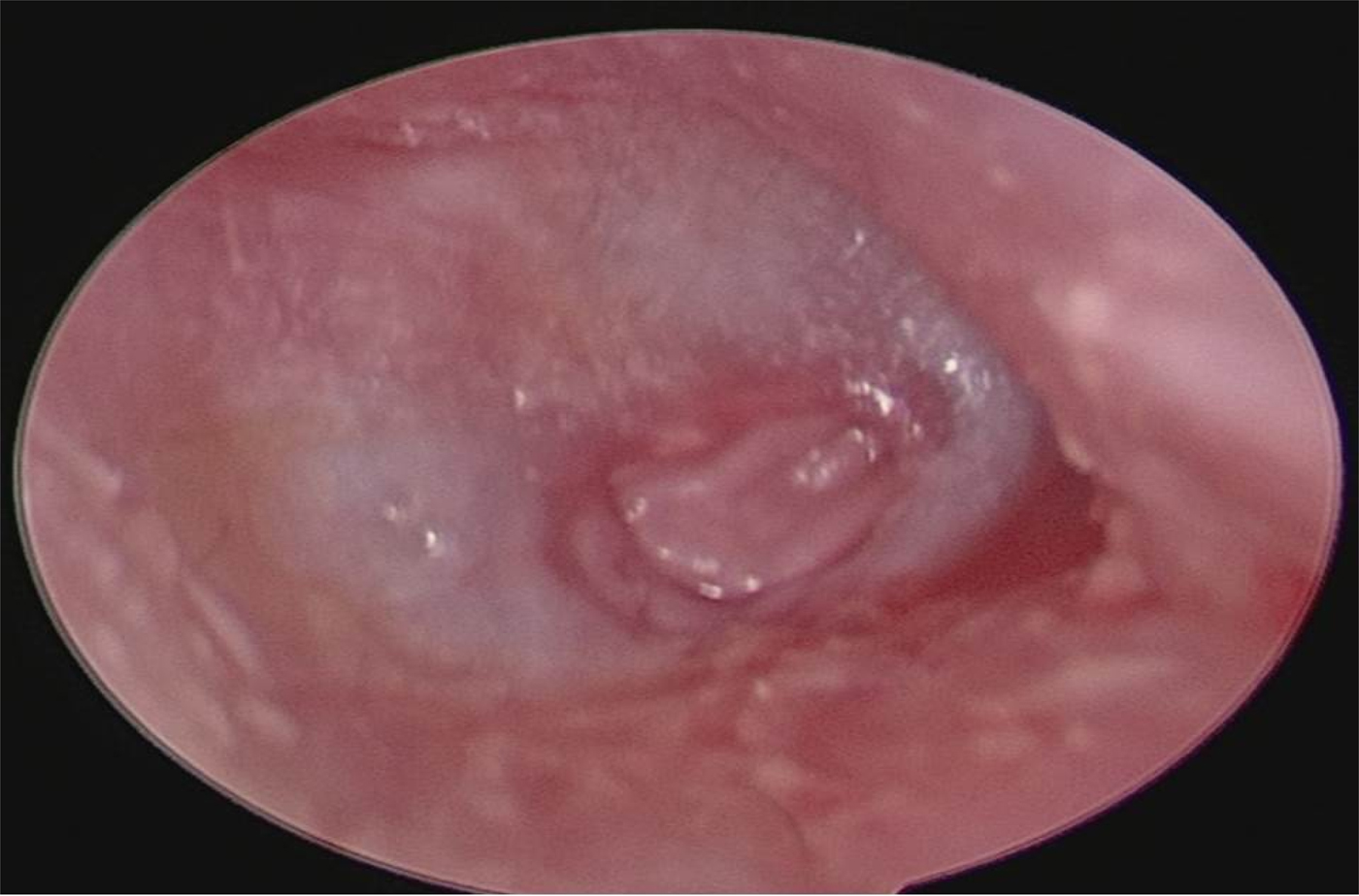
Under general anaesthesia with local injection of adrenaline 1:100 000 combined with 2 per cent lidocaine, the surgeon used a 0-degree, 3 mm diameter, 20 cm length rigid endoscope to visualise the perforation, and trimmed its edges with an angled pick. A tragal cartilage perichondrial graft was then harvested and the cartilage was freed from perichondrium on both sides. The middle ear was tightly packed with Gelfoam through the perforation, and the Eustachian tubal orifice was supported more tightly in order to prevent graft medialisation. The diameter of the perforation was measured using a sterile piece of aluminium foil. The cartilage graft, which was slightly larger than the perforation, was pushed through the perforation, and placed in an underlay fashion. The perichondrial layer was also pushed through the perforation, covering the cartilage graft completely (Figures 1–3). Finally, the external auditory canal was packed with Gelfoam to the level of the isthmus. A small dressing was applied to cover the auricle.Reference Celik, Samim and Oztuna2
Tympanic membrane perforation after its edges have been trimmed.

Placement of cartilage graft through the perforation using the endoscopic push-through technique.

Tympanic membrane perforation after placement of the perichondrial graft through the perforation.

Microscopic underlay myringoplasty
The patient was put under general anaesthesia with a local injection of adrenaline 1:100 000 combined with 2 per cent lidocaine in the post-auricular area, canal and tragus. A tragal cartilage perichondrial graft was harvested and prepared in the same manner as in the endoscopic transcanal push-through myringoplasty group. A post-auricular incision was made and a Palva flap was elevated to obtain access to the posterior canal wall. The posterior meatal skin was cut posteriorly 1 cm from the lateral end of the bony canal, and the edge of the perforation was trimmed. A tympanomeatal flap was elevated to the level of the annulus. If the anterior annulus was not in view, the bony canal was carefully widened using a diamond bur, with protection of the reflected canal skin. After elevation of the annular ring posteriorly and inferiorly, the cartilage perichondrial graft was placed in an underlay fashion, and was supported with Gelfoam in the middle ear, particularly near the Eustachian tube.Reference Glasscock and Gulya8
Post-operatively, patients in both groups were given oral amoxicillin/clavulanate potassium, 1 g twice daily for one week. Non-steroid anti-inflammatory agents were added depending on patients’ needs. Gelfoam in the external auditory canal was removed at three weeks post-operatively.
Outcome assessment
In both groups, the operative time (from the start of surgery (after anaesthesia induction) to the end of surgery) and intra-operative blood loss were recorded. At the end of the third month post-operatively, an endoscopic examination was conducted to assess graft take-up, and audiometric evaluation was carried out to measure air–bone gaps. In addition, any intra-operative or post-operative complications were recorded.
Data collection and allocation concealment
At the time of enrolment (day 0), each participant underwent a brief interview with the physician; a questionnaire was completed, and demographic and disease-related information were obtained.
Group assignments were placed into opaque sealed envelopes. The envelopes were opened by the otosurgeon just prior to surgery.
Operative time and blood loss were estimated by nursing staff in the operating theatre. At the end of the third post-operative month, ear endoscopy was carried out by a colleague otologist.
Statistical analysis
The data collected were analysed using SPSS statistical software, version 15 (SPSS, Chicago, Illinois, USA).
Ethical considerations
All participants included in the study were informed about the procedures and the expected results, and written informed consent was obtained. The local ethics committee approved the study.
Results
Fifty-six patients with a small anterior tympanic membrane perforation, with a dry ear for at least three months, were included in our study. They were randomly divided into two groups: endoscopic transcanal push-through myringoplasty (n = 28) and microscopic underlay myringoplasty (n = 28).
The patients’ mean age (± standard deviation (SD)) was 34 ± 12.9 years. There were 34 females and 22 males. Twenty-four patients had their right ear operated on and 32 their left ears.
The mean operative time for the endoscopic transcanal push-through myringoplasty group was three times shorter than that of the microscopic underlay myringoplasty group (p < 0.05) (Table I). The mean blood loss in the endoscopic transcanal push-through myringoplasty group was five times lower than that of the microscopic underlay myringoplasty group (p < 0.05) (Table II).
Comparison of operative times in both groups

* Statistically significant at the 95 per cent confidence level (t-test, 2-tailed). SD = standard deviation
Comparison of blood loss in both groups

* Statistically significant at the 95 per cent confidence level (t-test, 2-tailed). SD = standard deviation
By the end of the third post-operative month, the overall success rate was 89.2 per cent (50 out of 56 patients showed graft take-up without complications (namely, residual perforation, medialisation of the graft or ear discharge)). The success rate was 85.7 per cent (24 out of 28 patients) in the microscopic underlay myringoplasty group and it was 92.9 per cent (26 out of 28 patients) in the endoscopic transcanal push-through myringoplasty group. An example of a tympanic membrane graft on endoscopic examination three months’ post-operatively, in a patient who underwent endoscopic transcanal push-through myringoplasty, is shown in Figure 4.
Tympanic membrane graft three months’ post-operatively (endoscopic transcanal push-through myringoplasty group).

In the microscopic underlay myringoplasty group, two cases had medialisation of the graft and two other cases had residual perforation. In the endoscopic transcanal push-through myringoplasty group, there was only a small pin-point sized perforation at the anterosuperior edge of the graft in two patients.
The mean air–bone gap was measured in both groups pre-operatively and at three months post-operatively. The mean (± SD) pre-operative air–bone gap was 17.4 ± 3.7 dB in the endoscopic transcanal push-through myringoplasty group, while it was 18.5 ± 2.2 dB in the microscopic underlay myringoplasty group; there was no statistically significant difference between the two groups. At the end of the follow-up period, there was a marked improvement in air–bone gap for both groups, with values of 6.1 ± 3.7 dB in the endoscopic transcanal push-through myringoplasty group and 9.3 ± 5.5 dB in the microscopic underlay myringoplasty group at three months post-operatively, with no statistically significant difference between the two groups (Table III). There was also no statistically significant difference in terms of air–bone gap closure, which was 11.4 ± 2.3 dB in the endoscopic transcanal push-through myringoplasty group compared to 9.2 ± 5.1 dB in the microscopic underlay myringoplasty group (Table III).
Comparison of pre- and post-operative air–bone gaps, and air–bone gap closure, in both groups

ABG = air–bone gap; SD = standard deviation
Discussion
Visualisation of the tympanic membrane perforation's anterior edge and the anterior annulus in cases with a prominent anterior canal wall is a challenge during myringoplasty. In order to overcome this difficulty, some surgeons proceed to anterior canalplasty, which is considered an invasive procedure.Reference Celik, Samim and Oztuna2 Utilisation of an endoscope provides advantages to the surgeon in these circumstances because it magnifies the surgical field on a straight line. With its wider angle, and by getting the endoscope closer to the surgical area, deep corners can also be visualised.Reference Tarabichi1–Reference Yung9 During myringoplasty, the anterior annulus can easily be seen, and the operation can then be completed without performing anterior canalplasty. Manipulation of the endoscope is easier and faster compared to the microscope.Reference Pothier10 In the endoscopic push-through technique described in our study, the graft can be placed through the perforation by a transcanal underlay approach, without skin incisions or the preparation of a tympanomeatal flap.
Since the time when the endoscopic technique was introduced for middle-ear surgery, the concept of minimally invasive surgery has developed. The endoscopic technique could prevent external incisions, soft tissue dissection and bone drilling. Furthermore, operative times have been shown to markedly decrease with trained surgeons.Reference Presutti, Marchioni, Mattioli and Villari11
Canalplasty was performed in 10 of 28 patients (36 per cent) who underwent microscopic myringoplasty. However, none of the 28 patients who underwent the endoscopic procedure required such an intervention. This explains the shorter operative time and reduced blood loss in the endoscopic group.
Celik et al. reported a mean (± SD) operative time of 36.1 ± 5.8 minutes using the endoscopic push-through technique, which is similar to our results.Reference Celik, Samim and Oztuna2 In addition, Dündar et al. showed a statistically significant difference in operative time between endoscopic and microscopic techniques (51.37 vs 67.03 minutes, respectively).Reference Dündar, Kulduk, Soy, Aslan, Hanci and Muluk12 Choi et al. reported average times of 68.2 and 88.9 minutes for endoscopic and microscopic techniques respectively. They conducted canalplasty in 33.3 per cent of their patients when the microscope was used exclusively during surgery.Reference Choi, Noh, Park, Lee, Yook and Choi13 Nassif et al. reported average operative times of 80 and 90 minutes for endoscopic and microscopic techniques respectively.Reference Nassif, Berlucchi and Radaelli de Zinis14 Lade et al. found that the endoscopic tympanoplasty results were similar to those of the microscopic technique, but the former technique was more tolerated in terms of the cosmetic appearance. They concluded that endoscopic tympanoplasty is a potential alternative to microscopic tympanoplasty.Reference Lade, Choudhary and Vashishth15
Anterior tympanic membrane perforations are associated with increased failure rates after reconstruction. In this study, we preferred cartilage-perichondrium composite as a grafting material. Cartilage is durable, strong against absorption and resists negative pressure (such as that produced by sniffing). Tragal cartilage-perichondrium is particularly preferred because it is easy to harvest and it prevents post-operative scarring.
The overall graft success rate in our study was 89.2 per cent. In the endoscopic push-through myringoplasty group, the success rate was 92.9 per cent, with a mean (± SD) air–bone gap gain of 11.4 ± 2.3 dB. The graft success rate in the microscopic myringoplasty group was 85.7 per cent, with an air–bone gap gain of 9.2 ± 5.1 dB. Nassif et al. compared the results of endoscopic and microscopic myringoplasty in children and found no statistically significant differences either in success rates (90.9 per cent and 82.6 per cent respectively) or the last recorded air–bone gap between the endoscopic and microscopic groups (6.6 dB and 6.2 dB respectively).Reference Nassif, Berlucchi and Radaelli de Zinis14 Ayache reported a success rate of 96 per cent for endoscopic transcanal cartilage myringoplasty, with a non-significant decline in air–bone gap, from 17.7 dB to 7.9 dB. He concluded that endoscopic transcanal cartilage myringoplasty is an easy, reliable, feasible and less invasive procedure, which should be learned.Reference Ayache5
Dornhoffer studied cartilage graft and its use in the reconstruction of tympanic membrane in various pathologies over more than 20 years, and compared cartilage to perichondrium as graft material. The author found that cartilage had a 100 per cent success rate versus 85 per cent in the perichondrium group.Reference Dornhoffer16
The endoscopic technique has several disadvantages. The single-handed technique is tricky at first, until the surgeon becomes familiar with the instruments and the endoscope. Additionally, if external canal skin is injured, the patient will face significant bleeding. This will require frequent suctioning, contaminate the lens and add extra time to the procedure.
• Microscopic myringoplasty is the most frequently performed procedure for repairing tympanic membrane perforations
• The endoscopic transcanal approach bypasses the narrow ear canal segment and provides a wider view
• An open-label randomised clinical trial was conducted on 56 patients with small anterior tympanic membrane perforations
• Patients underwent endoscopic transcanal push-through myringoplasty or microscopic underlay myringoplasty
• The endoscopic push-through technique was as effective as microscopic underlay myringoplasty for anterior perforations
• Additionally, endoscopic transcanal push-through myringoplasty is less invasive and it takes less time to perform
Our study had only a short follow-up period. We plan to re-examine our patients after six months and again after one year. Having been encouraged by our results in small-sized perforations, we have started using endoscopic push-through cartilage myringoplasty in patients with larger perforations involving the anterior half of the tympanic membrane. The preliminary results, in terms of healing and hearing, seem to be comparable to those of the present study. This is the subject of our future research.
Conclusion
Myringoplasty performed using an endoscopic push-through technique for anterior tympanic membrane perforations is as effective as microscopic underlay myringoplasty. Furthermore, it is less invasive and is associated with a reduced operative time.
Competing interests
None declared.