Introduction
Inherited retinal disorders cause blindness which results in loss of quality of life and a high risk of accidental injury for affected individuals as well as tremendous socioeconomic costs for societies associated with generally normal life expectancies. There are currently no effective cures available. The ocular compartmentalization, the direct accessibility of the neuroretina to both clinical investigations and ocular surgical intervention, and the immune-privileged status of the eye, make the eye an attractive target organ for gene therapy, in particular gene augmentation. Over the last ten years, ocular gene augmentation therapy achieved significant progress in animal models. These studies led to the initiation of clinical trials for one form of retinal dystrophy, namely Leber congenital amaurosis (LCA) type 2 with adeno-associated viral (AAV) vectors. Several independent clinical trials indicated that AAV-mediated gene augmentation is safe and that visual function in patients was improved. Thus gene augmentation might be a promising tool for the development of therapeutics for retinal diseases (Boye et al., Reference Boye, Boye, Lewin and Hauswirth2013). However, a more recent study described that AAV treatment improved visual function, but the progressive retinal degeneration inherent to LCA2 was not rescued in the analyzed subset of patients (Cideciyan et al., Reference Cideciyan, Jacobson, Beltran, Sumaroka, Swider, Iwabe, Roman, Olivares, Schwartz, Komaromy, Hauswirth and Aguirre2013). Further, for many genes, viral-mediated gene augmentation is currently not feasible due to the large size of the cDNA and the insufficient packaging capacity of currently used viral vectors. Moreover, due to alternative splicing of several ocular disease genes, it is often elusive which isoform has to be replaced in the human retina for the restoration of the visual function. Therefore, the development of alternative gene-based strategies to cure the retinal dystrophy or to slow down or even stop the progression of the disease underlying retinal degeneration is necessary.
Translational read-through is an attractive alternative to gene augmentation therapy
Patient screening has provided detailed knowledge about mutations in the genes causing hereditary diseases, indicating that nonsense mutations are disease causing in numerous inherited disorders and several cancers (Peltz et al., Reference Peltz, Morsy, Welch and Jacobson2013). Although one-third of all inherited genetic diseases are caused by in-frame nonsense mutations, their incidence in individual cases of genetic disorders can range from 5 to 70% (Lee & Dougherty, Reference Lee and Dougherty2012). Therefore, a gene-based therapy that targets in-frame nonsense mutations may treat a substantial proportion of patients making the approach both practical and economical.
Recent studies demonstrated translational read-through (TR) as an attractive alternative to gene augmentation therapy for in-frame nonsense mutations in ocular and non-ocular disease genes (Hainrichson et al., Reference Hainrichson, Nudelman and Baasov2008; Linde & Kerem, Reference Linde and Kerem2008; Keeling & Bedwell, Reference Keeling and Bedwell2011; Overlack et al., Reference Overlack, Goldmann, Wolfrum, Nagel-Wolfrum and Ahuja2011). Compared to gene augmentation approaches, TR has several advantages: (i) no integration of genetic material into host genomic DNA, therefore it is no risk for random integration resulting in alterations of the genetic code sand risk of increased carcinogenic effects, (ii) the size of the gene is not crucial, (iii) targeted genes remain under endogenous control, therefore tissue specificity, timing, and duration of expression as well as alternative splicing are not altered. Thus, recessive genetic disorders caused by in-frame nonsense mutations are good targets for TR therapies, since small amounts of functionally active protein may be sufficient to be therapeutically relevant (Perez et al., Reference Perez, Rodriguez-Pombo, Ugarte and Desviat2012).
Research over the past two decades has made translational read-through inducing drugs (TRIDs) such as PTC124 (Ataluren), aminoglycoside antibiotics, and designer aminoglycosides promising candidates for targeting multiple ocular and non-ocular genetic diseases. A comprehensive list of all diseases which have been investigated or are currently in the focus of research has been recently provided by Lee and Dougherty (Reference Lee and Dougherty2012). The list of hereditary diseases includes cystic fibrosis and Duchenne/Becker muscular dystrophy, for which current clinical trials featuring the TRID PTC124 have been published with encouraging results (Peltz et al., Reference Peltz, Morsy, Welch and Jacobson2013). Further disorders currently investigated are ataxia telangiectasia, Rett syndrome, Hurler syndrome, hemophilia, methylmalonic aciduria, poor drug metabolism, spinal muscular atrophy, peroxisome biogenesis disorder, and cancer. In addition to these non-ocular diseases, efforts on TR treatment for hereditary ocular dystrophies, namely X-linked recessive choroideremia (Moosajee et al., Reference Moosajee, Gregory-Evans, Ellis, Seabra and Gregory-Evans2008), retinitis pigmentosa (RP) (Gregory-Evans et al., Reference Gregory-Evans, Po, Chang and Gregory-Evans2012), and most recently on congenital aniridia (Gregory-Evans et al., Reference Gregory-Evans, Wang, Wasan, Zhao, Metcalfe and Gregory-Evans2014) as well as a series of studies on treatment option for the ocular component for different subtypes of the human Usher syndrome (Nudelman et al., Reference Nudelman, Rebibo-Sabbah, Shallom-Shezifi, Hainrichson, Stahl, Ben Yosef and Baasov2006, Reference Nudelman, Rebibo-Sabbah, Cherniavsky, Belakhov, Hainrichson, Chen, Schacht, Pilch, Ben-Yosef and Baasov2009; Rebibo-Sabbah et al., Reference Rebibo-Sabbah, Nudelman, Ahmed, Baasov and Ben Yosef2007; Goldmann et al., Reference Goldmann, Rebibo-Sabbah, Overlack, Nudelman, Belakhov, Baasov, Ben Yosef, Wolfrum and Nagel-Wolfrum2010, Reference Goldmann, Overlack, Wolfrum and Nagel-Wolfrum2011, Reference Goldmann, Overlack, Moller, Belakhov, van Wyk, Baasov, Wolfrum and Nagel-Wolfrum2012) have led to promising results which will be reviewed in detail below.
Mechanism of translational read-through
In humans, 61 of the 64 triplet codons encode for specific amino acids, while three codons, UAA (“ochre”), UAG (“amber”), and UGA (“opal”), promote translation termination and are synonymously called noncoding, nonsense codons, or stop codons (Peltz et al., Reference Peltz, Morsy, Welch and Jacobson2013). Mutations within the gene coding sequence could alter the codon triplet normally encoding a specific amino acid to a premature “stop” signal which is transcribed into the mRNA. Such point mutations result in the generation of a premature termination codon and are termed in-frame nonsense mutations or premature termination codons (PTCs). In-frame nonsense mutations cause premature translational termination of the mRNA, and subsequently the lack of normal full-length protein expression (Fig. 1A). However, to some extend, the translational machinery ignores the nonsense mutation and occasionally overrides it. This phenomenon is called translational read-through (TR) of nonsense mutations. TR occurs when a near-cognate tRNA successfully competes with the termination factors at the site of the nonsense codon. Mispairing of a near-cognate aminoacyl tRNA to a stop codon results in the incorporation of an amino acid into the peptide chain and subsequently in the elongation of protein synthesis, leading to the translation of a full-length protein from mutant mRNA (Fig. 1A). TR is rather infrequent, but TRIDs, such as PTC124 or aminoglycosides advance TR, consequently increase the expression of full-length protein (Keeling et al., Reference Keeling, Wang, Conard and Bedwell2012).
Translational read-through drugs (TRIDs)-mediated translational read-through of in-frame nonsense mutations. (A) Scheme of TRID induced read-through of in-frame nonsense mutations. Translation of wildtype mRNAs results in the generation of full-length proteins. In-frame nonsense mutations introduce (PTCs; red X) in the mRNAs which result in truncated, non-functional proteins leading to disease phenotypes. TRIDs promote the incorporation of amino acids at PTCs of the mutant mRNAs and induce recovery of full-length proteins. Designer aminoglycosides (orange) act on the 40 s ribosomal subunit, whereas PTC124 (green) modulates 60 s ribosomal subunits. (B) Translational read-through of the human Usher syndrome disease causing USH1C p.R155X in-frame nonsense mutation in HEK293T cells. Indirect immunofluorescence revealed substantial harmonin a1 (USH1C) expression (red) cells was transfected with wildtype harm_a1 cells. Only a few positive cells in the harm_a1-p.R155X transfected controls were visible. NB84 and PTC124 treatment restored harmonin a1 (red) in p.R155X transfected cells. Nuclear DNA was stained by DAPI (blue). GFP (green) was used as transfection control. Scale bar, 10 µm.

As mentioned above, TR occurs by mispairing of a near-cognate aminoacyl tRNA with a stop codon. Previous studies suggest that glutamine is preferentially incorporated at UAG or UAA codons, whereas tryptophan is inserted at the UGA (Perez et al., Reference Perez, Rodriguez-Pombo, Ugarte and Desviat2012). Other studies describe that mispairing at the stop codon can occur with one of the three nucleotides. Consequently, six different amino acids could be incorporated at either the UAA or UGA codons, whereas seven different amino acids can be incorporated at a UAG codon. For all cases, one amino acid would represent the one present in the wildtype protein (Keeling et al., Reference Keeling, Wang, Conard and Bedwell2012). Further functional analysis of the resulting protein in preclinical studies might provide important information whether the altered amino acid is tolerated and the resulting protein is fully or at least partially functional (Zingman et al., Reference Zingman, Park, Olson, Alekseev and Terzic2007; Linde & Kerem, Reference Linde and Kerem2008; Goldmann et al., Reference Goldmann, Overlack, Wolfrum and Nagel-Wolfrum2011, Reference Goldmann, Overlack, Moller, Belakhov, van Wyk, Baasov, Wolfrum and Nagel-Wolfrum2012).
Over the last few years, various small molecule drugs, so-called TRIDs which act on the ribosome subunits and thereby facilitate the functional over-read of in-frame nonsense mutations in eukaryotic cells have been identified (Fig. 1A). In the following section, we will give a short summary on selected TRIDs.
Translational read-through inducing drugs
Aminoglycosides currently are the most studied TRIDs. They belong to the class of antibiotics which are clinically used to defend against bacterial infections. Aminoglycosides interact with the small ribosomal RNA subunit and thereby decrease translational accuracy leading to a deleterious protein synthesis in prokaryotes (Lee & Dougherty, Reference Lee and Dougherty2012). Already in 1985, the first TR of an in-frame nonsense mutation in mammalian cells was demonstrated with the application of the aminoglycosides G418 and paromomycin (Burke & Mogg, Reference Burke and Mogg1985). Since then aminoglycoside-mediated TR has been validated in numerous cell lines for diverse disease models and several aminoglycosides, including gentamicin, geneticin (G418), amikacin, kanamycin, paromomycin, and hygromycin (Lee & Dougherty, Reference Lee and Dougherty2012; Peltz et al., Reference Peltz, Morsy, Welch and Jacobson2013). Among those, gentamicin and G418 were the most commonly analyzed aminoglycosides for read-through therapy. Although G418 exhibits the highest TR activity among all aminoglycosides tested to date, due to its cytotoxicity in mammalian cells even at low doses, it cannot be applied for long-term treatment in human (Manuvakhova et al., Reference Manuvakhova, Keeling and Bedwell2000; Chernikov et al., Reference Chernikov, Terekhov, Krokhina, Shishkin, Smirnova, Kalashnikova, Adnoral, Rebrov, Denisov-Nikol’skii and Bykov2003; Floquet et al., Reference Floquet, Rousset and Bidou2011; Kandasamy et al., Reference Kandasamy, Atia-Glikin, Belakhov and Baasov2011). Thus, gentamicin was applied in the first clinical trials in Duchenne/Becker muscular dystrophy and cystic fibrosis patients (Finkel, Reference Finkel2010). However, the narrow therapeutic window between the dose sufficient to induce read-through expression and that which can cause nephrotoxicity and ototoxicity clearly limited the long-term use of gentamicin (Warchol, Reference Warchol2010; Lopez-Novoa et al., Reference Lopez-Novoa, Quiros, Vicente, Morales and Lopez-Hernandez2011).
Two major efforts were undertaken to identify new TRIDs with improved biocompatibility while still having sustained read-through activity: (i) design of aminoglycoside analogs by modification of their structure and (ii) high-throughput screening for novel read-through agents in small compound libraries. The generation of aminoglycoside analogs, so-called designer aminoglycosides, is based on the hypothesis that the separation of the structural elements of aminoglycosides inducing read-through from those causing toxicity should result in aminoglycoside analogs with improved read-through activity and reduced cytotoxicity. So far, designer aminoglycosides based on the modification of neomycin (“TC” derivates), kanamycin B (“JC” derivates), and paromomycin (“NB” derivates) have been developed (Lee & Dougherty, Reference Lee and Dougherty2012). In particular, NB compound analogs are of interest for the therapy of retinal dystrophies, since their improved retinal biocompatibility compared to “classical” aminoglycosides has already been demonstrated (Goldmann et al., Reference Goldmann, Rebibo-Sabbah, Overlack, Nudelman, Belakhov, Baasov, Ben Yosef, Wolfrum and Nagel-Wolfrum2010, Reference Goldmann, Overlack, Wolfrum and Nagel-Wolfrum2011, Reference Goldmann, Overlack, Moller, Belakhov, van Wyk, Baasov, Wolfrum and Nagel-Wolfrum2012). Another group of antibiotics, namely macrolides such as erythromycin might serve as an alternative to aminoglycoside antibiotics and are currently under investigation (Lee & Dougherty, Reference Lee and Dougherty2012).
In order to identify additional TRIDs, several high throughput screens were performed (Welch et al., Reference Welch, Barton, Zhuo, Tomizawa, Friesen, Trifillis, Paushkin, Patel, Trotta, Hwang, Wilde, Karp, Takasugi, Chen, Jones, Ren, Moon, Corson, Turpoff, Campbell, Conn, Khan, Almstead, Hedrick, Mollin, Risher, Weetall, Yeh, Branstrom, Colacino, Babiak, Ju, Hirawat, Northcutt, Miller, Spatrick, He, Kawana, Feng, Jacobson, Peltz and Sweeney2007; Du et al., Reference Du, Keeling, Fan, Liu and Bedwell2009; Gatti, Reference Gatti2012). Using a protein transcription/translation (PTT)-enzyme linked ELISA, two leading non-aminoglycoside compounds RTC13 and RTC14 were identified (Du et al., Reference Du, Keeling, Fan, Liu and Bedwell2009). RTC13 demonstrated higher biocompatibility than RTC14 and therefore efforts were undertaken to develop RTC13 analogs (Jung et al., Reference Jung, Ku, Du, Hu and Gatti2011). Subsequent studies demonstrated TR on ataxia and telangiectasia Duchenne muscular dystrophy in vitro and in vivo in the mdx mouse model for Duchene muscular dystrophy, respectively (Gatti, Reference Gatti2012; Kayali et al., Reference Kayali, Ku, Khitrov, Jung, Prikhodko and Bertoni2012).
In another approach, PTC therapeutics performed two high throughput screens for read-through mediators using a library containing 800,000 small molecules (Welch et al., Reference Welch, Barton, Zhuo, Tomizawa, Friesen, Trifillis, Paushkin, Patel, Trotta, Hwang, Wilde, Karp, Takasugi, Chen, Jones, Ren, Moon, Corson, Turpoff, Campbell, Conn, Khan, Almstead, Hedrick, Mollin, Risher, Weetall, Yeh, Branstrom, Colacino, Babiak, Ju, Hirawat, Northcutt, Miller, Spatrick, He, Kawana, Feng, Jacobson, Peltz and Sweeney2007; Lee & Dougherty, Reference Lee and Dougherty2012). Out of initial 3500 candidate hit compounds, the small molecule PTC124 (Ataluren, 3-[5-(2-fluorophenyl)-[1,2,4]oxadiazol-3-yl]benzoic acid) was identified for further investigations (Peltz et al., Reference Peltz, Morsy, Welch and Jacobson2013). PTC124 is an achiral, 284 Da compound with no structural similarity to aminoglycosides or other clinically used drugs. In contrast to aminoglycosides, chemical foot printing analyses revealed that PTC124 acts on the 60s ribosomal subunit (Finkel, Reference Finkel2010). In addition, PTC124 has been shown to be clinically favorable since it can be orally delivered and has great bioavailability. A clinical phase I trial applying PTC124 in doses from 10 to 50 mg/kg twice per day revealed no drug-related side effects in children or adults (Hirawat et al., Reference Hirawat, Welch, Elfring, Northcutt, Paushkin, Hwang, Leonard, Almstead, Ju, Peltz and Miller2007). Phase IIa/b and phase III clinical studies for cystic fibrosis and Duchenne/Becker muscular dystrophy have been initiated, ongoing or recruiting (Peltz et al., Reference Peltz, Morsy, Welch and Jacobson2013; http://clinicaltrials.gov/ct2/results?term=Ataluren&Search).
Mechanism of translation termination at normal versus premature stop codons
A major concern of read-through therapy is the potential risk to induce read-through at normal stop codons located at the 3′ end of a gene, resulting in proteins with C-terminal extensions. However, studies performed with gentamicin and PTC124 suggest that TR of normal stop codons does not occur to a significant amount (Keeling et al., Reference Keeling, Brooks, Hopwood, Li, Thompson and Bedwell2001; Welch et al., Reference Welch, Barton, Zhuo, Tomizawa, Friesen, Trifillis, Paushkin, Patel, Trotta, Hwang, Wilde, Karp, Takasugi, Chen, Jones, Ren, Moon, Corson, Turpoff, Campbell, Conn, Khan, Almstead, Hedrick, Mollin, Risher, Weetall, Yeh, Branstrom, Colacino, Babiak, Ju, Hirawat, Northcutt, Miller, Spatrick, He, Kawana, Feng, Jacobson, Peltz and Sweeney2007; Keeling et al., Reference Keeling, Wang, Conard and Bedwell2012). Research on translational control revealed that translational termination is not 100% efficient and that all stop codons whether normal or premature have low levels of read-through (Bidou et al., Reference Bidou, Allamand, Rousset and Namy2012). Previously published data suggest that 0.001–0.1% of read-through occurs at physiological stop codons, whereas the read-through at nonsense mutations is 10-fold higher, ranging from 0.01–1% (Manuvakhova et al., Reference Manuvakhova, Keeling and Bedwell2000; Keeling et al., Reference Keeling, Wang, Conard and Bedwell2012), suggesting mechanistic differences between both read-through processes.
In eukaryotes, translational termination is mediated by a release factor complex composed of eRF1, a class I release factor that recognizes all three stop codons and the class II release factor GTPase eRF3 (Keeling et al., Reference Keeling, Wang, Conard and Bedwell2012). If a stop codon enters the ribosomal A site, subsequently eRF1/eRF3-GTP and aminoacyl-tRNA randomly scan the recognition site (Keeling et al., Reference Keeling, Wang, Conard and Bedwell2012). The identification of the stop codon by eRF1/eRF3-GTP leads to its binding to the stop codon, and subsequently GTP hydrolysis triggers the release of the newly translated polypeptide (Jackson et al., Reference Jackson, Hellen and Pestova2012; Keeling et al., Reference Keeling, Wang, Conard and Bedwell2012). In the case of TR of a stop codon, a near-cognate aminoacyl tRNA is mispaired with the stop codon and an amino acid is added to the nascent polypeptide which results in progressing protein translation.
However, there is evidence that translational termination is more efficient at normal stop codons than at PTCs. First, ribosomal toeprinting indicates a shorter retention time of the ribosome at the termination stop codon making the mispairing of near-cognate aminoacyl tRNA unlikely (Amrani et al., Reference Amrani, Ganesan, Kervestin, Mangus, Ghosh and Jacobson2004; Keeling et al., Reference Keeling, Wang, Dai, Murugesan, Chenna, Clark, Belakhov, Kandasamy, Velu, Baasov and Bedwell2013). Second, it has been shown that close proximity of the stop codon with the 3′-poly A-tail associated poly(A)-binding proteins (PABP) increases translational termination (Keeling et al., Reference Keeling, Wang, Conard and Bedwell2012; Peltz et al., Reference Peltz, Morsy, Welch and Jacobson2013). And third, tandem stop codons are frequently present at the end of an open reading frame providing additional protection against faulty elongation and facilitating translation termination (Liang et al., Reference Liang, Cavalcanti and Landweber2005; Keeling et al., Reference Keeling, Wang, Dai, Murugesan, Chenna, Clark, Belakhov, Kandasamy, Velu, Baasov and Bedwell2013). These additional safeguards might protect normal stop codons from TR and guarantee accurate translational termination even in the presence of TRIDs.
Limitations and efforts to overcome
The process of nonsense-mediated mRNA decay (NMD) is a conserved eukaryotic quality-control surveillance pathway that targets PTC-containing mRNAs for degradation. Thereby, NMD prevents the synthesis of truncated proteins with potential dominant-negative or toxic gain-of-function activities (Wilschanski et al., Reference Wilschanski, Yahav, Yaacov, Blau, Bentur, Rivlin, Aviram, Bdolah-Abram, Bebok, Shushi, Kerem and Kerem2003; Keeling et al., Reference Keeling, Wang, Conard and Bedwell2012; Perez et al., Reference Perez, Rodriguez-Pombo, Ugarte and Desviat2012). NMD is induced by the interaction of proteins of the exon junction complex (EJC) with NMD factors which are deposited on mRNAs during the splicing process (Cohen et al., Reference Cohen, Bitner-Glindzicz and Luxon2007). With some exceptions, NMD is triggered by PTCs located 50–55 base pairs upstream of an EJC (Perez et al., Reference Perez, Rodriguez-Pombo, Ugarte and Desviat2012). NMD reduces the abundance of mRNAs containing nonsense mutations for translational read-through by TRIDs and represents a limitation to the efficacy of TRIDs induced read-through. Recent results suggested that NMD attenuation by co-administration of the NMD attenuator NMDI-1 and gentamicin significantly enhanced in vivo TR therapy in the IduaW392X mucopolysaccharidosis I-Hurler (MPS I-H) mouse model (Keeling et al., Reference Keeling, Wang, Dai, Murugesan, Chenna, Clark, Belakhov, Kandasamy, Velu, Baasov and Bedwell2013). Thus, the inhibition of NMD with specific drugs might be a potential therapeutic target, to increase the efficacy of TR.
On the other hand a variety of factors such as the applied TRIDs, the identity of stop codons, as well as upstream and downstream mRNA sequences may influence TR efficacy (Manuvakhova et al., Reference Manuvakhova, Keeling and Bedwell2000; Keeling & Bedwell, Reference Keeling and Bedwell2002). In particular, stop codon UGA followed by a cystein (C) is most amendable to aminoglycoside-mediated TR. Thus, independent analysis of each specific PTC might be necessary before TRIDs can be applied for therapy.
As for other drugs, the application and delivery mode as well as the therapeutic concentration of TRIDs has to be adjusted to the specific target tissue and cells. So far, for none of the existing TRIDs, the therapeutic concentration to induce TR in retinal cells has been determined. In current clinical trials for cystic fibrosis and Duchenne/Becker muscular dystrophy, PTC124 is systemically administered daily (Peltz et al., Reference Peltz, Morsy, Welch and Jacobson2013). To date, no evidence for PTC124 crossing the blood–brain barrier or the blood–retina barrier was demonstrated and therefore, systemic drug administration for treatments of brain disorders and retinal dystrophies is questionable. Specifically designed delivery formulations, e.g., coupling to functionalized nanoparticles or encapsulation in liposomes may overcome this limitation. However, data from several studies indicated that crossing the blood–brain or blood–retina barrier might not be an issue for aminoglycosides (Guerin et al., Reference Guerin, Gregory-Evans, Hodges, Moosajee, Mackay, Gregory-Evans and Flannery2008; Wang et al., Reference Wang, Zou, Shen, Song and Yang2012; Keeling et al., Reference Keeling, Wang, Dai, Murugesan, Chenna, Clark, Belakhov, Kandasamy, Velu, Baasov and Bedwell2013). In this context, topical ocular administration of TRIDs may be also an attractive option.
In a recent study, Gregory-Evans et al. (Reference Gregory-Evans, Wang, Wasan, Zhao, Metcalfe and Gregory-Evans2014) demonstrated successful disease reversal in the developmental eye defect of PAX6 mutant mice by topical administration of PTC124 to the eye. In this study, PTC124 was applied to the cornea within the novel drug formulation “START.” This formulation was designed to enhance particle dispersion and to increase suspension viscosity demonstrating therapeutic potential of PTC124 for retinal applications.
Translational read-through therapy for hereditary ocular dystrophies caused by in-frame nonsense mutations
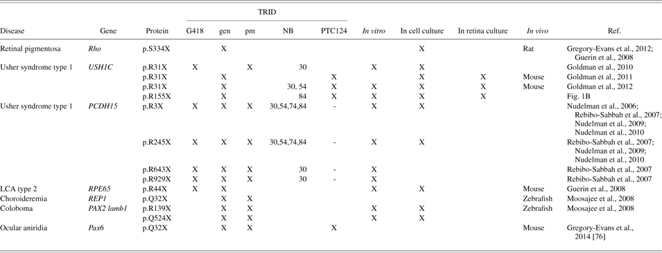
Over the last few years, the potential of TR as a treatment option for hereditary ocular dystrophies was intensively studied. In this paragraph, we provide a short summary of the heterogeneous group of ocular diseases addressed to date. In Table 1, we have compiled a list of approaches tackling nonsense mutations in retinal disease genes by TRIDs.
Translational read-through studies in the retina
Abbreviations: TRID: Translational read-through inducing drug; G418: geneticin; gen: gentamicin; pm: paromomycin; NB: designer aminoglycoside; LCA: Leber congenital amaurosis; REP1: Rab escort protein 1; PAX2: paired box 2; lamb1: laminin beta 1.
RP is the most common form of hereditary retinal degeneration with a prevalence of 1:4000 (Estrada-Cuzcano et al., Reference Estrada-Cuzcano, Roepman, Cremers, den Hollander and Mans2012). RP is a clinically and genetically heterogeneous group of hereditary retinal disorders characterized by rod photoreceptor dysfunction giving rise to night blindness, followed by tunnel vision, and eventually progressing to complete blindness due to cone photoreceptor dysfunction. Mutations in the rhodopsin gene are one of the most common diseases causing alterations (Athanasiou et al., Reference Athanasiou, Aguila, Bevilacqua, Novoselov, Parfitt and Cheetham2013). In 2008, Guerin and coworkers analyzed TR in the S334ter-4 rat model, which carries a nonsense mutation in the rhodopsin gene at residue 334 that leads to the translation of a truncated protein lacking the last 15 amino acids resulting in an autosomal dominant “gain of function.” The S334ter-4 rat is being used as a model for human autosomal dominant RP. Cell culture studies using luciferase assays demonstrated TR of the nonsense mutation in the rhodopsin. Systemic treatment with aminoglycoside increased the number of surviving photoreceptor cells and preserved retinal function (Guerin et al., Reference Guerin, Gregory-Evans, Hodges, Moosajee, Mackay, Gregory-Evans and Flannery2008). However, the major constrain of the systemic application of aminoglycoside is originated in the limited ocular penetration of TRIDs with increasing age of the animals.
In the same study, the rd12 transgenic mouse model was analyzed for the efficacy of TR in vivo. The rd12 mouse having a PTC in the retinal pigment epithelium-specific 65 kDa protein gene (RPE65) is an autosomal recessive model displaying retinal degeneration (Guerin et al., Reference Guerin, Gregory-Evans, Hodges, Moosajee, Mackay, Gregory-Evans and Flannery2008). In humans, mutations in RPE65 lead to LCA type 2. However, TR was not observed using luciferase reporter assays in cell culture or in vivo (Guerin et al., Reference Guerin, Gregory-Evans, Hodges, Moosajee, Mackay, Gregory-Evans and Flannery2008).
Further analysis of read-through in RP causing nonsense mutations was performed in a syndromic disease, in which RP is one of the clinical features, namely Usher syndrome (USH). USH is the most common cause of combined inheritable deaf-blindness (Wolfrum, Reference Wolfrum and Satpal2011; Yang, Reference Yang and Sadaf2012). Based on differences in clinical course, USH is divided into three clinical types (USH1-3) which are also genetically heterogeneous. The most severe form of the disease is USH1 which is characterized by profound prelingual hearing loss, vestibular areflexia, and prepubertal onset of RP. Several studies demonstrated TR of different PTCs in the protocadherin 15 (PCDH15) gene (USH1F) which was mediated by aminoglycosides and NB compounds in vitro translation assays and in transient transfected cells by Western blot analysis and using immunofluorescence (Nudelman et al., Reference Nudelman, Rebibo-Sabbah, Shallom-Shezifi, Hainrichson, Stahl, Ben Yosef and Baasov2006, Reference Nudelman, Rebibo-Sabbah, Cherniavsky, Belakhov, Hainrichson, Chen, Schacht, Pilch, Ben-Yosef and Baasov2009, Reference Nudelman, Glikin, Smolkin, Hainrichson, Belakhov and Baasov2010; Rebibo-Sabbah et al., Reference Rebibo-Sabbah, Nudelman, Ahmed, Baasov and Ben Yosef2007).
TR analysis of the p.R31X nonsense mutation in the USH1C gene revealed recovery of full-length harmonin protein in in vitro translation assays, in transient transfected cells, organotypic retina culture and in vivo following application of PTC124, aminoglycosides, and the NB compounds (Goldmann et al., Reference Goldmann, Rebibo-Sabbah, Overlack, Nudelman, Belakhov, Baasov, Ben Yosef, Wolfrum and Nagel-Wolfrum2010, Reference Goldmann, Overlack, Wolfrum and Nagel-Wolfrum2011, Reference Goldmann, Overlack, Moller, Belakhov, van Wyk, Baasov, Wolfrum and Nagel-Wolfrum2012). Importantly, functional activity of the different recovered harmonin isoforms a1 and b3 in TRIDs treated cells was successfully demonstrated in protein binding assays, namely by the interaction with the USH2A protein and in actin filament bundling assays, respectively (Goldmann et al., Reference Goldmann, Rebibo-Sabbah, Overlack, Nudelman, Belakhov, Baasov, Ben Yosef, Wolfrum and Nagel-Wolfrum2010, Reference Goldmann, Overlack, Wolfrum and Nagel-Wolfrum2011, Reference Goldmann, Overlack, Moller, Belakhov, van Wyk, Baasov, Wolfrum and Nagel-Wolfrum2012). In a comparative study analyzing PTC124, NB30, and NB54, the authors did not observe significant differences in read-through efficacy of TRIDs in transfected cells in retinal cultures and in vivo. Furthermore, toxicity assays on human and murine retinal explants revealed an excellent biocompatibility for NB54 and PTC124. Recently, we analyzed TR of p.R155X, a second in-frame nonsense mutation in the USH1C gene, in cell cultures. We observed recovery of full-length harmonin expression after applying PTC124 and NB84 (Fig. 1B).
In further studies, zebrafish were used to analyze TR in animal models for choroideremia (chm ru848; juvenile chorioretinal degeneration) and ocular coloboma (noi tu29a and gup m189; congenital optic fissure closure defects) (Moosajee et al., Reference Moosajee, Gregory-Evans, Ellis, Seabra and Gregory-Evans2008). Choroideremia is an X-linked retinopathy caused by mutations in the CHM gene that encodes the Rab escort protein 1 (REP1). The mutant zebrafish chm ru848 is currently the only existing animal model for choroideremia. The zebrafish chm ru848 has a recessive in-frame nonsense mutation in the rep1 gene resulting in a stop at amino acid position 32 (p.Q32X). Nonsense mutations account to over one-third of cases of choroideremia in humans.
Ocular coloboma is a significant cause of congenital eye anomaly. Nonsense mutations have been identified in many cases for example within the PAX2 gene causing renal-coloboma syndrome in humans. In zebrafish, two ocular coloboma models carrying nonsense mutations exist. The zebrafish model noi tu29a (no isthmus), which displays a mild coloboma phenotype, has a nonsense mutation within the paired box 2.1 (pax2.1) gene resulting in a stop (UGA) at amino acid position 139 (p.R139X). The gup m189 (grumpy) models, which display a severe coloboma phenotype, carry a nonsense mutation in the lamb1 gene encoding laminin β1. In luciferase assays in Cos-7 cells read-through was detected for all mutations by applying paromomycin and gentamicin. Of further importance, in vitro prenylation assays demonstrated the functionality of the recovered rep1 protein. Importantly, drug application to chm ru848, noi tu29a, and gup m189 mutant embryos significantly increased survival time compared to untreated mutant controls. Taken together, these findings indicated that the zebrafish models may prove to be suitable tools in preclinical drug development for novel TRIDs.
Congenital aniridia is a rare genetic disorder characterized by iris hypoplasia associated with additional ocular abnormalities. In humans, most cases of congenital aniridia are linked with PAX6 mutations (paired box 6) that result in PAX6 haploinsufficiency. Approximately 50% of all PAX6 disease-associated mutations are nonsense mutations. Pax6 is the master control gene for eye morphogenesis and encodes a transcription factor. Gregory-Evans and coworkers analyzed the efficacy of the read-through therapy in the semidominant Pax6-deficient transgenic mouse model of aniridia (Pax6Sey +/−) following systemic and topical application of PTC124 and gentamicin. For this study, the novel drug formulation “START” (0.9% sodium chloride, 1% Tween 80, 1% powdered PTC124, 1% carboxymethylcellulose) was used which was designed to enhance the particle dispersion and to increase suspension viscosity. Topical administration of PTC124 applied with “START” on Pax6Sey +/− mouse eyes represented the most promising results. PTC124/START therapy stopped disease progression, reversed corneal, lens, and retinal malformation, and restored electrophysiological function of the retina (Gregory-Evans et al., Reference Gregory-Evans, Wang, Wasan, Zhao, Metcalfe and Gregory-Evans2014). This study suggests that the eye retains significant developmental plasticity into the post-natal period and stays susceptible to molecular remodeling providing a window of opportunity to treat childhood blindness. All in all the present data from the different preclinical approaches emphasize that TR therapy is a feasible therapy option targeting a wide range of diverse hereditary retinal dystrophies caused by in-frame nonsense mutations.
Conclusions
In conclusion, the here described preclinical studies provide a proof of concept for using TRIDs to circumvent PTCs and thereby treat hereditary retinal dystrophies caused by in-frame nonsense mutations. The high ocular biocompatibility combined with the sustained read-through efficacies places PTC124 and designer aminoglycosides in the spotlight for treating PTC-based retinal disorders. Promising recent data on local topical drug administration further highlight the great potential of TRIDs for ocular therapies. Optimizing TR therapeutic strategies will be a crucial step to provide thousands of patients a feasible treatment option to cure hereditary retinal degenerations and blindness.
Acknowledgment
Funds were provided by the Deutsche Forschungsgemeinschaft (DFG GRK 1044 to UW); the ProRetina Stiftung (UW); the FAUN-Stiftung, Nurnberg (to KNW/UW); Foundation Fighting Blindness (FFB) grant TA-NMT-0611-0538-JGU (to KNW/UW); European Community FP7/2009/241955 (SYSCILIA) (to UW) and FP7/2009/242013 (TREATRUSH) (to UW); BMBF, grant 0314106 (HOPE2) (to UW) and under the frame of E-Rare-2, the ERA-Net for Research on Rare Diseases (EUR-USH) (to KNW). Authors thanks Prof. Dr. Timor Bassov for providing designer aminoglycosides and Dr. Christina Gewinner for critically reading the manuscript.
List of abbreviations
AAV: adeno-associated virus; chm: Choroideremia: Da: Dalton; EJC: exon-junction-complex; ELISA: enzyme linked immunosorbent assay; eRF1: eukaryotic release factor 1; eRF3: eukaryotic release factor 3; gup: grumpy; G418: geneticin; GTP: guanosine-5′-triphosphate; lamb1: laminin beta 1; LCA: leber congenital amaurosis; MPS I-H: mucopolysaccharidosis I-Hurler; NMD: nonsense-mediated mRNA decay; noi: no isthmus; PCDH15: protocadherin-15; PTC: premature termination codon; PTC124 : Ataluren, 3-[5-(2-fluorophenyl)-[1,2,4]oxadiazol-3-yl]benzoic acid; PAX2: paired box 2; PAX6: paired box 6; PTT: protein transcription/translation; rd12: retinal degeneration 12; Rep1: rab escort protein 1; Rho: rhodopsin; RP: retinitis pigmentosa; RPE65: retinal pigment epithelium-specific 65 kDa protein; RTC14: read-through compound 14; START: sodium chloride, Tween 80, powdered ataluren, carboxymethylcellulose; TC: termination codon; TR: translational read-through; TRID: translational read-through inducing drug; UAA: uracil, adenin, adenin; UAG: uracil, adenin, guanin; UGA: uracil, guanin, adenin; USH: Usher syndrome; USH1C: Usher syndrome type 1C; USH2A: Usherin.