

Introduction
Human language achieves an efficient mode of communication based on a precise mapping between sounds and meaning that is shared by all the members of the group. The power of this communication tool is based on elementary bricks that can be combined in multiple ways to convey new meanings. These elementary bricks (phonemes, syllables and words) are realized as a continuous speech signal that should be correctly segmented by the listeners in order to decipher the information. The human brain easily performs these complex operations in the left perisylvian regions. Since Broca’s princeps publication,Reference Broca1 numerous studies in neuro-psychology and neuro-imaging have questioned whether a particular organization of this part of the brain might explain the language faculty in our species. Indeed, leftward structural asymmetries are observed, both at the macroscopical and cytoarchitectonic levels, such as a longer sylvian fissure and a larger planum temporale Reference Geschwind and Levitsky2 and less frequently a larger inferior frontal region.Reference Knaus, Bollich, Corey, Lemen and Foundas3 The white matter volume underlying Heshl’s gyri is larger on the left than on the right side.Reference Penhune, Zatorre, MacDonald and Evans4 Bigger pyramidal cells are noted in the left auditory cortexReference Hutsler5 associated with thicker myelinated fibres.Reference Anderson, Southern and Powers6 The width of individual cortical columns, and the distance between those columns, are greater in the left superior temporal lobe.Reference Seldon7 It was argued that these structural features might allow the left hemisphere to code the rapid and complex acoustic transitions characterizing speech more accurately than the right.Reference Boemio, Fromm, Braun and Poeppel8, Reference Zatorre and Belin9 It thus would seem easy to attribute human speech processing capacities to these structural differences, all the more since the planum temporale is less asymmetric in groups of oral or written-language impaired children relative to the normal population.Reference Plante10
While these structural features may indeed be particularly adapted to process speech, a simple causal relationship between them and the language faculty might be an oversimplification. First, humans are heavily trained with speech stimuli, making it difficult to ascertain whether the observed cytoarchitectonic characteristics of the left temporal lobe are the consequence rather than the cause of speech processing in this hemisphere. Second, the neuronal responses in some of these regions depend more on the linguistic value of the stimuli than on their acoustical characteristics. For example, fMRI activations to phonetic contrasts, observed in both the posterior part of the superior temporal cortex and the supra-marginal gyrus, depend more on the subjects’ native language than on their acoustical features.Reference Dehaene-Lambertz, Dupoux and Gout11, Reference Jacquemot, Pallier, LeBihan, Dehaene and Dupoux12 Third, sign languages, which rely on spatial cues rather than on fast temporal cues as do oral languages, involve the same left perisylvian regions.Reference Damasio, Bellugi, Poizner and Gilder13, Reference Sakai, Tatsuno, Suzuki, Kimura and Ichida14 Finally, asymmetries are also observed in other animals. For example, great apes also exhibit a larger left planum temporale,Reference Cantalupo, Pilcher and Hopkins15, Reference Gannon, Holloway, Broadfield and Braun16 although cytoarchitectonic differences between both hemispheres are less salient than in humans.Reference Buxhoeveden, Switala, Litaker, Roy and Casanova17 As in humans, asymmetries seem to be linked to the communicative salience of the vocalizations rather than to their acoustic parameters.Reference Ehret18, Reference Petersen, Beecher, Zoloth, Green, Marler and Moody19 This asymmetry develops along with exposure and is not seen in infant rhesus monkeysReference Hauser and Andersson20 and infant sea-lionsReference Boye, Gunturkun and Vauclair21 while in harpy eagles, the active experience of hunting modifies the initial left side bias in response to the call of a prey toward the right side.Reference Palleroni and Hauser22
Another way to tackle this question is to examine the infant brain before intensive exposure to speech. Because research on infants during the last few decades has revealed that they are particularly responsive to speech from birth onwards, we may wonder whether it is the particular organization of the perisylvian regions in our species that favour language acquisition. Thanks to the development of non-invasive brain imaging techniques, it is now possible to study brain development in normal infants.
Structural asymmetries in infants
Genetic studies have revealed asymmetrical gene expression in the perisylvian regions, specific to the human lineage.Reference Sun, Collura, Ruvolo and Walsh23 These genes, which either regulate cell signalling or control other genes or protein expression, are expressed at an early developmental stage (especially between 12 and 14 weeks of gestation), a critical time for cortical regionalization. LM04, for example, is expressed more on the right side than on the left in humans. For this gene, asymmetry is also present in mice but not biased systematically to the same hemisphere across individuals as in humans.Reference Sun, Patoine, Abu-Khalil, Visvader, Sum and Cherry24
During the last trimester of human gestation, sulci appear first on the right hemisphere. The right superior frontal, superior temporal and Heschl’ gyri are detectable one or two weeks earlier than their left-sided homologous.Reference Chi, Dooling and Gilles25, Reference Dubois, Benders, Cachia, Lazeyras, Ha-Vinh Leuchter and Sizonenko26 This asymmetry in sulcation development is not reported in macaque fetuses.Reference Fukunishi, Sawada, Kashima, Sakata-Haga, Fukuzaki and Fukui27 At birth, grey and white matter volumes are larger in the left hemisphere contrarily to adults.Reference Gilmore, Lin, Prastawa, Looney, Vetsa and Knickmeyer28 The sylvian fissure is longer on the left sideReference Sowell, Thompson, Rex, Kornsand, Tessner and Jernigan29 and is associated with a larger left planum temporale Reference Chi, Dooling and Gilles25, Reference Witelson and Pallie30 while the superior temporal sulcus is larger on the right.Reference Dubois, Benders, Cachia, Lazeyras, Ha-Vinh Leuchter and Sizonenko26 Twin studies reveal a strong genetic influence in these areas,Reference Thompson, Cannon, Narr, van Erp, Poutanen and Huttunen31 with little influence of auditory stimulation, at least at the macroscopic level. The volumes of both left planum temporale and Heschl’s gyrus are similarly larger in hearing and congenitally deaf adults.Reference Emmorey, Allen, Bruss, Schenker and Damasio32 Contrary to the hypothesis of an equipotential brain at the beginning of life,Reference Lenneberg33 these observations point to evolutionary genetic changes in the human lineage which favoured a differential development between the left and right hemispheres in a systematic way across humans.
Functional asymmetries in infants
Although these asymmetries concern, in particular, the superior temporal regions, they may not be directly related to the development of language in our species, but two other results suggest that asymmetries in the perisylvian regions are not only structural but that they also reflect a different functional organization. With diffusion tensor imaging (DTI), it is now possible to tract the main white matter fascicles. Furthermore, because the indices measured with DTI are sensitive to the tract organization (their compactness and myelination) it becomes possible to follow tract maturation during development.Reference Dubois, Dehaene-Lambertz, Perrin, Mangin, Cointepas and Duchesnay34, Reference Dubois, Dehaene-Lambertz, Soares, Cointepas, Le Bihan and Hertz-Pannier35 A study in 23 two-month-old infants revealed that maturation in two fasciculi, the arcuate fasciculus and the cortico-spinal tract is more advanced on the left side.36 These two bundles are related to the two main lateralized functions in humans: language and handedness, the arcuate fasciculus connects auditory temporal areas to production frontal areas and the cortico-spinal tract is the main motor tract. Since the studied infants were both linguistically and manually limited, tract lateralization points to a differential maturation between left and right hemispheres that precedes any behavioural expression as overt speech or organized gestures.
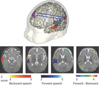
A second interesting element is given by brain functional images. During the first year of life, there is, at rest, no left–right difference in cerebral blood flow, even in linguistic regions (inferior frontal, superior temporal and plurimodal temporo-parietal regions).Reference Chiron, Jambaque, Nabbout, Lounes, Syrota and Dulac37 However, in response to auditory stimuli, asymmetrical responses favouring the left side are observed in fMRI and ERP studies. In fMRI, 20 second-long speech recordings were played either normally or backward. While forward and backward speech share common segmental features (fast temporal auditory transitions and phonetic information conveyed by temporally symmetrical phonemes), backward speech violates universal prosodic rules, affecting language discrimination capacities in infantsReference Mehler, Jusczyk, Lambertz, Halsted, Bertoncini and Amiel-Tison38 as in animals. Activation to both forward and backward stimuli was significantly larger in the left than in the right planum temporale in neonatesReference Pena, Maki, Kovacic, Dehaene-Lambertz, Koizumi and Bouquet39 and 3-month-old infants.Reference Dehaene-Lambertz, Dehaene and Hertz-Pannier40 In both studies, although the interaction between utterances type and hemisphere was not significant, activation in this left region reached significance only for forward speech, but not for backward speech (Figure 1). Thus, a significant leftward asymmetry is present for speech-like stimuli from birth on, but without statistical evidence of any specificity for linguistic stimuli over non-linguistic ones (possibly because of a lack of statistical power). Similarly, higher voltages were recorded over the left hemisphere in an event-related potentials study (ERPs) to CV syllables and tones in 4-month-olds, again with no stronger asymmetry for the linguistic stimuli.Reference Dehaene-Lambertz41 However, in a recent experiment, in which was studied the capacity in two-month-old infants to perceive a change of gender and of a vowel along the visual and the auditory dimensions, we observed that gender information was channelled toward the right hemisphere and linguistic information to the left.42 These results suggest for the first time a clear functional distinction between the two hemispheres from the first months of life onwards.
FMRI study of infants’ speech processing. Activations obtained in awake three-month-old infants listening to blocks of 20 s of normal (forward) or reversed (backward) speech relative to silence are projected on a 3D image of a three-month-old brain on the top line, and on axial slices on the bottom. Activations to forward speech (blue scale) are more dorsal and posterior along the superior temporal regions than those to backward speech (orange scale). However, the regions significantly more activated by forward than by backward speech are the left inferior parietal region and the right dorso-lateral prefrontal regions (blue-pink scale). This figure illustrates the complex cooperation between temporal, parietal and frontal regions to recognize the native language (forward speech) (adapted from Ref. Reference Dehaene-Lambertz, Dehaene and Hertz-Pannier40).

To summarize, the structural development of the left and right hemispheres is strongly asymmetric in our species, especially around the posterior part of the superior temporal region. These gross structural differences are also accompanied by microstructural differences in the tract organization of the fascicles that sustain handedness and language: the cortical-tract and the arcuate fasciculus. Finally, a leftward bias for the processing of linguistic stimuli is present very early. However, if a lesion occurs, the right perisylvian areas can take over language processing normally devoted to the left hemisphere (such as, for example, phonetic discrimination in infants,Reference Dehaene-Lambertz, Pena, Christophe, Charolais and Landrieu43 and even expressive and receptive language in older childrenReference Hertz-Pannier, Chiron, Jambaque, Renaux-Kieffer, Van de Moortele and Delalande44) showing that this leftward response is only a bias. Indeed, several studies have underscored the fact that language development usually remains within the normal range,Reference Bates and Roe45 after early brain lesion, whatever the side of the lesion.
Functional continuity from infanthood to adulthood: phoneme perception
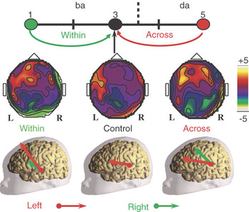
While the observed lateralization pattern is not as strong as in adults and consolidates during development and acquisition of more sophisticated language skills,Reference Holland, Plante, Weber Byars, Strawsburg, Schmithorst and Ball46 the brain regions involved when infants listen to speech are nevertheless close to those observed in adults. ERPs have been used to decipher the processing of brief sounds. By subtracting the response evoked by a stimulus preceded either by itself or by another close stimulus (e.g. da da da da versus ba ba ba da), it is possible to observe a mismatch response whose topography and latency depend on the changing feature. When the change in a series of CV syllables either concerns the voice or the phonemes, mismatch responses in infants show different scalp topographies as well as a different latencies, suggesting that, as in adults, different networks are involved in the representation of the various sound features.Reference Dehaene-Lambertz41, 42 Among these networks, one demonstrating phonetic properties, such as normalization across different speakersReference Dehaene-Lambertz and Pena47 and categorical perceptionReference Dehaene-Lambertz and Baillet48 was identified. Dipole modelling of the active regions coding a phonetic change suggests a more dorsal and posterior origin than that coding for a similar acoustical change (Figure 2). This shift is compatible with the involvement of posterior temporal and inferior parietal regions during phonetic processing, as demonstrated in adults.Reference Jacquemot, Pallier, LeBihan, Dehaene and Dupoux12, Reference Dehaene-Lambertz, Pallier, Serniklaes, Sprenger-Charolles, Jobert and Dehaene49
ERP study of infants’ phonetic processing. Categorical perception in three-month-old infants along a synthetic continuum /ba/ to /da/. Evoked responses to the last syllable of series of four syllables were recorded with 65 electrodes. In control trials, the same syllable /ba/ was repeated. In within-category change trials, it was preceded by another syllable belonging to the same category /ba/ while in across-category change trials, the previous syllable belonged to the /da/ category. However, the physical change measured on the synthetic continuum was similar in both cases (top row). The voltage cartographies to the same test syllable (/ba3/) were different in each condition (middle row). Although the acoustical change was similar in across- and within-category change, ERP to the test syllable was larger and more diffuse in the case of a phonetic change (across) than of a pure acoustical change (within), demonstrating categorical perception. Moreover, the dipole in the across-category change condition was more posterior and dorsal than in the within-category change condition (bottom row) (adapted from Ref. Reference Dehaene-Lambertz and Baillet48).

A hierarchically organized speech perception network
Using fMRI in infants, we showed that responses to forward and backward sentences elicit activations of the superior temporal gyrus, encompassing Heschl’s gyrus, and extending to surrounding areas of the superior temporal sulcus and the temporal pole (Figure 1).Reference Dehaene-Lambertz, Dehaene and Hertz-Pannier40 The angular gyrus and precuneus were more activated by forward speech than by backward speech, suggesting that the angular gyrus, which is involved in lexical storage in adults, also plays a role in the storage of the prosodic patterns that infants use to recognize their native language. Finally, only in awake babies was a significant activation observed for forward speech, in the right dorsolateral prefrontal cortex, which is involved in memory retrieval mechanisms in adults (Figure 1).
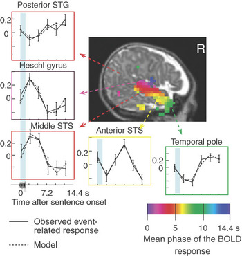
In a second study, we parsed the infant’s network of perisylvian responsive regions into functionally distinct regions based on their speed of activation and on their sensitivity to sentence repetition (Figure 3).Reference Dehaene-Lambertz, Hertz-Pannier, Dubois, Meriaux, Roche and Sigman50, Reference Dehaene-Lambertz, Dehaene, Anton, Campagne, Ciuciu, Dehaene, Denghein, Jobert, LeBihan, Sigman, Pallier and Poline51 We observed that the phase of the activation in response to a single sentence increases as one moves from the auditory primary cortex toward the posterior part of the superior temporal gyrus and toward the temporal poles and inferior frontal regions (Broca’s area). Given the size of the delays involved (several seconds), this organization is unlikely solely to reflect synaptic delays. Rather, this temporal gradient of activation might be the result of different cognitive operations that integrate over increasingly larger and possibly more abstract speech units, and may therefore require longer processing time or more sustained activity. This hierarchical architecture of the human temporal lobes presents homologies with that of the monkey brain.Reference Kaas and Hackett52, Reference Pandya and Yeterian53 Thus it is possible that human speech recycles a pre-existing primate system for hierarchical auditory representations.Reference Dehaene and Cohen54 Such a nested organization of processing units with a progressively longer temporal window of integration would provide infants with an adequate tool to segment the speech stream in its prosodic components. Auditory long-term memory, as suggested by Fritz et al.Reference Fritz, Mishkin and Saunders55 may be the essential difference between humans and other primates, allowing memorization of the different sub-units but also their recombination. The involvement of the left inferior frontal region in the speech perception network, which was observed when two-month old infants were engaged in a short-term verbal memory taskReference Dehaene-Lambertz, Hertz-Pannier, Dubois, Meriaux, Roche and Sigman50 may be an indicator of this early efficient speech coding system.
Phase measurement along the superior temporal region in three-month-old infants. The phase of the evoked fMRI responses to a single sentence was measured in 3-month-old infants (after Dehaene-Lambertz et al.Reference Dehaene-Lambertz, Hertz-Pannier, Dubois, Meriaux, Roche and Sigman50). A systematic gradient of response delays was found along both temporal regions, with fast on-line responses near Heschl gyrus, and increasingly slower responses as one moves either back into the planum temporale and Wernicke’s area on the left side or forward along the STS toward the temporal pole and Broca’s area. A similar arrangement exists in adults (Dehaene-Lambertz et al.Reference Dehaene-Lambertz, Dehaene, Anton, Campagne, Ciuciu, Dehaene, Denghein, Jobert, LeBihan, Sigman, Pallier and Poline51), where it cannot be attributed purely to synaptic or hemodynamic delays, but may reflect integration and closure of speech segments of different lengths (phoneme, syllable, word, whole phrase). The presence of this gradient in very young infants, prior to any babbling, and its similarity to the hierarchical organization of anatomical projections in other primates (Refs 52, 53), suggests that it may constitute an innate bias that constrains language acquisition to a nested hierarchical structure.

Our fMRI results also indicate that brain regions involved in receptive speech processing in infants are not limited to unimodal auditory regions. They extend to remote regions, including areas such as the frontal regions that are usually considered as barely functional at this age. Conversely, the infant’s brain does not respond diffusely to speech, as connectionist models would have predicted.Reference Elman, Bates, Johnson, Karmiloff-Smith, Parisi and Plunkett56 It is functionally structured, recruiting distant regions in cooperative networks. We do not know yet whether another structured stimulus, such as music for example, would activate the same networks. However, the infant’s brain processing properties (allowing categorical perception, normalization of the speech input, short-term memory, recognition and long-term storing of intonation contours) make it efficiently adapted to the most frequent auditory input encountered by the human infant, namely speech. Furthermore, the hierarchical temporal organization of the perisylvian areas may promote acquisition of the different nested levels of language (phonemes, syllables, words, constituents, phrases, sentences, and so on).
Conclusion
From the first weeks of life onwards, the human brain displays normalization and phonetic categorization capacities, rhythmic and prosodic sensitivity, which make it particularly adapted to processing speech. These capacities mostly rely on brain circuits close to those observed in adults, i.e. the left perisylvian areas. It seems unlikely that the influence of a pre- and postnatal auditory environment be sufficient by itself to generate this complex organization in a few weeks of exposure. On the contrary, the similarity between functionally immature infants and competent mature adults implies a strong genetic bias for speech processing in those areas. This ‘bias’ might partially result from recycling of auditory processes observed in other mammals (e.g. perceptive discontinuities along some acoustical dimension, rhythmic sensitivity, hierarchical temporal organization) but is not limited to them. The functional properties of the superior temporal areas and their connectivity with remote regions in humans might be crucial to ensure language learning. For example, connections with other brain areas, such as the motor or the visual system, which possess their own biases to compute conspecific representations, might be critical to reinforce linguistic representations and their shaping by the native language. While language acquisition studies have mostly focused on speech input analyses so far, a better understanding of the functional properties of the brain regions involved in speech processing in infants (e.g. preference for fast transitions, possibility of auditory long-term storing, etc) might help to specify the crucial parameters favouring language acquisition. These new insights will provide a strong basis for the study of early developmental disorders affecting language and communication in humans.
Acknowledgement
This paper is a revised version of G. Dehaene-Lambertz, L. Hertz-Pannier and J. Dubois (2006) Nature and nurture in language acquisition: anatomical and functional brain-imaging studies in infants. Trends in Neuroscience, 29(7), 367–373.
Ghislaine Dehaene-Lambertz is full-time researcher in CNRS. She is directing a team using brain imaging to study the cerebral bases of cognitive functions in infants.
Lucie Hertz-Pannier is the Director of the Biomedical research laboratory. She is working on brain imaging in normal and pathological children.
Jessica Dubois has a post-doctoral position in Neurospin, south of Paris. She is working on brain development.
Stanislas Dehaene is a cognitive neuroscientist, Professor at the Collège de France, Chair of Experimental Cognitive Psychology. He is directing the INSERM-CEA Cognitive Neuroimaging Unit and investigates high-level human cognitive functions such as language, mathematics, attention …, combining experimental psychology, neuropsychology and neuroimagery approaches.