Introduction
Entomopathogenic nematodes (EPNs) of the genus Steinernema are soil-dwelling organisms that serve as effective biological control agents against various agricultural pests. By targeting the larval stages of arthropods, they provide a sustainable alternative to chemical pesticides without posing risks to human health or the environment (Shapiro-Ilan et al Reference Shapiro-Ilan, Hazir, Glazer, Kogan and Higley2019). The efficacy of Steinernema species relies on a specialized symbiotic relationship with Xenorhabdus bacteria. The non-feeding infective juveniles (IJs) carry the bacteria in their gut, protecting them from environmental stress and vectoring them into the insect host. Upon entry, the infective juveniles release lethal venom proteins (Lu et al Reference Lu, Macchietto, Chang, Barros, Baldwin, Mortazavi and Dillman2017) and their bacteria into the host hemocoel. The combined action of the nematodes and their bacterial symbionts suppresses the host immune system, causes rapid mortality, typically within 48 h, and converts host tissues into a nutrient source for nematode reproduction (Çimen et al Reference Çimen, Touray, Gulsen and Hazir2022; Hazir et al Reference Hazir, Kaya, Touray, Çimen and Shapiro-Ilan2022).
Although EPNs have been isolated globally using insect-baiting techniques, their distribution is strongly influenced by soil composition, host availability, and climate (Stock Reference Stock and Campos-Herrera2015). As of the end of 2024, the genus Steinernema comprises 114 recognized species (Půža et al Reference Půža, Machado and Malan2025). In South Africa, recent surveys have significantly expanded the known diversity of this genus. Several species have been described from the region over the last decade, including S. jeffreyense Malan, Knoetze & Tiedt, 2015 (Malan et al Reference Malan, Knoetze and Tiedt2016a), S. fabii Abate, Malan, Tiedt, Wingfield, Slippers & Hurley, Reference Abate, Malan, Tiedt, Wingfield, Slippers and Hurley2016 (Abate et al Reference Abate, Malan, Tiedt, Wingfield, Slippers and Hurley2016), S. nguyeni Malan, Knoetze & Tiedt, 2016 (Malan et al Reference Malan, Knoetze and Tiedt2016b), S. beitlechemi Çimen, Půža, Nermut, Hatting, Ramakuwela, Faktorová & Hazir, 2016 (Çimen et al Reference Çimen, Půža, Nermuť, Hatting, Ramakuwela, Faktorova and Hazir2016a), S. biddulphi Çimen, Půža, Nermut’, Hatting, Ramakuwela & Hazir, 2016 (Çimen et al Reference Çimen, Půža, Nermuť, Hatting, Ramakuwela and Hazir2016b), S. bertusi Katumanyane, Malan, Tiedt & Hurley, Reference Katumanyane, Malan, Tiedt and Hurley2020 (Katumanyane et al Reference Katumanyane, Malan, Tiedt and Hurley2020), S. batswanae Didiza, Lephoto & Gray, Reference Didiza, Lephoto and Gray2021 (Didiza et al Reference Didiza, Lephoto and Gray2021), and S. bakwenae Malan, Knoetze, Kapp & Tiedt, Reference Malan, Knoetze, Kapp and Tiedt2023 (Malan et al Reference Malan, Knoetze, Kapp and Tiedt2023). Some of these indigenous isolates are currently being evaluated for integration into local integrated pest management programs, taking advantage of their specific adaptations to regional environmental conditions (Katumanyane et al Reference Katumanyane, Ferreira and Malan2018; Moore et al Reference Moore, Ehlers, Manrakhan, Gilbert, Kirkman, Daneel, De Waal, Nel, Sutton and Malan2024; Steyn et al Reference Steyn, Daneel and Malan2019).
Surveys were conducted during late winter in the Highveld region of the Mpumalanga Province, specifically in the area between the towns of Bethal and Standerton. The Highveld is a well-established biogeographic and topographic subregion of South Africa’s interior plateau, characterised by elevations generally exceeding 1,500 m above sea level, a grassland biome, a temperate semi-arid climate with cold dry winters, and soils derived predominantly from dolerite intrusions and sedimentary Karoo Supergroup rocks. It spans parts of several provinces, including the Free State, Gauteng, Mpumalanga, and the North West.
Morphological and morphometric characterization was performed using both light microscopy and scanning electron microscopy (SEM). In addition, molecular analyses targeting the ITS regions and the D2-D3 expansion segments of the 28S ribosomal DNA were performed to support its validity under the phylogenetic and evolutionary species concepts. Based on these combined data, the new species is herein described as Steinernema hattingi n. sp.
Materials and methods
Collection and examination of nematodes
Soil samples were collected from a maize field in the area between the towns of Bethal and Standerton, Mpumalanga Province, South Africa. Each sample consisted of 3–10 subsamples, that were taken randomly from the surface to a depth of 20 cm. Five late instar Galleria mellonella (L.) (Lepidoptera: Pyralidae) larvae were placed in a 600 mL plastic container filled with moistened soil obtained from samples (Bedding and Akhurst, Reference Bedding and Akhurst1975). Containers were kept at room temperature (22 ± 3 oC). Mortality of G. mellonella larvae was checked daily, and dead larvae were transferred to a modified White trap (Kaya and Stock Reference Kaya, Stock and Lacey1997) until emergence of infective juveniles (Hazir et al Reference Hazir, Kaya, Touray, Çimen and Shapiro-Ilan2022).
Steinernema hattingi n. sp. was reared in last-instar larvae of G. mellonella as described by Poinar (Reference Poinar1979). First- and second-generation adults were obtained by dissecting infected hosts about three and five days after death, respectively. Specimens of all stages were heat-killed, fixed in TAF (Courtney et al Reference Courtney, Polley and Miller1955), and processed to glycerine by the method of Seinhorst (Reference Seinhorst1959). Nematodes were mounted in fresh glycerine under a coverslip supported by stainless steel wires slightly exceeding body diameter. Morphometric data were taken from 20 adults of each generation and from IJs using light microscopy and the image analysis software Cell D (Olympus Soft Imaging Solutions GmbH, Germany).
Scanning electron microscopy
Adults of both generations and IJs were fixed in 4% formalin buffered with 0.1 M sodium cacodylate (pH 7.2) for 24 h at 4–6 °C, post-fixed in 2% osmium tetroxide for 12 h at 25 °C, dehydrated through a graded ethanol series, critical-point dried with liquid CO₂, mounted on SEM stubs, and sputter-coated with gold (Nguyen and Smart Reference Nguyen and Smart1995, Reference Nguyen and Smart1997). Specimens were examined with a Verios 5 UC scanning electron microscope (Thermo Fisher Scientific).
Molecular analysis
DNA was extracted from single females. Each specimen was placed in a sterile 0.5 mL Eppendorf tube containing 20 μL of extraction buffer (17.7 μL ddH₂O, 2 μL 10× PCR buffer, 0.2 μL 1% Tween, 0.1 μL proteinase K). Tubes were frozen at –20 °C for 20 min, then incubated at 65 °C for 1 h and at 95 °C for 10 min. Lysates were cooled on ice, centrifuged for 2 min at 9000 g, and 1 μL of supernatant was used as template for PCR. A fragment of rDNA comprising ITS1, 5.8S, and ITS2 regions was amplified with primers 18S (5′-TTGATTACGTCCCTGCCCTTT-3′; forward) and 28S (5′-TTTCACTCGCCGTTACTAAGG-3′; reverse) (Vrain et al Reference Vrain, Wakarchuk, Levesque and Hamilton1992). The D2–D3 expansion segment of the 28S rDNA was amplified using primers D2F (5′-CCTTAGTAACGGCGAGTGAAA-3′; forward) and 536 (5′-CAGCTATCCTGAGGAAAC-3′; reverse) (Nguyen Reference Nguyen, Nguyen and Hunt2007).
PCR reactions (10 μL total volume) contained 7.25 μL ddH₂O, 1.25 μL 10× PCR buffer, 1 μL dNTPs, 0.75 μL of each primer, 0.1 μL polymerase, and 1 μL DNA extract. Thermal cycling conditions for ITS were: 94 °C for 7 min; 35 cycles of 94 °C for 60 s, 50 °C for 60 s, 72 °C for 60 s; and final extension at 72 °C for 7 min (Nguyen, Reference Nguyen, Nguyen and Hunt2007). For 28S rDNA, conditions were: 94 °C for 7 min; 35 cycles of 94 °C for 60 s, 55 °C for 60 s, 72 °C for 60 s; and final extension at 72 °C for 10 min (Mráček et al., Reference Mráček, Půža and Nermuť2014). PCR products (2 μL) were electrophoresed for 45 min at 120 V in 1% agarose gel with TAE buffer and stained with ethidium bromide (20 μL ETB per 100 mL gel). Amplified fragments were sequenced by Eurofins Genomics (Ebersberg, Germany). The presence of a double peak in the otherwise flawless ITS sequence chromatogram suggested the presence of intra-individual variability (Půža et al., Reference Půža, Chundelová, Nermuť, Žurovcová and Mráček2015). Thus, the PCR products of two single females were cloned into the pGEM®-T Easy vector (Promega) following the manufacturer’s protocol. For each female, DNA from five recombinant clones was re-amplified and purified with Exo-SAP-IT (USB Corporation, Cleveland, OH) according to the manufacturer’s instructions and sequenced using the universal primers T7 (5′-GATAATACGACTCACTATAGGGCTTCTTGCATTCTG-3′) and SP6 (5′-TCATTTAGGTGACACTATAGGACTTCAGGTGGCTGGA-3′). Sequences were deposited in GenBank under accession numbers PZ103902, PZ103903, and PZ103904 (ITS) and PZ103905 (28S).
Sequences were edited and compared with those available in GenBank using the Basic Local Alignment Search Tool (BLAST) at the National Center for Biotechnology Information (NCBI). Alignments of the obtained sequences with members of the glaseri-group were generated for each DNA region using default ClustalW parameters in MEGA 7.0 (Kumar et al Reference Kumar, Stecher and Tamura2016) and refined manually in BioEdit (Hall Reference Hall1999). Pairwise distances were calculated in MEGA 7.0 using codon positions 1st + 2nd + 3rd + noncoding.
Phylogenetic relationships were inferred using the Minimum Evolution method (Rzhetsky and Nei Reference Rzhetsky and Nei1992) implemented in MEGA 7.0 (Kumar et al Reference Kumar, Stecher and Tamura2016). Steinernema affine (Bovien Reference Bovien1937) Wouts, Mráček, Gerdin & Bedding, 1982 and Steinernema akhursti Qiu, Hu, Zhou, Mei, Nguyen & Pang, 2005 were designated as outgroups. The Minimum Evolution tree was constructed with the Close-Neighbour-Interchange (CNI) algorithm (Nei and Kumar Reference Nei and Kumar2000), and the initial tree was generated by the Neighbour-Joining method (Saitou and Nei Reference Saitou and Nei1987). Evolutionary distances were computed using the p-distance model (Nei and Kumar Reference Nei and Kumar2000) and expressed as the number of base substitutions per site.
Entomopathogenic bacteria isolation and molecular characterisation
Genomic DNA was isolated from 48-hour bacterial cultures using the DNeasy Blood & Tissue Kit (Qiagen) according to the manufacturer’s protocol. For molecular identification, the 16S rRNA gene and two housekeeping genes, recA and gyrB, were amplified. The 16S rRNA gene was targeted using 10F and 1507R primers (Sandström et al Reference Sandström, Russell, White and Moran2001), while recA and gyrB genes were amplified using specific primer sets (RecA1F/RecA2R and 8SF/9Rev) as described by Tailliez et al (Reference Tailliez, Laroui, Ginibre, Paule, Pagès and Boemare2010). Each 12.5 μL PCR reaction contained 1 μL of DNA template, 0.75 μL of each primer, 0.5 μL BSA, 1 μL dNTPs, 1.25 μL 10× buffer, 0.1 μL polymerase, and 7.2 μL ddH₂O. The thermal cycling conditions for 16S rRNA included an initial denaturation at 94 °C (1 min), 33 cycles of 94 °C (60 s), 55 °C (60 s), and 72 °C (2 min), with a 3 min final extension. Specific cycling profiles for recA and gyrB were implemented as detailed in the previous methodology. The resulting sequences were registered in GenBank with accession numbers KU578109 (16S), KU578110 (recA), and KU578111 (gyrB).
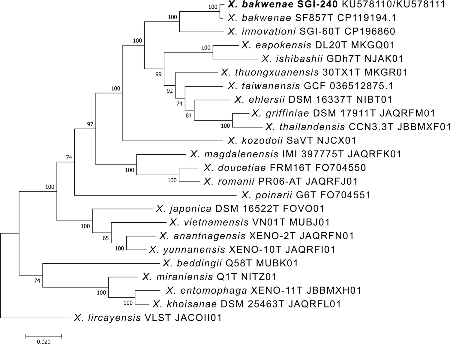
Phylogenetic relationships were inferred using the Maximum Likelihood method based on the Kimura 2-parameter model (Kimura Reference Kimura1980) implemented in MEGA 7 (Kumar et al Reference Kumar, Stecher and Tamura2016). The analysis was performed on a concatenated dataset of the recA and gyrB gene sequences representing a subset of species of the genus Xenorhabdus forming a derived clade that includes the studied strain. Gene sequences for the analysed taxa were retrieved from genome assemblies available in GenBank, while sequences of the studied strain were generated in this study. Xenorhabdus lircayensis was used as an outgroup. Initial tree(s) for the heuristic search were obtained automatically using neighbour-joining and BioNJ algorithms based on pairwise distances estimated by the Maximum Composite Likelihood approach, and the topology with the highest log likelihood was selected. Rate variation among sites was accounted for by including a proportion of invariable sites (+I). Branch support was assessed by bootstrap analysis, and values are indicated at the nodes. Branch lengths are expressed as the number of substitutions per site.
Results
Steinernema hattingi n. sp.
The species epithet hattingi is given in honor of the South African entomologist Dr. Justin Hatting. The true pioneering work on EPNs in South Africa predates Dr. Hatting’s surveys by several decades. Dr. Justin Hatting is credited with reviving entomopathogenic nematode research in South Africa in the early 2000s, following several decades of limited activity in this field.
Steinernema hattingi n. sp. A: First generation male, tail with spicules, lateral; B: Second generation male, tail with spicules, lateral; C: First generation male, tail with arrangement of genital papillae; D: First generation female, vulval region; E: Second generation female, vulval region; F: First generation female, tail; G: Second generation female, conical tail; H–I: Infective juvenile. H: Anterior region with excretory pore; I: Tail with anus and hyaline region. (Scale bar: A–J = 50 μm).

Figure 1. Long description
A multi-panel scientific illustration containing ten panels labeled A through J, each with a 50 mu m scale bar.
* Panel A: Lateral view of a first generation male tail showing curved spicules and a rounded terminus.
* Panel B: Lateral view of a second generation male tail, similar to A but with slightly different spicule curvature.
* Panel C: Ventral-lateral view of a first generation male tail highlighting the arrangement of genital papillae as small circular outlines along the cuticle.
* Panel D: Lateral view of a first generation female vulval region, densely packed with oval-shaped eggs.
* Panel E: Lateral view of a second generation female vulval region with fewer, more spaced-out eggs.
* Panel F: Tail of a first generation female showing a blunt, rounded terminus with a small mucro.
* Panel G: Conical tail of a second generation female, tapering more sharply than the first generation.
* Panel H: Anterior region of an infective juvenile showing the narrow head and the position of the excretory pore.
* Panel I: Tail of an infective juvenile showing the anus and a distinct hyaline region at the posterior tip.
* Panel J: A more distal view of the infective juvenile tail, showing the internal structure and tapering point.
Measurements
Morphometric characters of Steinernema hattingi n. sp. All measurements are in μm and in the form: mean ± S.D. (range)

Table 1. Long description
The table presents morphometric characters for Steinernema hattingi n. sp. with measurements in micrometers, expressed as mean plus or minus S. D. and range. The columns are divided into Males (Holotype, 1st generation Paratypes, 2nd generation Paratypes), Females (1st generation Paratypes, 2nd generation Paratypes), and Infective juveniles (Paratypes).
Key data points include:
* Total length (L): 1st generation males average 1789, 1st generation females average 5219, and infective juveniles average 1059.
* Maximum diameter: 1st generation females are the widest at 300, while infective juveniles are the narrowest at 33.
* Pharynx length (E S): Ranges from 145 in males and juveniles to 192 in 2nd generation females.
* Tail length (T): Infective juveniles have the longest tails at 66, while 2nd generation males have the shortest at 26.
* Spicule length (S L): Measured in males only, averaging 83 in the 1st generation and 65 in the 2nd generation.
* Gubernaculum length (G L): Averages 50 in 1st generation males and 37 in 2nd generation males.
* Ratios: D percent (E P / E S times 100) is highest in 1st generation males (86) and lowest in 2nd generation females (52). E percent (E P / T times 100) is highest in 1st generation females (429). H percent (H T / T times 100) is specific to infective juveniles at 44.
a Measured along the chord.
Description
Infective juvenile (IJ)
Body slender, slightly C-shaped when heat-killed, averaging 1059 μm in length. Head region rounded, continuous with body contour, slightly truncate at the anterior end. Lip region with four cephalic papillae and a pair of amphidial apertures. Cuticle distinctly annulated, striations ca 2 μm wide at mid-body (clearly visible under SEM). Lateral fields with eight ridges at mid-body (nine lines), the outermost pair less prominent. Field beginning anteriorly with a single incisure on the 4th annulus; at about the 14th annulus, 2 ridges appear, increasing to 6 ridges (7 incisures) near the excretory pore. Two additional ridges arise at the level of the basal bulb, forming eight ridges in total. Posteriorly, the number of ridges decreases gradually; at the level of the anus, adjacent ridges fuse to form four ridges, and two ridges remain at the level of the phasmid. Lateral field formula: 2, 6, 8, 4, 2. Deirids not observed. Hemizonid distinct, located at the mid-basal bulb level. Pharynx with slender corpus, cylindrical; isthmus narrow, surrounded by nerve ring. Excretory pore situated posterior to mid-pharynx (D% = 57). Cardia conspicuous. Bacterial vesicle typically well developed, ellipsoidal, about 10 μm wide and 23 μm long. Tail conoid, tapering gradually, with dorsal and ventral depressions near the beginning of the hyaline region. Hyaline portion comprising about 45% of tail length. Anus sickle-shaped, approximately 4 μm wide. Phasmids distinct, positioned near the mid-tail, ventral to the lateral field.
First-generation male
Body J-shaped in heat-killed specimens, ventrally curved posteriorly, and tapering anteriorly. Cuticle finely annulated with faint transverse striations. Stoma short and wide, reduced. Excretory pore situated posterior to the nerve ring, at about 86% of the distance from the anterior end to the base of the pharynx. Spicules paired, curved, brown, about 80 μm long; tip pointed. Manubrium elongated, nearly twice as long as wide (ratio length/width = 1.8:1). Calomus distinct but short; lamina curved, bearing two internal ribs. Thin velum extending from calomus to approximately 80% of lamina length. Gubernaculum boat-shaped, anterior end pointed. Tail short, dorsally convex, conoid with bluntly rounded terminus; mucron absent. Genital papillae 23 in total, arranged as 11 pairs and one single midventral papilla anterior to the cloaca. Paired papillae are distributed as follows: six subventral precloacal pairs, one sublateral precloacal pair, one subventral adcloacal pair, two sublateral postcloacal pairs, and one subdorsal postcloacal pair.
Second-generation male
General morphology similar to that of the first-generation male but smaller. Tail either non-mucronate or bearing a short (ca 1.5 μm) papilla-like mucron (Figure 2H).
Steinernema hattingi n. sp. SEM of infective juvenile, female, and male. A–C: Infective juvenile. A: Head region with four papillae and amphid openings (a); B: Lateral field in mid-body (ridges numbered 1–8); C: Tail region with anus and phasmid opening (arrow); D: Second generation female, tail with postanal swelling; E–F: First generation male. E: Tail with genital papillae (numbered), ventro-lateral; F: Tail with spicules and part of genital papillae (numbered), dorso-lateral; G–H: Second generation male. G: Tail with genital papillae (numbered), ventral; H: Tail tip with part of genital papillae (numbered) and spicules.

Figure 2. Long description
A collection of eight S E M micrographs labeled A through H.
Panel A shows the head of an infective juvenile with a scale bar of 4 micrometers. The rounded anterior end features four small papillae and a lateral amphid opening labeled a.
Panel B displays the mid-body lateral field of an infective juvenile with a scale bar of 5 micrometers. Eight longitudinal ridges are numbered 1 through 8.
Panel C shows the tapered tail of an infective juvenile with a scale bar of 10 micrometers. An arrow points to the phasmid opening labeled p h.
Panel D depicts the tail of a second-generation female with a scale bar of 10 micrometers, showing a distinct postanal swelling.
Panel E and F show the tail of a first-generation male. Panel E, with a 50 micrometer scale bar, shows the ventro-lateral view with genital papillae numbered 1 through 11. Panel F, with a 30 micrometer scale bar, shows a dorso-lateral view focusing on the spicules and papillae 6 through 11.
Panel G and H show the tail of a second-generation male. Panel G, with a 20 micrometer scale bar, provides a ventral view of the curved tail with papillae 1 through 11. Panel H, with a 5 micrometer scale bar, is a high-magnification view of the tail tip showing the mucro labeled m, spicules, and papillae 6, 8, 9, 10, and 11.
Steinernema hattingi n. sp. LM of female, male, and infective juvenile. A, C: First-generation female. A: Tail region; C: Vulval region. B, D: Second-generation female; B: Tail region; D: Vulval region. E: First-generation male, tail with spicules and gubernaculum; F: Second-generation male, tail with spicules and gubernaculum. G, H: Infective juvenile. G: Anterior portion showing head and excretory pore (arrow); H: Tail with anus and hyaline region.

Figure 3. Long description
A collection of eight light micrographs arranged in a two-column grid.
* Panel A (Top-Left): Tail region of a first-generation female showing a rounded terminus with a small mucro. Scale bar 60 micrometers.
* Panel B (Top-Right): Tail region of a second-generation female, appearing more slender and pointed than the first generation. Scale bar 60 micrometers.
* Panel C (Upper-Middle Left): Vulval region of a first-generation female with slightly protruding lips. Scale bar 50 micrometers.
* Panel D (Upper-Middle Right): Vulval region of a second-generation female showing the mid-body section. Scale bar 50 micrometers.
* Panel E (Lower-Middle Left): Tail of a first-generation male, curved ventrally, containing dark, curved spicules and a gubernaculum. Scale bar 50 micrometers.
* Panel F (Lower-Middle Right): Tail of a second-generation male, also showing the spicule and gubernaculum structure within the curved posterior. Scale bar 50 micrometers.
* Panel G (Bottom-Left): Anterior portion of an infective juvenile. A black arrow points to the excretory pore located behind the head. Scale bar 30 micrometers.
* Panel H (Bottom-Right): Tail of an infective juvenile showing the anus and a distinct, clear hyaline region at the very tip. Scale bar 30 micrometers.
First-generation female
Body C-shaped or strongly spiral in heat-killed specimens. Cuticle faintly annulated, visible under SEM. Head bluntly rounded, slightly tapering anteriorly, continuous with body contour, bearing six labial and four cephalic papillae. Excretory pore situated anterior to the basal bulb. Vulval opening slightly posterior to mid-body, transverse, moderately protruding from body contour, slightly asymmetrical; epiptygma small. Vagina short, leading to paired uteri. Rectum narrow; anal opening distinct, without postanal swelling. Tail of mature females obese, bluntly conoid, shorter than the anal body diameter.
Second-generation female
Morphologically similar to first-generation females but smaller. Vulva asymmetrical, distinctly protruding from body contour. Tail conical, about 1.5× anal body diameter, with a pointed tip. Postanal swelling slightly developed.
Type material
Holotype first-generation male, paratype males (25 slides with 50 specimens), paratype females (10 slides with 40 specimens), and IJs (10 slides with 115 specimens) deposited at the EPN collection in the Laboratory of Entomopathogenic nematodes, Faculty of Agriculture and Technology, University of South Bohemia, Česke Budějovice, Czech Republic. Five slides with paratype IJs (3 slides with 60 specimens), males and females (F1, 10 slides with 30 specimens; F2, 5 slides with 25 specimens; M1, 3 slides with 28 specimens; and M2, 3 slides with 24 specimens), deposited at the Department of Biology, Faculty of Arts and Science, Adnan Menderes University, Turkey. One slide with IJs (1 slide with 20 specimens), males and females (F1, 3 slides with 10 specimens; F2, 3 slides with 20 specimens; M1, 3 slides with 20 specimens; and M2, 3 slides with 20 specimens), also deposited at the Agricultural Research Council (ARC): The Biosystematics Division, South Africa.
Molecular characterisation and phylogenetic analysis
Cloning of the PCR products of the ITS region revealed three haplotypes differing by 2–6 bp. This level of variation is minimal compared with that reported for some other species in the glaseri-group (Půža et al Reference Půža, Chundelová, Nermuť, Žurovcová and Mráček2015), and similar to the variation observed in Steinernema pwaniense (Půža et al Reference Půža, Nermut, Mráček, Gengler and Haukeland2017).
Overall, the molecular data strongly support S. hattingi n. sp. as a distinct species. The ITS region of S. hattingi n. sp. differs from most related glaseri-group species by 83–161 bp (Table 4), corresponding to 81–91% sequence similarity. The region containing the D2 and D3 expansion segments of the 28S rRNA gene differs by 13–36 bp (Table 4), corresponding to 95–98.3% similarity. In general, the ITS sequence of S. hattingi n. sp. is more similar to species from the Longicaudum-clade, whereas the D2–D3 sequence shows greater similarity to species of the Khoisanae-clade.
Pairwise nucleotide similarities (below diagonal) and number of base differences (above diagonal) of ITS and D2D3 regions among species of the khoisanae clade of the glaseri-group and selected other glaseri-group species

Table 4. Long description
The table is split into two vertical sections.
Top Section: I T S region comparisons for 16 species.
* Species 1 to 3 are variants of Steinernema hattingi, showing high similarity (99.5 to 99.8 percent) and low base differences (2 to 6).
* Species 4 through 16 include S. bakwenae, S. innovationi, S. jeffreyense, S. khoisanae, S. tophus, S. keralense, S. hermaphroditum, S. lamjungense, S. pui, S. adamsi, S. longicaudum, S. guangdongense, and S. taiwanensis.
* Similarity values below the diagonal range from 79.5 percent (between S. tophus and S. keralense) to 97.7 percent (between S. adamsi and S. guangdongense).
* Base differences above the diagonal range from 19 to 164.
Bottom Section: D 2 D 3 region comparisons for 13 species.
* Species list is similar but excludes S. pui and specific clades of S. hattingi.
* Similarity values below the diagonal range from 94.9 percent to 99.5 percent (between S. tophus and S. innovationi).
* Base differences above the diagonal range from 4 to 37.
* S. hattingi (Row 1) shows the highest divergence from S. lamjungense (36 base differences) and S. adamsi (34 base differences).
Phylogenetic analyses based on both genes demonstrate a clear separation of S. hattingi n. sp. from all other glaseri-group species (Figures 4 and 5). Both analyses place S. hattingi n. sp. within the Khoisanae-clade, together with S. bakwenae, S. innovationi, S. jeffreyense, S. khoisanae, and S. tophus.
Phylogenetic relationships of Steinernema hattingi n. sp. and other members of the glaseri-group of Steinernema based on analysis of ITS rDNA regions. Steinernema affine and S. akhursti were used as outgroup taxa. The percentage of replicate trees in which the associated taxa clustered together in the bootstrap test (10,000 replicates) is shown next to the branches. Branch lengths indicate evolutionary distances and are expressed in units of the number of base differences per site.

Figure 4. Long description
A rectangular cladogram with horizontal branches. A scale bar at the bottom indicates 0.050 base differences per site. The tree is rooted by S. affine (A Y 2 3 0 1 5 9) at the bottom and S. akhursti (D Q 3 7 5 7 5 7) just above it.
Moving from top to bottom, the terminal taxa are:
* A clade containing three specimens of S. hattingi n. sp. (P Z 1 0 3 9 0 2, P Z 1 0 3 9 0 3, P Z 1 0 3 9 0 4) with bootstrap values of 55 and 100.
* S. khoisanae (D Q 3 1 4 2 8 7) and S. jeffreyense (K C 8 9 7 0 9 3).
* S. bakwenae (M N 9 1 3 4 9 1).
* A sister pair of S. tophus (K J 7 0 1 2 4 1) and S. innovationi (K J 5 7 8 7 9 3) with a bootstrap of 100.
* S. keralense (M T 1 3 9 6 4 6).
* A cluster containing S. hermaphroditum, S. lamjungense, S. pui, and S. adamsi.
* A cluster containing S. longicaudum, S. guangdongense, and S. taiwanensis.
* A cluster containing S. diaprepesi, S. australe, and S. brazilense.
* A cluster containing S. boemarei, S. arenarium, S. apuliae, and S. riojaense.
* A cluster containing S. phyllophagae, S. glaseri, S. khuongi, and S. cubanum.
* A cluster containing S. aciari, S. thanhi, S. loci, and S. leizhouense.
* A cluster containing S. pwaniense, S. litchii, S. indicum, S. karii, and S. ethiopiense.
Major nodes show bootstrap support values ranging from 56 to 100, with the main glaseri-group branch supported by a value of 95.
Phylogenetic relationships of Steinernema hattingi n. sp. and other members of the glaseri-group of Steinernema based on analysis of D2-D3 expansion segments of the 28S rDNA. Steinernema affine was used as an outgroup taxon. The percentage of replicate trees in which the associated taxa clustered together in the bootstrap test (10,000 replicates) is shown next to the branches. Branch lengths indicate evolutionary distances and are expressed in units of the number of base differences per site.

Figure 5. Long description
A horizontal phylogenetic tree with branch lengths representing evolutionary distances. At the bottom, Steinernema affine (A F 3 3 1 8 9 9) serves as the outgroup.
Moving from top to bottom, the terminal taxa are:
- S. hattingi n. sp. (P Z 1 0 3 9 0 5) and S. jeffreyense (K P 1 6 4 8 6 6) cluster together with a bootstrap value of 82.
- A clade containing S. bakwenae, S. khoisanae, S. tophus, and S. innovationi.
- A clade containing S. pwaniense, S. litchii, S. karii, and S. indicum.
- S. ethiopiense, S. keralense, and S. hermaphroditum appear as individual branches.
- A clade containing S. guangdongense, S. longicaudum, and S. taiwanensis.
- S. lamjungense (H M 0 0 0 1 0 2) branches independently.
- A clade containing S. australe, S. brazilense, S. puertoricense, and S. diaprepesi.
- A clade containing S. khuongi, S. cubanum, S. glaseri, and S. phyllophagae.
- A clade containing S. riojaense, S. apuliae, S. vulcanicum, S. arenarium, S. boemarei, and S. akhursti.
- S. aciari and S. leizhouense form a basal group within the main cluster.
Bootstrap values ranging from 53 to 100 are positioned at the nodes. A scale bar at the bottom left indicates 0.01 base differences per site.
The molecular data confirm the status of S. hattingi n. sp. as a new species according to the phylogenetic and evolutionary species concept (Adams Reference Adams1998).
Ecological characterisation
The natural host is unknown in nature. The type isolate, SGI-240, was recovered from a maize field in the area between the towns of Bethal and Standerton, Mpumalanga Province, South Africa (26°46′43.8″S, 29°18′83.7″E). A temperate, subtropical highland climate with dry, cold winters and significant diurnal temperature fluctuations characterizes the climatic conditions in this region.
Life cycle
The life cycle of S. hattingi n. sp. is relatively rapid. Following experimental infection of G. mellonella with 50 IJs per insect at room temperature (22 °C), most insects died within 24 h. The cadavers became flaccid and light brown in colour. Males and young females were observed after 48 h, while second-generation adults appeared 5–6 days after infection. Infective juveniles (IJs) emerged approximately 2 days later.
Bacterial symbiont
Sequence comparisons via BLAST revealed that the closest relative for all examined markers is Xenorhabdus bakwenae (99.58% similarity for 16S rDNA, 99.76% for recombinase A gene, and 99.52% for gyrase B gene). This bacterium was originally isolated from S. bakwenae (Malan et al Reference Malan, Knoetze, Kapp and Tiedt2023), which is also native to South Africa, and a member of the glaseri-group. Within the Khoisanae-clade, the identity of the symbiont is known for all species except S. tophus. Xenorhabdus species associated with nematodes of the khoisanae group belong to two relatively distant clades. Symbionts of S. bakwenae and S. innovationi (X. bakwenae and X. innovationi) are members of the most derived Xenorhabdus clade (Figure 6), whereas symbionts of S. khoisanae and S. jeffreyense (X. khoisanae) group with X. beddingi, X. miraniensis, and X. entomophaga.
Phylogenetic relationships of the Xenorhabdus bakwenae SGI-240 isolated from Steinernema hattingi n. sp., with other closely related species of Xenorhabdus based on concatenated recA and gyrB gene sequences. Bootstrap support values (1,000 replicates) are shown next to branches. Xenorhabdus lircayensis was used as an outgroup. Branch lengths represent evolutionary distances in substitutions per site.

Figure 6. Long description
A horizontal phylogenetic tree diagram. At the bottom left, a scale bar indicates 0.020 substitutions per site. The tree is rooted by the outgroup Xenorhabdus lircayensis V L S T J A C O I I 01 at the bottom.
Moving from top to bottom, the terminal nodes are grouped as follows:
- A top cluster includes X. bakwenae S G I 240 K U 578110/K U 578111 and X. bakwenae S F 857 T C P 119194.1 with a bootstrap value of 100, closely related to X. innovationi S G I 60 T C P 196860.
- The next clade contains X. eapokensis D L 20 T M K G Q 01 and X. ishibashii G D h 7 T N J A K 01.
- A large central clade includes X. thuongxuanensis, X. taiwanensis, X. ehlersii, X. griffiniae, and X. thailandensis.
- Below this is X. kozodoii S a V T N J C X 01.
- A distinct group follows containing X. magdalenensis, X. doucetiae, and X. romanii.
- Further down are X. poinarii, X. japonica, X. vietnamensis, X. anantnagensis, and X. yunnanensis.
- The final group before the outgroup includes X. beddingii, X. miraniensis, X. entomophaga, and X. khoisanae.
Bootstrap support values are indicated at each node, with many major nodes showing values of 100, 97, or 74.
The association of S. hattingi n. sp. with X. bakwenae thus represents another case of a Khoisanae-clade nematode hosting a symbiont from the most derived Xenorhabdus clade. Together with the observation that nematodes of the closely related Longicaudum-clade are also associated with Xenorhabdus species from this clade (Půža and Machado Reference Půža and Machado2024), this may indicate that such an association is ancestral, whereas the occurrence of X. khoisanae in some Khoisanae-clade species likely reflects a secondary symbiont switch. However, further studies are required to confirm this hypothesis and to better understand the evolutionary history of these associations.
Diagnosis and relationships
Morphological and morphometric characters place the new species within the glaseri-group, characterised by large IJs with average body length exceeding 1000 μm. The IJ of S. hattingi n. sp. has a mean body length of 1059 μm (909–1151), with lateral fields composed of eight equally spaced ridges (nine incisures) at mid-body. The excretory pore is located posterior to mid-pharynx (D% = 57 (51–63)). Tail relatively short, 66 μm (59–92), with the hyaline region occupying less than half of the total tail length. Male spicules curved, brown, with an elongate manubrium (length/width ratio = 1.8:1). Male tail non-mucronate in both generations, though a short papilla-like mucron may occur in some second-generation males. Genital papillae 23 in total, comprising 11 pairs and one single precloacal papilla. First-generation female with moderately protruding vulva, without postanal swelling. Second-generation female with a distinctly protruding vulva and slightly developed postanal swelling.
Discussion
Steinernema hattingi n. sp. is distinguished from other Steinernema species by a combination of morphological and morphometric features of the IJs and males (Tables 2 and 3). The IJ body length exceeding 1000 μm, the presence of eight ridges in the lateral fields, and the characteristic shape of the male spicules place S. hattingi n. sp. within the glaseri-group (sensu Spiridonov et al., Reference Spiridonov, Reid, Podrucka, Subbotin and Moens2004). With nearly 30 described species, this is the largest phylogenetic assemblage within the genus Steinernema, comprising taxa distributed across the Americas, Eurasia, and Africa. Molecular data indicate that within the glaseri-group, S. hattingi n. sp. belongs to the Khoisanae-clade (sensu Spiridonov & Subbotin, Reference Spiridonov, Subbotin, Hunt and Nguyen2016), which includes species confined to South Africa (Půža et al. Reference Půža, Machado and Malan2025). Diagnostic comparison is therefore focused on members of this clade, namely S. bakwenae, S. innovationi Çimen, Lee, Hatting, Hazir & Stock, Reference Çimen, Lee, Hatting, Hazir and Stock2014, S. jeffreyense, S. khoisanae Nguyen, Malan & Gozel, Reference Nguyen, Malan and Gozel2006, and S. tophus Çimen, Lee, Hatting, Hazir & Stock, Reference Çimen, Lee, Hatting, Hazir and Stock2014.
Comparative morphometrics of first-generation males of Steinernema hattingi n. sp. and other species of the glaseri-group. All measurements are in μm as mean (range). Abbreviations are as defined in Table 1

Table 2. Long description
The table consists of seven columns: Species, S L, G L, W, D%, S W%, and G S%. All measurements are in micrometers and presented as mean followed by the range in parentheses.
* S. hattingi n. sp.: S L 83 (76–92), G L 50 (45–60), W 118 (82–156), D% 86 (81–92), S W% 173 (133–243), G S% 61 (55–69).
* S. aciari: S L 86 (75–95), G L 56 (48–65), W 104 (88–125), D% 76 (69–88), S W% 204 (180–240), G S% 65 (57–77).
* S. adamsi: S L 62 (44–69), G L 37 (28–59), W 70 (56–90), D% 68 (61–80), S W% 127 (93–143), G S% 61 (43–100).
* S. apuliae: S L 72 (64–80), G L 50 (46–54), W 104 (86–124), D% 81 (75–92), S W% 160 (150–180), G S% 70 (60–80).
* S. arenarium: S L 76 (63–93), G L 55 (49–60), W 188 (184–219), D% 93 (83–102), S W% 210, G S% 65 (60–66).
* S. australe: S L 72 (55–78), G L 45 (36–51), W 75 (61–97), D% 71 (59–87), S W% 172 (118–196), G S% 62 (46–71).
* S. bakwenae: S L 80 (66–89), G L 51 (44–56), W 104 (72–126), D% 89 (67–104), S W% 194 (129–228), G S% 63 (56–79).
* S. boemarei: S L 79 (64–96), G L 52 (43–65), W 96 (70–122), D% 85 (68–99), S W% 170 (120–240), G S% 70 (50–90).
* S. brazilense: S L 83 (75–89), G L 47 (41–56), W 94 (78–99), D% 65 (57–80), S W% 192 (158–208), G S% 57 (48–65).
* S. caudatum: S L 75, G L 52, W 91, D% 71, S W% 222, G S% 70.
* S. cubanum: S L 58 (50–67), G L 39 (37–42), W 97 (77–117), D% 70, S W% 141, G S% 67.
* S. diaprepesi: S L 79 (71–90), G L 54 (45–61), W 113 (90–145), D% 80 (66–86), S W% 180 (150–200), G S% 69 (59–79).
* S. ethiopiense: S L 73 (69–77), G L 73 (69–77), W 92 (81–99), D% 57 (54–61), S W% 164 (154–175), G S% 67 (63–70).
* S. glaseri: S L 77 (64–90), G L 46 (44–59), W 72 (54–92), D% 70 (60–78), S W% 205 (164–243), G S% 70 (64–84).
* S. guangdongense: S L 86 (80–94), G L 64 (47–73), W 112 (90–135), D% 70 (67–78), S W% 175 (152–216), G S% 75 (59–82).
* S. hermaphroditum: S L 68 (65–70), G L 48 (47–50), W data not available, D% 48 (44–50), S W% 150 (140–160), G S% 71 (70–75).
* S. indicum: S L 66 (60–70), G L 51 (43–58), W 125 (82–188), D% 72 (62–79), S W% 182 (152–380), G S% 78 (65–88).
* S. innovationi: S L 81 (74–91), G L 58 (54–63), W 139 (100–171), D% 82 (67–95), S W% 140 (110–190), G S% 70 (60–80).
* S. jeffreyense: S L 87 (79–95), G L 57 (51–61), W 139 (94–167), D% 47 (34–68), S W% 215 (171–295), G S% 65 (61–71).
* S. karii: S L 83 (73–91), G L 57 (42–64), W 136 (107–166), D% 66 (53–78), S W% 151, G S% 70 (50–80).
* S. keralense: S L 68 (60–72), G L 55 (50–63), W 138 (120–156), D% 76 (62–89), S W% 136 (114–169), G S% 82 (72–92).
* S. khoisanae: S L 85 (70–88), G L 56 (50–63), W 108 (90–132), D% 85 (71–99), S W% 203 (167–227), G S% 70 (60–80).
* S. khuongi: S L 79 (64–99), G L 55 (50–62), W 94 (66–111), D% 68 (60–83), S W% 120 (100–150), G S% 70 (50–100).
* S. kushidai: S L 63 (48–72), G L 44 (39–60), W 97 (75–156), D% 51 (42–59), S W% and G S% data not available.
* S. lamjungense: S L 87 (81–94), G L 57 (50–66), W 154 (116–205), D% 61 (44–91), S W% 173 (151–193), G S% 65 (56–77).
* S. leizhouense: S L 82 (75–93), G L 55 (50–60), W 104 (75–143), D% 75 (67–83), S W% 213 (177–251), G S% 67 (60–74).
* S. litchii: S L 86 (76–96), G L 65 (58–72), W 99 (83–119), D% 80 (70–95), S W% 204 (175–230), G S% 76 (71–83).
* S. loci: S L 71 (60–80), G L 46 (40–52), W 85 (60–100), D% 73 (61–80), S W% 190 (170–210), G S% 70 (50–80).
* S. longicaudum: S L 91 (72–108), G L 60 (54–65), W 136 (86–194), D% 75 (56–92), S W% 161 (116–225), G S% 66 (56–88).
* S. phyllophagae: S L 72 (65–77), G L 51 (46–56), W 65 (57–79), D% 75 (68–80), S W% 187 (136–223), G S% 71 (61–77).
* S. puertoricense: S L 78 (71–88), G L 40 (36–45), W 101 (67–148), D% 77, S W% 152, G S% 51.
* S. pui: S L 84 (78–88), G L 62 (58–65), W 137 (118–180), D% 77 (70–91), S W% 152 (140–184), G S% 74 (69–81).
* S. pwaniense: S L 92 (80–97), G L 60 (56–64), W 158 (109–196), D% 85 (71–98), S W% 146 (110–185), G S% 77 (73–83).
* S. riojaense: S L 78 (72–83), G L 48 (43–51), W 127 (98–172), D% 90 (80–105), S W% 155 (115–179), G S% 61 (55–70).
* S. taiwanensis: S L 94 (89–99), G L 68 (65–70), W 112 (92–141), D% 70 (66–75), S W% 213 (210–214), G S% 73 (71–74).
* S. tophus: S L 79 (70–91), G L 59 (47–65), W 162 (112–185), D% 92 (71–103), S W% 120 (100–160), G S% 70 (60–90).
* S. vulcanicum: S L 71 (46–88), G L 50 (32–65), W 110 (48–159), D% 70 (42–90), S W% 140 (90–210), G S% 70 (60–90).
– Data not available.
Comparative morphometrics of infective juveniles of Steinernema hattingi n. sp. and other species of the glaseri-group. All measurements are in μm as mean (range). Abbreviations are as defined in Table 1

Table 3. Long description
A technical table presenting mean measurements and ranges in micrometers for various nematode species.
Columns from left to right are:
* Species
* L (Total length)
* W (Maximum width)
* E P (Distance from anterior end to excretory pore)
* N R (Distance from anterior end to nerve ring)
* E S (Distance from anterior end to esophagus base)
* T (Tail length)
* a (Ratio of L over W)
* b (Ratio of L over E S)
* C (Ratio of L over T)
* D percent (E P over E S times 100)
* E percent (E P over T times 100)
Key data points for Steinernema hattingi n. sp. include:
* L: 1059 (909 to 1151)
* W: 33 (29 to 38)
* E P: 83 (75 to 91)
* N R: 125 (110 to 137)
* E S: 145 (130 to 156)
* T: 66 (59 to 92)
* a: 33 (27 to 36)
* b: 7.3 (6 to 8)
* C: 16 (12 to 19)
* D percent: 57 (51 to 63)
* E percent: 126 (98 to 143)
Other species listed include S. aciari, S. adamsi, S. apuliae, S. arenarium, S. australe, S. bakwense, S. boemarei, S. brazilense, S. caudatum, S. cubanum, S. diaprepesi, S. ethiopiense, S. glaseri, S. guangdongense, S. hermaphroditum, S. indicum, S. innovationi, S. jeffreyense, S. karii, S. keralense, S. khoisanae, S. khuongi, S. kushidai, S. lamjungense, S. leizhouense, S. litchii, S. loci, S. longicaudum, S. puertoricense, S. pui, S. pwaniense, S. riojaense, S. taiwanensis, S. tophus, and S. vulcanicum. S. kushidai is the smallest species listed with a mean length of 589 micrometers, while S. australe is the largest at 1316 micrometers.
– Data not available.
Steinernema hattingi n. sp. can be distinguished from its phylogenetically closest relatives as follows: The infective juveniles (IJs) of S. hattingi n. sp. differ from those of S. bakwenae by having a shorter body (mean 1059 μm vs. 1106 μm), shorter tail (66 (59–92) μm vs. 87 (76–103) μm), and a shorter hyaline region (44% of tail length vs. 61% in S. bakwenae). Furthermore, the ‘a’ and ‘c’ ratios of S. hattingi n. sp. are higher (33 (27–36) and 16 (12–19) vs. 28 (25–31) and 12 (11–15), respectively). Males of S. hattingi n. sp. can be separated from those of S. bakwenae by a greater maximum body width (118 (82–156) μm vs. 104 (72–126) μm).
The IJs of S. hattingi n. sp. differ from those of S. innovationi by a longer distance from the anterior end to the nerve ring (125 (110–137) μm vs. 108 (97–117) μm) and a shorter tail (66 (59–92) μm vs. 76 (66–80) μm). Males of S. hattingi n. sp. differ from those of S. innovationi by a shorter gubernaculum (50 (45–60) μm vs. 58 (54–63) μm), higher SW% ratio (173 (133–243) vs. 140 (110–190)), and lower GS% ratio (61 (55–69) vs. 70 (60–80)).
From S. jeffreyense, the IJs of S. hattingi n. sp. differ by a longer body (1059 (909–1151) μm vs. 926 (784–1043) μm) and shorter tail (66 (59–92) μm vs. 81 (50–96) μm). The body of S. hattingi n. sp. is relatively thinner (‘a’ ratio 33 (27–36) vs. 26 (20–35)), and the tail is shorter relative to body length (‘c’ ratio 16 (12–19) vs. 12 (10–19)). The IJs also differ in D% and E% ratios (57 (51–63) vs. 66 (57–85) and 126 (98–143) vs. 109 (86–169), respectively). Males of S. hattingi n. sp. possess a shorter gubernaculum (50 (45–60) μm vs. 57 (51–61) μm) and have the excretory pore located more posteriorly (D% 86 (81–92) vs. 47 (34–68)). The two species also differ in SW% ratio (173 (133–243) vs. 215 (171–295)).
Compared with S. khoisanae, the IJs of S. hattingi n. sp. differ by a more anterior position of the excretory pore (D% 57 (51–63) vs. 68 (60–73)), shorter tail (66 (59–92) μm vs. 85 (76–97) μm), and a shorter hyaline region (44 (36–51)% of tail length vs. 54 (49–58)%). The tail of S. hattingi n. sp. is also shorter relative to body length (‘c’ ratio 16 (12–19) vs. 13 (11–14)). Males of S. hattingi n. sp. have a larger maximum body diameter (118 (82–156) μm vs. 108 (90–132) μm) and a lower SW% ratio (173 (133–243) vs. 203 (167–227)).
The IJs of S. hattingi n. sp. differ from those of S. tophus by a longer distance from the anterior end to the nerve ring (125 (110–137) μm vs. 105 (98–111) μm) and a relatively thinner body (‘a’ ratio 33 (27–36) vs. 28 (21–34)). Males of S. hattingi n. sp. have a smaller maximum body diameter (118 (82–156) μm vs. 162 (112–185) μm) and a shorter gubernaculum (50 (45–60) μm vs. 59 (47–65) μm). They also differ by a higher SW% ratio (173 (133–243) vs. 120 (100–160)).
Acknowledgements
This work was supported by the Faculty of Agriculture and Technology, University of South Bohemia, the Czech Science Foundation (Grant No. 23-06457S), and Aydin Adnan Menderes University (Project No. FEF-14001).
Competing interests
The authors declare none.