Introduction
Melioidosis is a severe, multisystem, infectious disease caused by the bacterium Burkholderia pseudomallei.Reference Zulkiflee, Prepageran and Philip1 It is uncommon in the western world, and is known as ‘the great mimicker’ as it can mimic virtually any other disease. This makes its diagnosis particularly challenging.Reference Inglis, Rolim and Rodriguez2, Reference Ma, Huang, Tang and Chang3
We report a case of melioidosis presenting as a discharging neck wound. We also describe a novel approach to the management of neck melioidosis, using a vacuum-assisted closure device. From the limited number of cases reported in the literature, it appears that vacuum-assisted closure pumps are under-utilised in the management of head and neck wounds.Reference Andrews, Smith, Goldstein and Funk4–Reference Rosenthal, Blackwell, McGrew, Carroll and Peters6 We believe that the success of such treatment in the reported case warrants further exploration of the role of vacuum-assisted closure pumps in head and neck surgery.
Case report
A 44-year-old, Afro-Caribbean woman presented with an enlarging, right-sided neck swelling causing compressive symptoms of the airway and dysphagia. There was no preceding history of note. She had multiple co-morbidities including diabetes mellitus type II and vasculitis. Her travel history included visits to Jamaica and Thailand many years ago.
On examination, the patient was apyrexial. There was a firm, tender mass in the right anterior triangle of the neck, discharging pus through two sinuses.
Fibre-optic pharyngolaryngoscopy revealed a mass effect pushing the pharynx and larynx to the left.
On admission, the patient's erythrocyte sedimentation rate was 74 mm/h. The rest of her blood test results were unremarkable. Empirical, intravenous co-amoxiclav and dexamethasone treatment was commenced, but the patient showed no signs of improvement.
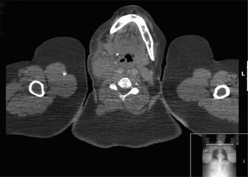
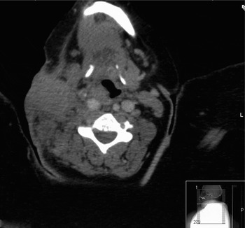
A neck ultrasound and computed tomography scan confirmed the presence of a large, heterogeneous, soft tissue mass in the right side of the neck.
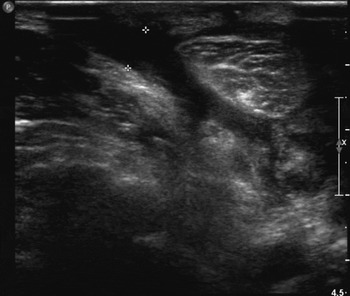
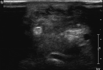
Intra-operatively, this mass was found to consist of multiple superficial and deep abscesses. The collections were small (less than 1 cm) and sited in close proximity to the great vessels (Figures 1 to 4). Incision and drainage yielded small amounts of pus.
Ultrasound scan showing irregular sinuses discharging through the right side of the neck. Distance between two markers = 0.647 cm. P = posterior

Ultrasound scan showing clearly defined, superficial collection of mixed heterogeneity. Beyond this, there appears to be a further collection extending around the great vessels and muscles of the neck. P = posterior

Axial computed tomography scan demonstrating a large, heterogeneous mass within the right side of the neck, extending across level II from the right carotid sheath to the deep lobe of the parotid gland. The mass is obliterating the parapharyngeal fat space and is contiguous with the right sternocleidomastoid muscle. L = left

Axial computed tomography scan demonstrating a large, soft tissue density area within the right side of the neck on the sternocleidomastoid muscle, displacing the right common carotid artery medially. It is obliterating the right parapharyngeal fat space, and indenting and displacing the pharynx to the left. L = left; P = posterior

Initial microbiology results identified coliforms, and intravenous meropenem was commenced. However, post-operative wound dehiscence occurred, with copious purulent discharge. Another change of antibiotic therapy, to ciprofloxacin, again failed to provide any benefit. Surgical debridement of the wound provided further specimens for histological and microbiological analysis. The results demonstrated growth of burkholderia species with sensitivity to ciprofloxacin. Further results confirmed the species as pseudomallei.
Intravenous antibiotics were continued, with daily neck dressing changes. However, clinical progress was slow.
Following discussion, it was decided to use a vacuum-assisted closure dressing to promote healing. This dressing remained in situ for seven days, and proved beneficial. On subsequent inspection, there was considerably less discharge and the wound edges were obviously healing, with visible granulation tissue.
Following removal of the vacuum-assisted closure dressing and pump, intravenous antibiotic therapy was continued for another two weeks. The patient made good progress and was discharged from the hospital on oral antibiotics (ciprofloxacin), which was continued for three months, until full clinical and radiological resolution of disease was achieved.
At the time of writing, the patient was asymptomatic but remained under regular follow up.
Discussion
Melioidosis
Melioidosis, also known as Whitmore's disease, is a severe, multisystem, infectious disease.Reference Zulkiflee, Prepageran and Philip1 It is also known as pseudoglanders, Vietnamese time bomb and Rangoon beggar's disease.Reference Thomas, Jayachandran, Shenoy Chandrasekhara, Lakshmi and Narsimulu7 Its main endemic foci are Northern Australia and Southeast Asia.Reference Zulkiflee, Prepageran and Philip1, Reference Gibney, Cheng and Currie8
The causative organism is Burkholderia pseudomallei, a Gram-negative, aerobic bacillus. Its natural habitat is soil and water in the tropics and subtropics.Reference Zulkiflee, Prepageran and Philip1, Reference Thomas, Jayachandran, Shenoy Chandrasekhara, Lakshmi and Narsimulu7, Reference Raja9Burkholderia pseudomallei is a very resilient micro-organism, able to survive inside host cells for many years – hence the long latency period between infection and onset of clinical features.Reference Zulkiflee, Prepageran and Philip1, Reference Gibney, Cheng and Currie8, Reference Aldhous10
Melioidosis can be acquired via ingestion, inhalation and direct inoculation.Reference Ma, Huang, Tang and Chang3 The commonest mode is percutaneous inoculation.Reference Cheng and Currie11 Once infected, patients may be asymptomatic (i.e. subclinical melioidosis) or may develop acute or chronic suppurative disease, or rapidly progress to fulminant sepsis.Reference Zulkiflee, Prepageran and Philip1, Reference Raja9
Risk factors for melioidosis include diabetes mellitus, mycobacterial disease and steroid use.Reference Thomas, Jayachandran, Shenoy Chandrasekhara, Lakshmi and Narsimulu7, Reference Borgherini, Poubeau, Paganin, Picot, Michault and Thibault12, Reference Currie13 The association with diabetes mellitus is particularly strong (conferring a 100-fold increased risk of infection).Reference Currie13, Reference Muthusamy, Waran and Puthucheary14
Melioidosis has been termed ‘the great mimicker’ due to the exceptionally broad spectrum of clinical syndromes with which it can present.Reference Ma, Huang, Tang and Chang3 Unless there is a high clinical index of suspicion, diagnosis is very difficult and can only be confirmed by isolation of B pseudomallei from body fluids.Reference Inglis, Rolim and Rodriguez2, Reference Cheng and Currie11
Mortality varies widely amongst different countries (19–60 per cent).Reference Chan, Low, Raghuram, Fook-Chong and Kurup15–Reference Tan, Ang and Ong17 The overall mortality rate is currently 45 per cent, half what it was prior to the advent of antibiotics.Reference Thomas, Jayachandran, Shenoy Chandrasekhara, Lakshmi and Narsimulu7, Reference Kumar, Raj, Chacko, Lalitha, Chacko and Rajshekhar18 It is this very high mortality, in conjunction with the disease's mode of transmission, which have led to concerns that B pseudomallei could be used as a biological warfare weapon.Reference Zulkiflee, Prepageran and Philip1
Melioidosis can affect any organ. The commonest presentation is pneumonia, accounting for almost half of all cases. It can also cause acute respiratory distress syndrome, septic shock, abscesses, peritonitis, septic arthritis and osteomyelitis. Skin infections can have a very rapid rate of development, presenting similarly to necrotising fasciitis.Reference Zulkiflee, Prepageran and Philip1, Reference Thomas, Jayachandran, Shenoy Chandrasekhara, Lakshmi and Narsimulu7, Reference Raja9, Reference Cheng and Currie11, Reference Chan, Low, Raghuram, Fook-Chong and Kurup15, Reference Huis in 't, Wuthiekanun, Cheng, Chierakul, Chaowagul and Brouwer19 The central and peripheral nervous systems can be affected (with abscesses, encephalitis, meningitis, sagittal sinus thrombosis and Guillain–Barre syndrome), as can the cardiovascular system (with pyopericardium, endocarditis and mycotic aneurysms).Reference Cheng and Currie11, Reference Muthusamy, Waran and Puthucheary14, Reference Chan, Low, Raghuram, Fook-Chong and Kurup15, Reference Kumar, Raj, Chacko, Lalitha, Chacko and Rajshekhar18, Reference Woods, Currie, Howard, Tierney, Watson and Anstey20, Reference Baumann and Morita21 Ocular involvement can lead to corneal ulcers, subconjunctival abscesses and hypopyon.Reference Cheng and Currie11
In the head and neck, melioidosis has several modes of presentation. Examples include parotid abscess, thyroid abscess, chronic otitis media, cutaneous neck infections, cervical lymphadenopathy, sinusitis and periorbital cellulitis.Reference Zulkiflee, Prepageran and Philip1, Reference Ma, Huang, Tang and Chang3, Reference Kumar, Raj, Chacko, Lalitha, Chacko and Rajshekhar18, Reference Currie, Fisher, Howard, Burrow, Lo and Selva-Nayagam22, Reference Kiertiburanakul, Sungkanuparph, Kositchiwat and Vorachit23
Melioidosis causing suppurative cervical lymphadenitis can be very difficult to distinguish from tuberculosis or lymphoma on clinical grounds. Hence, a good (travel) history and high clinical index of suspicion are vital. Missing the diagnosis can have disastrous consequences. Melioidosis should always be considered in the differential diagnosis of a neck abscess where there is a relevant travel history.Reference Zulkiflee, Prepageran and Philip1, Reference Ma, Huang, Tang and Chang3
The management of melioidosis remains a challenge.Reference Sivalingam, Sim, Jasper, Wang, Liu and Ooi24 It consists of two phases: initial eradication therapy with intravenous antibiotics (ceftazidime or a carbapenem for 10–14 days), followed by maintenance oral antibiotic therapy (high-dose trimethoprim–sulfamethoxazole for three months).Reference Gibney, Cheng and Currie8, Reference Kumar, Raj, Chacko, Lalitha, Chacko and Rajshekhar18, Reference Sivalingam, Sim, Jasper, Wang, Liu and Ooi24 Long-term follow up, possibly life-long, is essential.Reference Zulkiflee, Prepageran and Philip1
In summary, melioidosis is a severe, multisystem, infectious disease caused by the bacterium B pseudomallei.Reference Zulkiflee, Prepageran and Philip1 It constitutes a particular diagnostic challenge as it can mimic virtually any disease.Reference Inglis, Rolim and Rodriguez2, Reference Ma, Huang, Tang and Chang3 In the western world, where it is uncommon, melioidosis is often not considered in the differential diagnosis. If not diagnosed early and treated with appropriate antibiotics, the prognosis is very poor.Reference Thomas, Jayachandran, Shenoy Chandrasekhara, Lakshmi and Narsimulu7
Vacuum-assisted closure devices
Negative pressure dressings, also known as vacuum- assisted closure devices, were first brought to prominence in 1991.Reference Andrews, Smith, Goldstein and Funk4,Reference Argenta and Morykwas25, Reference Morykwas, Argenta, Shelton-Brown and McGuirt26 The negative pressure applies suction across the wound, aiding closure and promoting local blood supply.Reference Rosenthal, Blackwell, McGrew, Carroll and Peters6 Although use of these devices has been reported in the management of head and neck wounds; they are primarily used on the trunk and limbs.Reference Andrews, Smith, Goldstein and Funk4, Reference DeFranzo, Argenta, Marks, Molnar, David and Webb27–Reference Hopf, Humphrey, Puzziferri, West, Attinger and Hunt29
Vacuum-assisted closure pumps were initially employed in general surgery to aid the closure of dehiscent abdominal wounds.Reference Rosenthal, Blackwell, McGrew, Carroll and Peters6 Their uses today are extensive, and include the management of soft tissue lesions such as leg ulcers, infected wounds, necrotising fasciitis, denuded bone, fistulae and skin graft donor sites.Reference Rosenthal, Blackwell, McGrew, Carroll and Peters6, Reference Mendez-Eastman30–Reference Song, Wu, Lohman, Gottlieb and Franczyk32
Negative pressure is believed to have multiple healing effects. The interstitial fluid is sucked out of the wound, decompressing the local microvasculature and thus increasing regional blood flow. This increases wound oxygenation, which, in addition to the mechanical debridement produced by the negative pressure, decreases the bacterial load. Moreover, the vacuum-assisted closure device produces a homogeneous distribution of force in the wound edges, promoting healing through granulation tissue formation.Reference Marathe and Sniezek5, Reference Morykwas, Argenta, Shelton-Brown and McGuirt26
In the presented case, the negative pressure dressing was prepared at a pressure of −75 mmHg and left in situ for seven days (therapy times of 3–12 days have been reported).Reference Rosenthal, Blackwell, McGrew, Carroll and Peters6 It was continuously applied via a portable device which allowed the patient full mobility. When removed, the purulent discharge had significantly decreased, with evidence of granulation tissue covering the wound surface. Following the application of the negative pressure dressing, wound management consisted of dry dressings and oral antibiotics.
• Melioidosis is a severe, multisystem, infectious disease with a high mortality rate
• Diagnosis is particularly challenging
• Melioidosis should be considered in the differential diagnosis of a neck abscess, particularly where there is a relevant travel history
• Vacuum-assisted closure pumps appear to be under-utilised in the management of head and neck wounds
• This is the first report of a vacuum-assisted closure device used in the management of melioidosis of the neck
• The successful outcome warrants further exploration of the role of vacuum-assisted closure pumps in head and neck surgery
From the limited number of reported cases, it would appear that vacuum-assisted closure pumps are under-utilised in the management of head and neck wounds.Reference Andrews, Smith, Goldstein and Funk4–Reference Rosenthal, Blackwell, McGrew, Carroll and Peters6 The largest case series, reported by Rosenthal et al., comprised 23 applications in the head and neck over three years. Some of the cases reported were managed solely by vacuum-assisted closure pumps.Reference Rosenthal, Blackwell, McGrew, Carroll and Peters6
Negative pressure dressings can be beneficial adjuncts in the management of head and neck wounds. We believe that the successful use of a vacuum-assisted closure device in the presented case warrants further exploration of the role of vacuum-assisted closure pumps in head and neck surgery.