




No CrossRef data available.
Article contents
Testing the prenatal hormone hypothesis of tic-related disorders: Gender identity and gender role behavior
Published online by Cambridge University Press: 01 June 2004
Abstract
The hypothesis that prenatal masculinization of the brain increases risk of tic disorders in postnatal life was tested by measuring gender and gender role behavior in 89 children and adults with a clinical diagnosis of Tourette syndrome or obsessive compulsive disorder and 67 healthy, unaffected children and adults. Consistent with this hypothesis, a tic disorder in females was associated with more gender dysphoria, increased masculine play preferences, and a more typically “masculine” pattern of performance on two sex-typed spatial tasks. Males with tic disorders reported increased masculine play preferences, and the strength of these preferences was positively associated with the severity of tic symptoms. In addition, unlike their female counterparts, males with tic disorders showed a relative impairment in mental rotation ability. These behavioral profiles are consistent with those of children who have verifiable elevations in prenatal androgen levels. These findings therefore support the hypothesis that an altered androgen-dependent process of sexual differentiation during prenatal life may contribute to the development of tic-related disorders.This work was supported by National Institutes of Health Grants MH01232 (B. P.) and MH59139 (B. P.) and the Suzanne Crosby Murphy endowment in Pediatric Neuropsychiatry, College of Physicians and Surgeons, Columbia University. The authors thank Amy Basile and Shibani Mukerji for their valuable assistance with behavioral testing.
Information
- Type
- Research Article
- Information
- Copyright
- © 2004 Cambridge University Press
In many mammalian species, including nonhuman primates, hormones play a major role in directing the development of brain systems that subserve sex-typed behaviors (Arnold & Breedlove, 1985; Breedlove, Cooke, & Jordan, 1999). In these species, sex steroid levels in prenatal life promote sex differences in the expression of reproductive behavior (Gorski, 1979), sex-typical play (Meaney, 1988), and certain learning and memory functions (Williams & Meck, 1991). Human research measuring similar behaviors in individuals who are known to have atypical levels of sex steroids in prenatal life supports the hypothesis that hormones also influence the development of a masculine or feminine gender role. Girls with congenital adrenal hyperplasia (CAH), for example, are exposed to androgen levels more typical of the prenatal hormonal environment in normal males (Carson, Okuno, Lee, Stetton, Didolkar, & Migeon, 1982). In postnatal life these girls tend to show more aggression (Berenbaum & Resnick, 1997), enhanced (i.e., more masculine) visuospatial abilities (Hampson, Rovet, & Altmann, 1998; Resnick, Berenbaum, Gottesman, & Bouchard, 1986), more masculine occupational preferences (Berenbaum, 1999), and increased bisexual or homosexual sexual preferences in fantasy or behavior (Zucker, Finegan, Doering, & Bradley, 1983).
Recent elaborations of theories pertaining to the postnatal effects of the brain's sexual differentiation during prenatal life include the development of sex differences in the prevalence or natural history of developmental disorders (Baron–Cohen & Hammer, 1997; King, Barkley, Delville, & Ferris, 2000; Peterson, Leckman, Scahill, Naftolin, Keefe, Charest, King, Hardin, & Cohen, 1994). For example, a strong male/female sex ratio (4:1 or higher) in autism (Rutter & Garmezy, 1983) and Tourette Syndrome (TS) (Peterson, Pine, Cohen, & Brook, 2001) suggests that increased levels of prenatal androgens may produce an excessive masculinization of the brain, thereby increasing the risk for the development of these disorders in postnatal life (Baron–Cohen & Hammer, 1997; Peterson et al., 1992). Within this conceptual framework of central nervous system (CNS) masculinization, important common features of the TS phenotype (disinhibition of sexual or aggressive impulses) and several core phenotypic features of autism (impaired socialization and language) are viewed as “hypermasculine” traits. This categorization of symptoms is based on other evidence that the behavioral phenotypes of typically developing males include more frequent aggressive and sexual behaviors (Collaer & Hines, 1995), a relative disadvantage in the processing of facial expressions (McClure, 2000), and poorer language skills (Hyde & Linn, 1988) than typically developing females. These typical gender differences in behavioral phenotypes and the hypothesis of excessive CNS masculinization in some childhood illnesses is consistent with a gradient model of hormone influences on behavior (Collaer & Hines, 1995), in which varying degrees of exposure of the developing CNS to sex steroid hormones promotes phenotypic differences between and within the sexes.
Several additional clinical features of TS implicate sex steroid levels in the determination of the severity of tic symptoms (Peterson et al., 1992). First, tics typically increase in severity during the prepubertal years, when gonadal androgen production markedly increases in both males and females. Second, androgen administration in adults has been reported to exacerbate tic symptoms (Leckman & Scahill, 1990). Third, blockade of the androgen receptor attenuates tic symptoms in some individuals (Peterson, Leckman, Scahill, Naftolin, Keefe, Charest, King, Hardin, & Cohen, 1994; Peterson, Zhang, Anderson, & Leckman, 1998). These clinical features most clearly suggest the importance of postnatal hormone levels in the pathophysiology of TS (i.e., activational influences). They also suggest the importance of prenatal hormone effects in TS (i.e., organizational influences) because prenatal sex steroid hormones are thought to predispose brain areas to respond to changes in postnatal hormone levels, thereby inducing a behavioral sensitivity to levels of sex steroid hormones in postnatal life (Collaer & Hines, 1995). Research of the activational influences of hormones on human behavior typically measures the association between changing hormone levels (e.g., at puberty or across the menstrual cycle) and behavior. Research of the organizational influences of hormones on human behavior typically measures behaviors that show large sex differences and emerge prior to puberty (e.g., gender identity, gender-linked play) in individuals who are presumed to have experienced different prenatal hormonal environments. It is this last approach that is applied in the present investigation to study the prenatal hormone hypothesis of tic-related disorders.
We know of no previous investigations assessing the prevalence of behavioral markers for prenatal androgen effects in individuals who have tic-related disorders. However, if tic disorders are indeed associated with exposure to increased androgen levels during prenatal development, then women and girls who have tic disorders should, like females with CAH, exhibit more male-typical cognitive and behavioral attributes than do unaffected females. The goal of this study was therefore to test the hypothesized role of androgens in the development and maintenance of tic disorders by comparing measures of sexually dimorphic attributes in children and adults with and without tic disorders. Measures of sex-typed behaviors were selected on the basis of extensive prior research on hormone–behavior correlates in humans. One of our comparison groups included typically developing children and adults. We also included a second comparison group of children and adults with obsessive compulsive disorder (OCD) because TS has been repeatedly shown to share a common genetic vulnerability with childhood-onset OCD, and because TS frequently co-occurs with OCD in both clinical and epidemiological samples.
Method
Participants
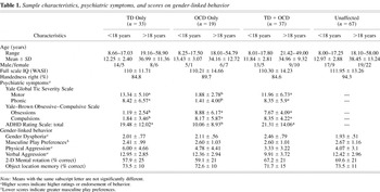
We recruited 89 children and adults with a clinical diagnosis of TS or OCD from the Tic and Obsessive–Compulsive Disorders Specialty Clinic at the Child Study Center of Yale University in New Haven, CT. DSM-IV clinical diagnoses were determined by the consensus of two experienced clinicians after reviewing clinical records and semi-structured diagnostic interviews. In addition, 67 healthy, unaffected children and adults were recruited as a comparison group through announcements and telemarketing lists. All participants were part of a larger study of the neurobiological basis of TS and OCD. Table 1 summarizes the characteristics of participants, including their preferred hand for writing, full scale IQ as measured by the Wechsler Abbreviated Scale of Intelligence (WASI; Wechsler, 1999), and the relevant psychiatric symptoms as measured by the Yale Global Tic Severity Scale (YGTSS; Leckman, Riddle, Hardin, Ort, Swartz, Stevenson, & Cohen, 1989), Yale–Brown Obsessive–Compulsive Scale (Scahill et al., 1997), and Attention-Deficit/Hyperactivity Disorder (ADHD) Rating Scale (DuPaul, 1991).
Sample characteristics, psychiatric symptoms, and scores on gender-linked behavior

Materials and procedures
Gender group identification and gender role behavior
One measure of identification with gender group was the gender label assigned by the subject in the Draw-A-Person Task (Zucker, Finegan, Doering, & Bradley, 1983). Typically, when asked to draw a person, children and adults draw figures that they later identify as someone from their own gender group. However, children who have higher frequencies of cross-gender play or gender dysphoria are more likely than controls to draw figures representing the opposite gender (Green, Fuller, & Rutley, 1972; Skilbeck, Bates, & Bentler, 1975; Zucker et al., 1983).
The second measure of identification with gender group was the Recalled Childhood Gender Identity Scale (Zucker et al., 1996). This 23-item questionnaire, in which each item is scored on a range of 1–5, was administered to both adolescents and adults. Stronger male-typical attributes are represented by lower scores on items assessing play preference (e.g., the stated sex of usual playmates or best friends, or preference for gender-typical toys or play styles), and greater gender dysphoria is represented by higher scores on items assessing gender identification (e.g., in contentment with gender or in comparisons of self with other individuals). The original scoring of the questionnaire excludes responses indicating no participation in the activity (e.g., “I did not play with other children”) or uncategorized responses (e.g., “My favorite toys and games were neither “masculine” or “feminine” or “I did not feel masculine or feminine”). Consequently, the number of items contributing to our two summary measures (Gender-Typical Play and Gender Dysphoria) varied across subjects. For this reason, and because such responses seem to reflect, in part, an undifferentiated development of gender role, a separate analysis also examined group differences in the number of these uncategorized responses.
The Reinisch Revision of the Leifer–Roberts Response Hierarchy (Reinisch, 1981) was used to measure each individual's potential for responding to interpersonal conflict with aggressive behavior. Six situations of interpersonal conflict were each presented six times, followed by one forced-choice alternative. The 36 forced-choice alternatives represented all possible combinations of four classes of responses—a physically aggressive response (e.g., “Hit them”), a verbally aggressive response (e.g., “Yell at them”), a response indicating withdrawal (e.g., “Go inside the house”), or a nonaggressive response (e.g., “Tell an adult”). Children and adults were asked to choose the one response from each pair that was most consistent with what they would do in that situation. Adults were asked to respond as they believed they would have at 10 years of age. Responses were summed to yield measures of verbal aggression, physical aggression, withdrawal, or nonaggressive coping. In previous research, men and boys were more likely than women and girls to report that they would respond with physical aggression (Cohen's d > 0.7; Reinisch & Sanders, 1986; Reinisch, 1981).
Sex-linked spatial abilities
Participants also completed two tasks of spatial ability. The Two-Dimensional Mental Rotation Task (Collins & Kimura, 1997) contains 24 items, each depicting an abstract figure in a random orientation and four other figures representing the target figure in other orientations. A small arrow placed directly above the target figure represents the hour hand of a clock at 12:00, indicating that the figure is at a 12:00 orientation. The task is to identify which of the four other figures represents the target figure orientated to a specified time (ranging between 2:00 and 10:00). Subjects are given 5 min to complete the measure, and most subjects fail to complete all 24 items. Males have previously been shown to have a large advantage on this task (Cohen's d > .80). The “Hard” and “Easy” forms were administered to adults and to children/adolescents, respectively.
The second spatial task was the Object Location Memory Task of Silverman and Eals, a task in which females have previously been shown to have an advantage (Eals & Silverman, 1994; Silverman & Eals, 1992). The task has one stimulus card and two response cards, each depicting a spatial array of common objects. The stimulus card depicts a spatial array of 27 common objects, including nature items (i.e., cat, elephant, bird, flower), household items (i.e., phone, iron, teapot, plant), and work-related items (i.e., briefcase, hat, cane). One response card measuring memory for object identities depicts an array of the original 27 objects in their original locations and 20 added objects. A second response card measuring memory for object location depicts the original 27 objects, with an exchange between the positions of 7 pairs of objects. Participants were presented with a stimulus card for 1 min and were told that they would be asked to remember what objects were on the page. After 1 min, the stimulus card was removed from sight and participants were presented with the first response card. After marking the added objects, the response card was removed. Participants were presented next with the second response card and asked to mark the objects that were moved to new locations or places on the card.
For both the object identification and location portions of this task, errors included total omissions (false negatives) and total commissions (false positives). As the female advantage in memory for object location is largest for objects in the right visual hemispace (Alexander, Packard, & Peterson, 2002), memory scores for object identities were derived separately for objects left and right of veridical center. For each task and for each side, accuracy was defined as 1 − (omissions + commissions)/N, where N is the number of objects.
Statistical analyses
Subject characteristics were analyzed using analysis of variance (ANOVA) or chi-square analyses. Chronic motor tics were identified in seven individuals with a primary diagnosis of OCD. In view of phenomological, natural history, and neurobiological evidence indicating that chronic tics and TS differ from each only in the duration of the symptoms (Peterson, Pine, et al., 2001), we first analyzed our data using the presence of any tic disorder (TD) as a grouping factor. Because tic disorders frequently co-occur with OCD in both clinical and epidemiological samples, and because of the limitations of analysis of covariance (ANCOVA; Miller & Chapman, 2001), a 2 (male vs. female) × 2 (TD vs. no TD) × 2 (OCD vs. no OCD) full factorial design was used to compare behavior across individuals with these tic-related disorders. All primary analyses using ANOVA, therefore, included comparisons between four groups derived from the coding for the presence (1) or absence (0) of a tic disorder or OCD; namely, an unaffected comparison group, a TD only group, an OCD only group, and a TD plus OCD group. The full factorial design has an advantage of identifying main effects of the disorders and any dissociation (i.e., interaction effect) between two disorders on the behavioral measures of interest (Willcutt, Pennington, Boada, Ogline, Tunick, Chhabildas, & Olson, 2001). All statistical models were hierarchically well formulated (i.e., the inclusion of interactions required inclusion of lower order terms that were a component of that interaction). Any significant main effects or interaction effects were followed by post hoc comparisons using Newman–Keuls tests. Individuals not meeting the full criteria of TS (5 males and 2 females with a chronic motor tic) were eliminated from a second analysis using TS, OCD, and sex as grouping factors. We detail the findings from that second analysis only when they differed from those of the first analysis.
The number of participants across a wide age range (8–60 years of age) precluded a detailed analysis of any age-dependent effects on the behavioral variables of interest. However, wherever possible, we compared behavior between individuals over 18 years of age (adults) and individuals younger than 18 years of age (children/adolescents).
Results
Gender effects on subject characteristics and diagnostic variables
No group or sex effects were found for age or for general cognitive function as measured by the WASI. A trend for increased left-hand preference was detected in individuals with a tic-related disorder, consistent with previous findings (Comings & Comings, 1987). Further analysis showed that the number of males with a left-hand preference was similar across the diagnostic groups. However, left-hand preferences were reported by a greater number of women and girls with tic symptoms (23.8%) compared to women and girls without tic symptoms (5.4%), χ2 (1) = 4.27, p < .05.
A series of multivariate ANOVAs (MANOVAs; sex by diagnostic group) were conducted to test whether sex was associated with the diagnostic variables within the three clinical groups (TD only, OCD only, TD plus OCD). This analysis showed the expected group differences in diagnostic variables (see Table 1 for a summary of these group differences). No sex differences were found on the YGTSS or the ADHD Rating Scale. However, an analysis of the severity of OCD symptoms also showed a main effect of sex, multivariate F (2, 75) = 7.76, p < .01, that was significant for both obsessions and compulsions, Fs (1, 76) = 13.26 and 11.23, respectively, ps < .05. A comparison of means indicated the presence of more severe OCD symptoms in our sample of women with OCD (Mcomp = 10.47, SD = 3.5; Mobsess = 10.43, SD = 4.0) compared to men with OCD (Mcomp = 6.42, SD = 4.8; Mobsess = 6.54, SD = 4.9). Including age grouping in the model did not alter these findings.
Gender group identification
Draw-a-Persons were completed by 67 individuals in the unaffected comparison group (36 males, 31 females), 33 individuals in the TD only group (22 males, 11 females), 24 individuals in the OCD only group (12 males, 12 females), and 32 individuals in the TD plus OCD group (21 males, 11 females). Gender labels assigned to the Draw-A-Persons were gender consistent for the majority of males (83.3%) and females (77.4%) in the unaffected comparison group. Gender-consistent labels were also applied to drawings by the majority of males within each of the three primary diagnostic groups (TD only: 95.2%; OCD only: 91.7%; TD plus OCD: 100%). In contrast, the percentage of drawings labeled as female was variable across women and girls from the different diagnostic groups (TD only: 81.8%; OCD only: 58.3%; TD plus OCD: 27.3%), χ2 (3) = 18.43, p < .01. Further analysis showed that significantly fewer females with a diagnosis of OCD (8/18 adults, 2/6 children/adolescents) drew a female figure, χ2 (1) = 7.64, p < .05, suggesting that females with a diagnosis of OCD may have had a less consolidated identification for gender than did the other females of this sample.
Recalled gender dysphoria
Children under 13 years of age did not complete the Recalled Childhood Gender Identity Scale. The group of adolescents and adults completing the questionnaire included 49 individuals in the unaffected comparison group (28 males, 21 females), 21 individuals in the TD only group (12 males, 9 females), 19 individuals in the OCD only group (10 males, 9 females), and 19 individuals in the TD plus OCD group (11 males, 8 females).
Preliminary analysis of the number of undifferentiated responses on the questionnaire showed a main effect of OCD, F (1, 100) = 3.99, p < .05, such that a diagnosis of OCD was associated with a greater number of undifferentiated responses (M = 2.22, SD = 1.82 vs. M = 1.55, SD =1.45). In addition, a significant three-way interaction was detected between TD, OCD, and sex, F (1, 100) = 7.48, p < .01. Post hoc comparisons showed that males having a diagnosis of TD plus OCD reported a greater number of undifferentiated responses (M = 2.54, SD = 1.91) compared with males having a diagnosis of TD only (M = 0.83, SD = 0.84). Undifferentiated responses in women, in contrast, were also highest in the OCD only group (M = 2.60, SD = 2.17) but not significantly higher than the number of undifferentiated responses in the unaffected comparison group (M = 1.47, SD = 1.28), in the TD only group (M = 2.22, SD = 1.64), or in the TD plus OCD group (M = 1.75, SD = 1.38). These results indicate that a diagnosis of OCD was associated with a greater number of undifferentiated responses on the questionnaire measure, particularly so for males with the disorder.
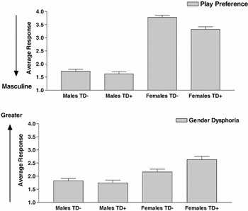
A MANOVA for gender-typical play preferences and gender dysphoria showed both main effects of sex, TD, and a sex by TD interaction, multivariate Fs (2, 99) = 374.61, 3.93, and 3.10, respectively, ps < .05, but no main effects or interaction effects of OCD on the two measures. Univariate analysis showed the expected overall sex differences in play preferences, such that males compared to females reported stronger preferences for masculine play and less gender dysphoria, Fs (1, 100) = 428.59 and 33.01, respectively, ps < .001. A tic disorder in both males and females was associated with stronger preferences for masculine play, F (1, 100) = 7.82, p < .01. However, the presence of a tic disorder in females but not in males was associated with greater gender dysphoria, F (1, 100) = 5.98, p < .05. Including in the model age as a grouping factor (i.e., adults vs. adolescents) or the symptom severity ratings as covariates did not show additional effects of age group or change the general significance level or the direction of these results (Figure 1). These findings indicate that masculine play preferences are increased in both males and females with a tic disorder relative to other males and females. In addition, they suggest that females with a tic disorder report greater gender dysphoria than do females without a tic disorder.

The main effect of tic disorder and gender by tic-disorder interaction effect on gender-linked behavior is illustrated by the marginal means for self-reports of gender dysphoria and masculine play preferences in males and females with and without a tic disorder. The group without a tic disorder includes individuals with no tic disorder or OCD (i.e., an unaffected comparison group) and a group of individuals with a diagnosis of OCD only (a clinical comparison group). Compared to individuals without a tic disorder, males and females with a tic disorder report stronger preferences for male-typical play (top; p < .01). Compared to females without a tic disorder, females with a tic disorder report more gender dysphoria (bottom; p < .05).
Reinish aggression inventory
A MANOVA for measures of physically aggressive, verbally aggressive, withdrawn, and nonaggressive coping, in which TD, OCD, and sex were grouping factors, showed only the expected sex difference in response, multivariate F (4, 124) = 9.49, p < .001, and no other main or interaction effects. Males compared to females reported more physical aggression, less withdrawal, and less nonaggression in response to conflict situations, Fs (1, 127) = 32.33, 17.50, and 5.83, respectively, ps < .01. Including age grouping (adults vs. children/adolescents) in the analysis replicated these general findings and showed additionally that adults compared to children/adolescents reported higher levels of verbal aggression (M = 12.28, SD = 3.12 vs. M = 10.77, SD = 3.36). However, no other age group effects on reports of coping style were detected.
Sex-typed spatial abilities
Analysis of the number of correct responses on the mental rotation task also showed the expected sex difference in performance and a sex by TD interaction, Fs (1, 139) = 10.87 and 9.84, respectively, ps < .01. Further analysis showed that males with a tic disorder had fewer correct responses on the mental rotation task (M = 8.8, SD = 3.7) than did males without a tic disorder (M = 11.9, SD = 3.7, p < .01). In contrast, females with a tic disorder had a greater number of correct responses on the mental rotation task (M = 8.4, SD = 4.7) than did females without a tic disorder (M = 7.2, SD = 3.5, p < .05). Including as covariates in the model IQ, severity ratings of inattention, or ratings of hyperactivity (both measures of the ADHD rating scale) did not affect the general significance levels or the direction of these group differences. These findings indicated a dissociation between the effects of TD on mental rotation ability in males and females. Compared to individuals without tic symptoms, mental rotation ability in individuals with tic symptoms was impaired in males but enhanced in females.
We used object memory scores as covariates in a conservative analysis of group and sex differences in memory for object location, as recommended by previous researchers (James & Kimura, 1997). An ANCOVA for object location scores, with side (left or right) as a repeated measure, and sex, OCD, and TD as grouping factors, showed the expected female advantage in performance, F (1, 142) = 4.75, p < .05, and a three-way interaction between side, sex, and TD, F (1, 142) = 6.96, p < .01. In general, object locations were recalled more accurately by females (M = 78.0%, SD = 13%) than by males (M = 74.3%, SD = 11%). Post hoc tests showed that for females without a TD, object locations were recalled more accurately in the right visual hemispace (M = 86%, SD = 11%) than in the left hemispace (M = 68%, SD = 10%, p < .05). In contrast, for females with a TD, the percentages of correct responses for objects in the right hemispace (M = 82%, SD = 11%) or left hemispace (M = 76%, SD =12%) were not significantly different (p = .44). Memory scores in males did not differ across groups or between right or left hemispace. These results suggest that a female pattern of response for object locations in each visual hemispace is reduced in females with a chronic TD, and so is more similar to the pattern demonstrated by normal males.
Not unexpectedly, adults were more accurate than children/adolescents on the mental rotation task (M = .65, SD = .22 vs. M = .53, SD = .19, F (1, 133) = 6.42, p < .01). However, there were no other effects of age group on sex-typed spatial abilities.
Within-group analyses
An examination of simple correlations between age, ratings of symptom severity, and measures of gender role development within each sex showed only that for males, preferences for masculine play were positively associated with the severity of motor and phonic tics, rs (23) = .68 and 61, respectively, ps < .01. All other correlations were of small magnitude (r < .25) and not statistically significant.
Discussion
Group comparisons across a variety of sex-typed behaviors in this study were remarkably consistent in demonstrating a systematic shift toward more male-typical responses in women and girls who have tic symptoms. The presence of a tic disorder in women and girls was associated with increased gender dysphoria, increased preferences for play styles typically preferred by boys, and a more typically “masculine” pattern of performance on two sex-typed spatial tasks. We also found a reduced prevalence of right-handedness in females who have tics. In the general population, more men than women are left-handed, and men more often show less consistent right-handed preferences across a variety of manual tasks (Hines & Gorski, 1985). Thus, in this respect, women and girls with tic symptoms in our sample were again more “masculine” in their behavioral attributes. One exception to this pattern of results was that we found no differences in aggressive responses across diagnostic groups, although we did replicate across our entire sample the expected greater endorsement of aggressive responses in males.
Greater gender dysphoria in women and girls has been associated in other human research with exposure to high levels of prenatal androgens (Ehrhardt & Baker, 1974; Ehrhardt, Epstein, & Money, 1968). Girls exposed prenatally to increased endogenous adrenal androgens (Berenbaum & Hines, 1992; Hines & Kaufman, 1994) or to exogenous androgenic progestins (Ehrhardt & Money, 1967) also exhibit increased male-typical play preferences, and some studies (Hampson et al., 1998; Hines, Fane, Pasterski, Mathews, Conway, & Brooke, 2003; Resnick et al., 1986) have reported that these girls have enhanced spatial abilities as well. The similarity of our findings to those reported for girls with unequivocal exposure to male-typical androgen levels during prenatal life is consistent with the hypothesis that an altered androgen-dependent process of sexual differentiation of the brain may contribute to the development of tic-related disorders (Peterson et al., 1992).
Changes in the brain organization subserving sex-linked behaviors in tic-related disorders are also suggested by our finding that women and girls with tic symptoms did not exhibit the typical female advantage for recall of object locations in the right visual hemispace, an advantage that is thought to reflect a greater specialization of the left cerebral hemisphere for spatial memory in females (Alexander et al., 2002). A reduced functional lateralization for spatial memory in women and girls who have tic symptoms is consistent with previous reports of reduced functional (Yazgan, Peterson, Wexler, & Leckman, 1995) and anatomical asymmetries (Peterson, Riddle, Cohen, Katz, Smith, Hardin, & Leckman, 1993; Singer et al., 1993) in individuals with TS and with more recent findings that volumes of the parieto-occipital cortex, a region thought to subserve the processing of spatial information, were reduced (i.e., masculinized) in girls and women with TS (Peterson, Staib, Scahill, Zhang, Anderson, Leckman, Cohen, Gore, Albert, & Webster, 2001).
Reduced functional asymmetries in women and girls with tic symptoms are also suggested by our finding of a reduced prevalence of right-handedness in females who have tics. Although left-hand preference may be a postnatal marker for a prenatal exposure of the developing fetal brain to a variety of physiological stressors (Bakan, Dibb, & Reed, 1973), left handedness may also be influenced by hormonal factors (Hines, 2002). Exposure to a synthetic estrogen (diethylstilbestrol) in the first trimester of fetal development, for example, increases left-handed representation in females (Smith & Hines, 2000). During fetal brain development, androgens are aromatized to estrogens, which then masculinize brain structure and function. Therefore, changes in sex-typed behaviors, functional cerebral asymmetries, and increased left hand preferences in females with tic symptoms are all consistent with the hypothesized increase in androgen (or aromatized estrogen) levels in the CNS during early prenatal life.
Notably, men with tic symptoms also reported elevated male-typical play preferences relative to other groups of men, and their preference for male-typical play correlated positively with the severity of their tic symptoms. Unlike their female counterparts, however, men and boys with tic symptoms were relatively impaired on a mental rotation task in which males usually have a large performance advantage. This latter result suggests that deficits in spatial processing observed previously in children with TS (Schultz, Carter, Gladstone, Scahill, Leckman, Peterson, Heping, Cohen, & Pauls, 1998) may be more pervasive in males than in females who have tic symptoms. The disadvantage in spatial processing in males could also be regarded as typically “hypomasculine” in pattern and therefore as potentially inconsistent with the hypothesized increase in prenatal androgens in individuals with tic-related disorders. This challenge to our hypothesis in males, however, assumes that androgen levels are positively and linearly associated with spatial abilities in males, and this assumption has proved to be untenable in correlational studies (Gouchie & Kimura, 1991; Moffat & Hampson, 1996). Further, hypomasculine patterns of spatial abilities in males with CAH have been reported in the same studies in which performance advantages have been reported in females with CAH (Hampson et al., 1998; Hines et al., 2003). Finally, prenatal administration of androgens to male rats amplified deficits on a spatial task in an animal model of ADHD (King et al., 2000). Taken together, these findings suggest that prenatal androgens may have sex-specific effects on spatial abilities (i.e., higher levels in females enhance spatial ability whereas higher levels in males impair spatial ability), consistent with our findings.
We argue that our results are consistent with the prenatal hormonal hypothesis of tic-related disorders. An alternative explanation for our findings, however, may be that other codeterminants of gender-linked behaviors, such as socialization and the development of gender schemas during early childhood (Ruble & Martin, 1998), are altered in individuals with tic disorders. The presence of a tic, for example, could interfere directly with typical gender role development, a possibility that is consistent with the temporal association between the onset of motor tics in early childhood, their later peak in severity at age 11 years (Peterson, 1996), and the concurrent emergence of gender segregation and gender subcultures (Maccoby, 1998). This cognitive–social explanation for the greater representation of masculinized behaviors in girls with tics is analogous to the argument made for girls with CAH, that genital virilization (which occurs to variable degrees in girls with CAH) alters the cognitive–social influences on the development of male-typical gender roles (Quadagno, Briscoe, & Quadagno, 1977). Unlike genital virilization, however, the development of motor tics does not precede early gender socialization in infancy or the emergence of gender identity and gender-linked play preferences. Girls with tic symptoms, therefore, would not be expected in the cognitive–social model to differ from other unaffected girls in their early play preferences or level of gender satisfaction. Moreover, in contrast to the social experiences associated with genital virilization in CAH, tics seem unlikely to be regarded as a particularly “masculine” trait. Furthermore, our finding that a diagnosis of OCD in females was associated with measures of gender dysphoria (in the Draw-A-Person Task) and with an “undifferentiated” gender role (in the response questionnaire)—but not with more male-typical responses on other measures—is consistent with the general conclusion from studies in other species that masculinization and feminization of behavior are independent processes (i.e., they are not simply extremes on a single continuum of behavior; Collaer & Hines, 1995; Hines, 2002). Therefore, given the independence of masculinization and feminization of behavior, the possibility that symptoms associated with TD (or OCD) could impede the acquisition of a feminine gender role in girls does not explain the development of a more masculine gender role in women and girls with TD. Finally, the hypothesis that the presence of tics promotes the social experiences that drive the development of a masculine gender role would not account for our finding of impaired mental rotation in males with tics. Nonetheless, in view of the unexplored possibility that tics are behaviors that alter gender role development, it would be important in future research to evaluate gender-linked behavior in larger numbers of children with tic-related disorders using a longitudinal design.
Clearly, this research is only a first step in establishing an association between gender-linked behavior and tic disorders. The findings of this study demonstrate the utility of exporting methodologies, previously employed in human investigations of the organizational influences of hormones on behavior, to the study of hormone effects on sex-typed developmental disorders (Alexander & Peterson, 2001). However, an obvious limitation of our methods is the inability to document prenatal androgen levels in our participants. Converging evidence for an altered androgen-dependent process of sexual differentiation in individuals with tic-related disorders may come from future research measuring steroid hormones during prenatal development (e.g., Wudy, Dorr, Sollender, Djalali, & Homoki, 1999) or using measures of other markers of prenatal androgen exposure. For example, we know of no evidence that genital virilization is more prevalent in females with tic disorders, yet androgen exposure early in prenatal development can produce subtle changes in external genitalia (e.g., a reduced ratio of anus to vagina length or vagina to clitoris length) (Herman et al., 2000) that could be measured in individuals with neuropsychiatric disorders that have prenatal masculinization of brain development as a putative feature of their etiologies. One other retrospective marker of elevated prenatal androgen levels is the ratio of the lengths of the second and fourth digits, which is usually smaller in males than in females (Manning, Scott, Wilson, & Lewis–Jones, 1998). Digit ratios are reduced in children who have CAH (Brown et al., 2002) or autism (Manning, Baron–Cohen, Wheelwright, & Sanders, 2001), consistent with the hypothesized increased masculinization of brain structure and function in these conditions. It may be informative to assess such retrospective markers of prenatal androgen levels and their association with sex-typed behaviors in individuals with tic-related disorders.
Finally, it may be significant that common components of “masculine” attributes, such as movement in space (Eals & Silverman, 1994; Silverman & Eals, 1992), propulsion (Benenson, Liroff, Pascal, & Cioppa, 1997), and activity level (Campbell & Eaton, 1999; Eaton & Enns, 1986) each involve motor activity. It seems possible, therefore, that the brain circuits subserving the motor activity of tics may also influence the motor activities and behavioral tendencies that characterize a masculine gender role. Addressing this hypothesis in future research on the neuroanatomical basis of tic behaviors may advance a more general understanding of the biological influences on typical gender role development.
References
REFERENCES
Alexander, G. M.,
Packard, M. G., &
Peterson, B. S.
(2002).
Sex and spatial position effects on object location memory
following intentional learning of object identities.
Neuropsychologia
40,
1516–1522.CrossRefGoogle Scholar
Alexander, G. M., &
Peterson, B. S.
(2001).
Sex steroids and human behavior: Implications for developmental
psychopathology.
CNS Spectrum
6,
75–88.CrossRefGoogle Scholar
Arnold, A. P., &
Breedlove, S. M.
(1985).
Organizational and activational effects of sex steroids on brain
and behavior: A reanalysis.
Hormones & Behavior
19,
469–498.CrossRefGoogle Scholar
Bakan, P.,
Dibb, G., &
Reed, P.
(1973).
Handedness and birth stress.
Neuropsychologia
11,
363–366.CrossRefGoogle Scholar
Baron–Cohen, S., &
Hammer, J.
(1997).
Is autism an extreme form of the male brain?
Advances in Infancy Research
11,
193–217.Google Scholar
Benenson, J. F.,
Liroff, E. R.,
Pascal, S. J., &
Cioppa, G. D.
(1997).
Propulsion: A behavioural expression of masculinity.
British Journal of Developmental Psychology
15,
37–50.CrossRefGoogle Scholar
Berenbaum, S. A.
(1999).
Effects of early androgens on sex-typed activities and interests in
adolescents with congenital adrenal hyperplasia.
Hormones & Behavior
35,
102–110.CrossRefGoogle Scholar
Berenbaum, S. A., &
Hines, M.
(1992).
Early androgens are related to childhood sex-typed toy preferences.
Psychological Science
3,
203–206.CrossRefGoogle Scholar
Berenbaum, S. A., &
Resnick, S. M.
(1997).
Early androgen effects on aggression in children and adults with
congenital adrenal hyperplasia.
Psychoneuroendocrinology
22,
505–515.CrossRefGoogle Scholar
Breedlove, S. M.,
Cooke, B. M., &
Jordan, C. L.
(1999).
The orthodox view of brain sexual differentiation.
Brain, Behavior & Evolution
54,
8–14.CrossRefGoogle Scholar
Brown, W. M.,
Hines, M.,
Fane, B. A., &
Breedlove, M.
(2002).
Masculinized finger length patterns in human males and females with
congenital adrenal hyperplasia.
Hormones & Behavior
42,
380–386.CrossRefGoogle Scholar
Campbell, D. W., &
Eaton, W. O.
(1999).
Sex differences in the activity level of infants.
Infant & Child Development
8,
1–17.3.0.CO;2-O>CrossRefGoogle Scholar
Carson, D. J.,
Okuno, A.,
Lee, P. A.,
Stetton, G.,
Didolkar, S. M., &
Migeon, C. J.
(1982).
Amniotic fluid steroid levels: Fetuses with adrenal hyperplasia,
46, XXY fetuses, and normal fetuses.
American Journal of Diseases of Children
136,
218–222.CrossRefGoogle Scholar
Collaer, M. L., &
Hines, M.
(1995).
Human behavioral sex differences: A role for gonadal hormones
during early development?
Psychological Bulletin
118,
55–107.Google Scholar
Collins, D. W., &
Kimura, D.
(1997).
A large sex difference on a two-dimensional mental rotation task.
Behavioral Neuroscience
111,
845–849.CrossRefGoogle Scholar
Comings, D. E., &
Comings, B. G.
(1987).
A controlled study of Tourette syndrome. VI. Early development,
sleep problems, allergies, and handedness.
American Journal of Human Genetics
41,
822–838.Google Scholar
DuPaul, G. J.
(1991).
Parent and teacher ratings of ADHD symptoms: Psychometric
properties in a community-based sample.
Journal of Clinical Child Psychology
20,
245–253.CrossRefGoogle Scholar
Eals, M., &
Silverman, I.
(1994).
The hunter-gatherer theory of spatial sex differences: Proximate
factors mediating the female advantage in location memory.
Ethology and Sociobiology
15,
95–105.CrossRefGoogle Scholar
Eaton, W. O., &
Enns, L. R.
(1986).
Sex differences in human motor activity level.
Psychological Bulletin
100,
19–28.CrossRefGoogle Scholar
Ehrhardt, A. A., &
Baker, S. W.
(1974).
Fetal androgens, human central nervous system differentiation, and
behavior sex differences. In
R. C. Friedman,
R. M. Richart, &
R. L. van the Wiele (Eds.),
Sex differences in behavior (pp.
33–52).
New York:
Wiley.
Ehrhardt, A. A.,
Epstein, R., &
Money, J.
(1968).
Fetal androgens and female gender identity in the early-treated
adrenogenital syndrome.
Johns Hopkins Medical Journal
122,
165–167.Google Scholar
Ehrhardt, A. A., &
Money, J.
(1967).
Progestin-induced hermaphroditism: IQ and psychosexual identity in
a study of ten girls.
The Journal of Sex Research
3,
83–100.CrossRefGoogle Scholar
Gorski, R. A.
(1979).
The neuroendocrinology or reproduction: An overview.
Biology of Reproduction
20,
111–127.Google Scholar
Gouchie, C., &
Kimura, D.
(1991).
The relationship between testosterone levels and cognitive ability
patterns.
Psychoneuroendocrinology
16,
323–334.CrossRefGoogle Scholar
Green, R.,
Fuller, M., &
Rutley, B.
(1972).
It-Scale for children and Draw-A-Person Test: 30 feminine vs. 25
masculine boys.
Journal of Personality Assessment
36,
349–352.CrossRefGoogle Scholar
Hampson, E.,
Rovet, J. F., &
Altmann, D.
(1998).
Spatial reasoning in children with congenital adrenal hyperplasia
due to 21-hydroxylase deficiency.
Developmental Neuropsychology
14,
299–320.CrossRefGoogle Scholar
Herman, R. A.,
Jones, B.,
Mann, D. R., &
Wallen, K.
(2000).
Timing of prenatal androgen exposure: Anatomical and endocrine
effects on juvenile male and female rhesus monkeys.
Hormones and Behavior
38,
52–66.CrossRefGoogle Scholar
Hines, M.
(2002).
Sexual differentiation of the human brain and behavior. In
D. W. Pfaff,
A. P. Arnold,
A. M. Etgen,
S. E. Fahrback, &
R. T. Ruben (Eds.),
Hormones, brain and behavior
(Vol. 4, pp.
425–462).
San Diego, CA:
Academic Press.
Hines, M.,
Fane, B. A.,
Pasterski, V. L.,
Mathews, G. A.,
Conway, G. S., &
Brooke, C.
(2003).
Spatial abilities following prenatal androgen abnormality:
Targeting and mental rotations performance in individuals with
congenital adrenal hyperplasia.
Psychneuroendocrinology
28,
1010–1026.CrossRefGoogle Scholar
Hines, M., &
Gorski, R. A.
(1985).
Hormonal influences on the development of neural asymmetries. In
D. F. Benson &
E. Zaidel (Eds.),
The dual brain: Hemispheric specialization in humans (pp.
75–96).
New York:
Guilford Press.
Hines, M., &
Kaufman, F. R.
(1994).
Androgen and the development of human sex-typical behavior:
Rough-and-tumble play and sex of preferred playmates in children with
congenital adrenal hyperplasia (CAH).
Child Development
65,
1042–1053.CrossRefGoogle Scholar
Hyde, J. S., &
Linn, M. C.
(1988).
Gender differences in verbal ability: A meta-analysis.
Psychological Bulletin
104,
53–69.CrossRefGoogle Scholar
James, T. W., &
Kimura, D.
(1997).
Sex differences in remembering the locations of objects in an
array: Location-shifts versus location-exchanges.
Evolution & Human Behavior
18,
155–163.CrossRefGoogle Scholar
King, J. A.,
Barkley, R. A.,
Delville, Y., &
Ferris, C. G.
(2000).
Early androgen treatment decreases cognitive function and
catecholamine innervation in an animal model of ADHD.
Behavioral Brain Research
107,
35–43.CrossRefGoogle Scholar
Leckman, J. F.,
Riddle, M. A.,
Hardin, M. T.,
Ort, S. I.,
Swartz, K. L.,
Stevenson, J., &
Cohen, D. J.
(1989).
The Yale Global Tic Severity Scale: Initial testing of a
clinician-rated scale of tic severity.
Journal of the American Academy of Child & Adolescent
Psychiatry
28,
566–573.CrossRefGoogle Scholar
Leckman, J. F., &
Scahill, L.
(1990).
Possible exacerbation of tics by androgenic steroids
[Letter].
New England Journal of Medicine,
322, 1674.Google Scholar
Maccoby, E. E.
(1998).
The two sexes: Growing up apart, coming together.
Cambridge, MA:
Belknap Press/Harvard University Press.
Manning, J. T.,
Baron–Cohen, S.,
Wheelwright, S., &
Sanders, G.
(2001).
The 2nd to 4th digit ratio and autism.
Developmental Medicine & Child Neurology
43,
160–164.CrossRefGoogle Scholar
Manning, J. T.,
Scott, D.,
Wilson, J., &
Lewis–Jones, D. I.
(1998).
The ratio of 2nd to 4th digit length: A predictor of sperm numbers
and concentrations of testosterone, luteinizing hormone and
oestrogen.
Human Reproduction
13,
3000–3004.CrossRefGoogle Scholar
McClure, E. B.
(2000).
A meta-analytic review of sex differences in facial expression
processing and their development in infants, children, and
adolescents.
Psychological Bulletin
126,
424–453.CrossRefGoogle Scholar
Meaney, M. J.
(1988).
The sexual differentiation of social play.
Trends in Neuroscience
11,
54–58.CrossRefGoogle Scholar
Miller, G. A., &
Chapman, J. P.
(2001).
Misunderstanding analysis of covariance.
Journal of Abnormal Psychology
110,
40–48.CrossRefGoogle Scholar
Moffat, S. D., &
Hampson, E.
(1996).
A curvilinear relationship between testosterone and spatial
cognition in humans: Possible influence of hand preference.
Psychoneuroendocrinology
21,
323–337.CrossRefGoogle Scholar
Peterson, B. S.
(1996).
Considerations of natural history & pathophysiology in the
psychopharmacology of Tourette's syndrome.
Journal of Clinical Psychiatry,
57(Suppl. 9),
24–34.Google Scholar
Peterson, B. S.,
Leckman, J. F.,
Scahill, L.,
Naftolin, F.,
Keefe, D.,
Charest, N. J., &
Cohen, D. J.
(1992).
Steroid hormones and CNS sexual dimorphisms modulate symptom
expression in Tourette's syndrome.
Psychoneuroendocrinology
17,
553–563.CrossRefGoogle Scholar
Peterson, B. S.,
Leckman, J. F.,
Scahill, L.,
Naftolin, F.,
Keefe, D.,
Charest, N. J.,
King, R. A.,
Hardin, M. T., &
Cohen, D. J.
(1994).
Steroid hormones and Tourette's syndrome: Early experience
with antiandrogen therapy.
Journal of Clinical Psychopharmacology
14,
131–135.Google Scholar
Peterson, B. S.,
Pine, D. S.,
Cohen, P., &
Brook, J.
(2001).
A prospective, longitudinal study of tic,
obsessive–compulsive, and attention deficit-hyperactivity
disorders in an epidemiological sample.
Journal of the American Academy of Child & Adolescence
Psychiatry
40,
685–694.CrossRefGoogle Scholar
Peterson, B. S.,
Riddle, M. A.,
Cohen, D. J.,
Katz, L. D.,
Smith, J. C.,
Hardin, M. T., &
Leckman, J. F.
(1993).
Reduced basal ganglia volumes in Tourette's syndrome using
three-dimensional reconstruction techniques from magnetic resonance
images.
Neurology
43,
941–949.CrossRefGoogle Scholar
Peterson, B. S.,
Staib, L.,
Scahill, L.,
Zhang, H.,
Anderson, C.,
Leckman, J. F.,
Cohen, D. J.,
Gore, J. C.,
Albert, J., &
Webster, R.
(2001).
Regional brain and ventricular volumes in Tourette syndrome.
Archives of General Psychiatry
58,
427–440.CrossRefGoogle Scholar
Peterson, B. S.,
Zhang, Z.,
Anderson, G. A., &
Leckman, J. F.
(1998).
A randomized, placebo-controlled, crossover trial of the use of an
adrogen receptor antagonist in the treatment of Tourette's
syndrome.
Journal of Clinical Psychopharmacology
18,
324–331.CrossRefGoogle Scholar
Quadagno, D. M.,
Briscoe, R., &
Quadagno, J. S.
(1977).
Effects of perinatal gonadal hormones on selected nonsexual
behavior patterns: A critical assessment of the nonhuman and human
literature.
Psychological Bulletin
84,
62–80.CrossRefGoogle Scholar
Reinisch, J. M., &
Sanders, S. A.
(1986).
A test of sex differences in aggressive response to hypothetical
conflict situations.
Journal of Personality and Social Psychology
50,
1045–1049.CrossRefGoogle Scholar
Reinisch, J. M.
(1981).
Prenatal exposure to synthetic progestins increases potential for
aggression in humans.
Science
211,
1171–1173.CrossRefGoogle Scholar
Resnick, S. M.,
Berenbaum, S. A.,
Gottesman, I. I., &
Bouchard, T. J.
(1986).
Early hormonal influences on cognitive functioning in congenital
adrenal hyperplasia.
Developmental Psychology
22,
191–198.CrossRefGoogle Scholar
Ruble, D. N., &
Martin, C. L.
(1998).
Gender development. In
N. Eisenberg (Ed.),
Handbook of child psychology
(5th ed.,
Vol. 3, pp.
933–1016).
New York:
Wiley.
Rutter, M., &
Garmezy, N.
(1983).
Developmental psychopathology. In
E. M. Hetherington (Ed.),
Socialization, personality and social development
(Vol. 4).
New York:
Wiley.
Scahill, L.,
Riddle, M. A.,
McSwiggin–Hardin, M.,
Ort, S. I.,
King, R. A.,
Goodman, W. K.,
Cicchetti, D., &
Leckman, J. F.
(1997).
Children's Yale–Brown Obsessive Compulsive Scale:
Reliability and validity.
Journal of the American Academy of Child & Adolescent
Psychiatry
36,
844–852.CrossRefGoogle Scholar
Schultz, R. T.,
Carter, A. S.,
Gladstone, M.,
Scahill, L.,
Leckman, J. F.,
Peterson, B. S.,
Heping, A.,
Cohen, D. J., &
Pauls, D.
(1998).
Visual–motor integration functioning in children with
Tourette syndrome.
Neuropsychology
12,
134–145.CrossRefGoogle Scholar
Silverman, I., &
Eals, M.
(1992).
Sex differences in spatial abilities: Evolutionary theory and
data. In
J. H. Barkow,
L. Cosmides, &
J. Tooby (Eds.),
The adapted mind (pp.
533–549).
New York:
Oxford University Press.
Singer, H. S.,
Reiss, A. L.,
Brown, J. E.,
Aylward, E. H.,
Shih, B.,
Chee, E.,
Harris, E. L.,
Reader, M. J.,
Chase, G. A.,
Bryan, R. N., &
Denckla, M. B.
(1993).
Volumetric MRI changes in basal ganglia of children with
Tourette's syndrome.
Neurology
43,
950–956.CrossRefGoogle Scholar
Skilbeck, W. M.,
Bates, J. E., &
Bentler, P. M.
(1975).
Human figure drawings of gender-problem and school-problem boys.
Journal of Abnormal Child Psychology
3,
191–199.Google Scholar
Smith, L. L., &
Hines, M.
(2000).
Language lateralization and handedness in women prenatally exposed
to diethylstilbestrol (DES).
Psychoneuroendocrinology
25,
497–512.CrossRefGoogle Scholar
Wechsler, D.
(1999).
Wechsler Abbreviated Scale of Intelligence (WASI).
San Antonio, TX:
Psychological Corporation/Harcourt Brace.
Willcutt, E. G.,
Pennington, B. F.,
Boada, R.,
Ogline, J. S.,
Tunick, R. A.,
Chhabildas, N. A., &
Olson, R. K.
(2001).
A comparison of cognitive deficits in reading disability and
attention-deficit/hyperactivity disorder.
Journal of Abnormal Psychology
110,
157–172.CrossRefGoogle Scholar
Williams, C. L., &
Meck, W. H.
(1991).
The organizational effects of gonadal steroids on sexually
dimorphic spatial ability.
Psychoneuroendocrinology
16,
155–176.CrossRefGoogle Scholar
Wudy, S. A.,
Dorr, H. G.,
Sollender, C.,
Djalali, M., &
Homoki, J.
(1999).
Profiling steroid hormones in amniotic fluid of midpregnancy by
routine stable isotope dilution/gas chromatography-mass
spectrometry: Reference values and concentrations in fetuses at risk
for 21-hydroxylase deficiency.
Journal of Clinical Endocrinology and Metabolism
84,
2724–2728.Google Scholar
Yazgan, M. Y.,
Peterson, B.,
Wexler, B. E., &
Leckman, J. F.
(1995).
Behavioral laterality in individuals with Gilles de la
Tourette's syndrome and basal ganglia alterations: A preliminary
report.
Biological Psychiatry
38,
386–390.CrossRefGoogle Scholar
Zucker, K. J.,
Bradley, S. J.,
Oliver, G.,
Blake, J.,
Fleming, S., &
Hood, J.
(1996).
Psychosexual development of women with congenital adrenal
hyperplasia.
Hormones and Behavior
30,
300–318.CrossRefGoogle Scholar
Zucker, K. J.,
Finegan, J. K.,
Doering, R. W., &
Bradley, S. J.
(1983).
Human figure drawings of gender-problem children: A comparison to
sibling, psychiatric and normal controls.
Journal of Abnormal Child Psychology
11,
287–298.CrossRefGoogle Scholar
Sample characteristics, psychiatric symptoms, and scores on gender-linked behavior
The main effect of tic disorder and gender by tic-disorder interaction effect on gender-linked behavior is illustrated by the marginal means for self-reports of gender dysphoria and masculine play preferences in males and females with and without a tic disorder. The group without a tic disorder includes individuals with no tic disorder or OCD (i.e., an unaffected comparison group) and a group of individuals with a diagnosis of OCD only (a clinical comparison group). Compared to individuals without a tic disorder, males and females with a tic disorder report stronger preferences for male-typical play (top; p < .01). Compared to females without a tic disorder, females with a tic disorder report more gender dysphoria (bottom; p < .05).
 You have
Access
- 45
- Cited by