Introduction
The bone-anchored hearing aid (BAHA) is a well established method of treating hearing loss in patients who are unable to benefit from traditional hearing aids.Reference House and Kutz1, Reference Ricci, Della Volpe, Faralli, Longari, Gulla and Mansi2 The device consists of a titanium, screw-like fixture which is surgically implanted into the temporal bone, behind the external auditory meatus, and connected to a sound processor via a percutaneous titanium abutment. Bone-anchored hearing aids have been in use for over 30 years, and are generally highly successful in improving hearing in both childrenReference Doshi, McDermott, Reid and Proops3, Reference Dun, de Wolf, Mylanus, Snik, Hol and Cremers4 and adults.Reference De Wolf, Shival, Hol, Mylanus, Cremers and Snik5
Complications associated with BAHAs include skin overgrowing the abutment, implant extrusion and infection.Reference House and Kutz1 However, the reported incidence of failure resulting in extrusion is variable; for example, various authors have cited 6.76, 17 and 3.4 per cent.Reference House and Kutz1 The most common complications involve local infection and inflammation at the implant site,Reference Holgers and Ljungh8, Reference Lustig, Arts, Brackmann, Francis, Molony and Megerian9 with as many as 33 per cent of all patients having skin infection or inflammation requiring treatment.Reference Holgers, Paulsson, Bjursten, Tjellström and Ljungh6, Reference Gillett, Fairley, Chandrashaker, Bean and Gonzalez7
It has been suggested that such inflammatory reactions may be related to inadequate cleaning of the implant site and consequent accumulation of cellular debris and micro-organisms in the surrounding tissues. Indeed, in a few cases, inflammation was reduced by simple cleansing of the site with soap and water.Reference Holgers and Ljungh8 However, in other cases the skin reaction had no obvious cause. Recently, Grant et al. Reference Grant, Monksfield, Proops, Brine, Addison and Sammons10 demonstrated that clinical signs of peri-BAHA inflammation were associated with increased fluid exudate in the peri-BAHA tissues, and with elevated levels of key biomarkers (of inflammation and other processes) consistent with increased tissue and bone remodelling.
In the current study, we used scanning electron microscopy to examine implants that had been either extruded or surgically removed from patients, because of failure to osseointegrate or due to adverse skin reactions, after a period ranging from a few months to several years. The implants had been collected over the years and stored in sealed universal containers for future research interest; some were stored dry and others in formalin. Some cases were of known provenance, while in others this could not be traced.
We report, for the first time, the appearance of biofilm-like coatings on all aspects of the failed BAHA fixtures examined, and also, surprisingly, on the threads of eight of the 16 internal connecting screws examined. Microbial culture of an internal screw obtained from a removed, failing fixture (disassembled under aseptic conditions) grew staphylococcus.
These observations suggest that microleakage may occur into the space around the threads of the BAHA internal screws, enabling colonisation by micro-organisms, which could potentially form an infection source or inflammatory trigger within the peri-implant tissues.
Methods
Scanning electron microscopy of failed fixtures
Nine failed BAHA fixtures, with their attached abutments, had been collected and stored in formalin or 6 per cent paraformaldehyde in phosphate-buffered saline. These fixtures were dehydrated in ethanol, critical point dried with liquid CO2 and gold sputter coated.
We also examined nine internal screws from additional fixtures, which were either received dry or in formalin.
Specimens were examined with a Jeol 5300lv or Jeol 840 scanning electron microscope (Jeol, (UK) Ltd., Welwyn Garden City, UK) operating at 10 kV accelerating voltage.
To obtain higher resolution images from the cultured screw (see below), we used a Philips X30 field emission gun environmental scanning electron microscope (FEI/Philips Electron Optics, Eindhoven, the Netherlands) operating in high vacuum mode at an accelerating voltage of 10–20 kV.
Images were stored using SemAfore software (Rimppi J. Oy, Ojakkala, Finland).
Microbiology of internal screw
One further BAHA fixture had failed to osseointegrate but had no documented problems regarding the surrounding tissue. This implant (with abutment) was removed 15 days after placement and transported to the laboratory in a sterile container.
The implant was dismantled in a class II biological safety cabinet (Bio-air Aura B3; Bio-air, Milan, Italy), using sterilised instruments, and the internal screw removed aseptically. This screw was then transferred to 1 ml sterile tryptone soya broth and incubated at 37°C anaerobically for 72 hours. Samples of the broth culture were then subcultured on blood agar plates, which were incubated both aerobically and anaerobically.
The resultant colonies were Gram-stained. Gram-positive bacteria underwent catalase testing to identify staphylococci.
The screw was then fixed and dehydrated as above, gold sputter coated and observed under environmental scanning electron microscopy to assess biofilm formation.
Scanning electron microscopy of unused internal screw
Images were also obtained of the internal screw of a new, unused, sterile flange fixture from a 3 mm BAHA abutment (lot number 609129; Cochlear Bone Anchored Solutions, Goteborg, Sweden), as a control.
Results
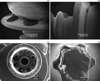
Scanning electron microscopy revealed debris and signs of cellular and bacterial colonisation on the titanium fixture screws and abutments of all nine implants examined (Figure 1). Debris appeared as a thick encrustation between the threads of the fixture and on the external parts of the abutment and the internal screw tightening notch. It was unsurprising to find such material on the fixtures, as they had been in contact with human tissue.
Representative scanning electron micrographs of bone-anchored hearing aid implants and components. (a) Junction between the bone-anchoring fixture and the abutment, showing dried biofilm on the fixture flange (in the circular depressions in this design) and at the junction with the abutment. (b) Another implant design with a solid flange, shown lying on its side, demonstrating deposited material at the junction with the abutment and attached to the screw threads of the fixture. (c) The top of an abutment, showing deposited material around the rim; the central hole gives access to the internal screw head connecting the abutment to the fixture. (d) The top of an internal screw, showing biofilm in the screw driver engagement notch.

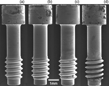
However, unexpectedly, contamination was also observed on six of the internal screws from these nine implants. Ten additional, used screws were then examined to investigate this further. Scanning electron micrographs for four of the retrieved screws are shown in Figure 2. All sixteen screws examined showed contamination of the screw head. This was expected, since the screw head is exposed to the external environment and the top tightening notch of the screw head may be contaminated, as seen in Figure 1(d). However, eight screws showed evidence of biofilm extending down the shaft and into the space between the screw threads, where it was several micrometres thick (see Figure 2d).
Representative scanning electron micrographs of four internal screws, showing contamination of all screw heads and extensive biofilm formation on the shaft and between the screw threads (d).

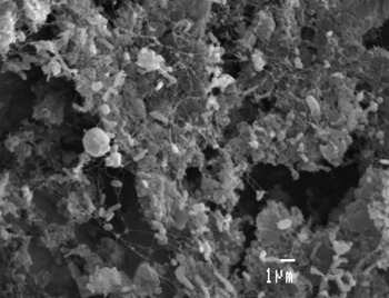
In most cases, because the samples had been simply allowed to dry and placed in saline or formalin, rather than being prepared for scanning electron microscopy using appropriate fixatives and dehydration, individual bacteria could not be distinguished clearly. However, in some cases (e.g. the sample shown in Figure 2d) the bacterial morphology was sufficiently well preserved to distinguish between cocci and rods; Figure 3 shows a mixed population of both morphological types.
Higher magnification scanning electron micrograph of the biofilm on the threads of the screw depicted in Figure 2(d), showing a thick encrustation with a mixed bacterial population of rods and cocci of various sizes. (Bar = 1 µm)

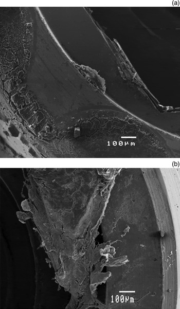
As all 16 internal screw heads examined showed evidence of contamination, it is reasonable to contend that this is the most likely portal of entry for fluid and micro-organisms. To investigate whether bacteria could also gain entry or exit between the abutment and the fixture, we examined six ‘upturned’ abutments for any evidence of biofilm between the abutment and the fixture. In most cases, we found deposited material only on the rim of the abutment (where it would have been exposed to minimal contamination by fluid from the surrounding tissues), although the encrustation was quite heavy (Figure 4a). The abutment from which we obtained the heavily contaminated screw shown in Figure 2(d) showed biofilm extending into the screw hole, suggesting that this could indeed be a portal of entry or exit for micro-organisms, connecting the spaces around the internal screw threads to the surrounding tissues (Figure 4b).
Representative scanning electron micrographs of ‘upturned’ abutments after disassembly, showing the surface that was in contact with the fixture. Biofilm can be seen in both micrographs: in (a) it is mostly confined to the outer rim, while in (b) (the connecting abutment of the screw shown in Figure 2d) the material is much thicker and occupies the area within the rim.

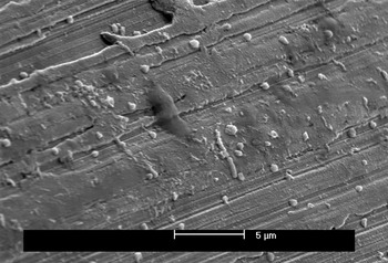
To determine whether viable organisms could be cultured from a failed BAHA, a freshly retrieved fixture that had failed to osseointegrate after 15 days was transported to the laboratory in a sterile container. The fixture was immediately placed in sterile saline to preserve biofilm structure and microbial viability. It was then disassembled under aseptic conditions, and the internal screw incubated anaerobically in liquid broth culture. After 24 hours, this yielded a culture of micro-organisms which, when subcultured on blood agar, grew in both aerobic and anaerobic conditions. The colonies were homogeneous, small and white, and consisted of Gram-positive cocci. They were identified as staphylococci by a positive catalase test, but the species was not determined. Subsequent scanning electron microscopy of the cultured screw revealed clusters of cocci, as expected, but occasional bacilli were also seen, and some areas of the screw were covered in a biofilm-like matrix (Figure 5).
Field emission gun environmental scanning electron micrograph of biofilm on a retrieved bone-anchored hearing aid screw, following microbial culture. Cocci and rod-shaped organisms can be seen, in addition to amorphous deposited material. (Bar = 5 µm)

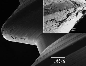
We also undertook scanning electron microscopy of a new, sterile fixture. This confirmed the absence of any biofilm-like debris on any part of the fixture or abutment, including the internal screw, as expected. However, the surface showed machining marks, and there was obvious metal machining swarf on the threads of the internal screw (Figure 6).
Scanning electron micrograph of a new, unused bone-anchored hearing aid internal screw, showing machining marks, scratches and debris on the surface (bar = 100 µm). Insert shows a higher magnification view of the debris (bar = 10 µm).

Discussion
Bacterial biofilms have been widely reported to occur in otorhinolaryngological disease, and to be present on devices used to treat otorhinolaryngological conditions. They have been implicated in diseases such as cholesteatoma, chronic otitis media, chronic rhinosinusitis and tonsillitis. They have also been found on indwelling devices such as tracheostomy tubes, speech restoration valves, cochlear implants and ventilation tubes (see reviews by PostReference Post11 (2007) and Macassey and DawesReference Macassey and Dawes12 (2008)).
Bacteria residing within a biofilm are resistant to phagocytosis and to antibiotics and disinfectants.Reference Fey13, Reference Fux, Stoodley, Hall-Stoodley and Costerton14, Reference Stewart and Franklin15 The reasons for this resistance to antimicrobial agents are not entirely clear. However, there is considerable metabolic heterogeneity within a biofilm, and some individual bacteria may be in a dormant state in which they are not actively synthesising cell wall components or nucleic acids, and are therefore not susceptible to the antibiotics which target these processes.Reference Stewart and Franklin15 Alternatively, antimicrobial agents may be inactivated as they infiltrate the biofilm.
Whatever the reasons for biofilm-related bacterial resistance, biofilms are responsible for the difficulty encountered when attempting to eradicate infections associated with implanted biomaterials. This explains why, in most cases, an infected implant must be removed before the infection can be treated.Reference Wilson16, Reference Cos, Tote, Horemans and Maes17
The current study aimed to investigate evidence of biofilm formation on retrieved BAHAs, focussing especially on the internal connecting screws. We found contamination on the heads of all 16 screws examined; furthermore, eight screws showed evidence of thick biofilm formation on the shaft and between the screw threads. In at least one case, the biofilm appeared also to exist in the space between the abutment and the fixture, where it could potentially shed micro-organisms into the surrounding tissue. In the one case in which individual bacteria could be clearly distinguished, the biofilm consisted of a mixed population of bacterial types. The suggestion that viable bacteria may colonise the BAHA internal screw was supported by culture of staphylococcus from the internal screw of a BAHA unit that had been placed for only 15 days but had failed to osseointegrate (and was then removed and disassembled under aseptic conditions). Scanning electron micrographs of the contaminated screw revealed other bacteria which may have coexisted with the staphylococcus, and which were presumably not detected by culturing because of specific growth requirements (as is the case with many anaerobic species). (In future studies, the use of molecular methods would facilitate the identification of such uncultivatable bacteria.)
Bacterial penetration into the screw space and biofilm formation after such a relatively short time period (following placement) is perhaps surprising. However, it has been demonstrated that bacterial microleakage may occur from the inner part of a dental implant to the exterior within 48 hours.Reference do Nascimento, Barbosa, Issa, Watanabe, Ito and Albuquerque18
In our institution, in all adult cases and many paediatric cases, the BAHA is inserted in a single-stage procedure under aseptic conditions in the operating theatre. The BAHA screw fixture is supplied already assembled, with the abutment connected to it by an internal screw tightened to a controlled torque, and is inserted as a sterile unit. Contamination of the internal screw threads during placement is therefore unlikely.
Penetration of micro-organisms into the spaces surrounding the internal screw thread is most likely to occur after implant placement, with the most likely portal of entry being the head of the screw, since this is exposed to the external environment and tissue exudate may collect around it. Scanning electron micrographs revealed contamination of the screw access hole of several abutments, as well as all the screw heads. Moreover, a new BAHA screw examined by scanning electron microscopy revealed no evidence of bacterial presence (Figure 6). However, whilst less likely, micro-organisms could possibly also gain entry between the abutment and the bone-anchoring fixture itself, at their point of apposition.
Scanning electron microscopy of the unused fixture screw revealed that the surfaces of the threads were pitted, with machining debris attached to several threads. In some cases, this machining debris was several micrometres in width or length, and could be sufficient to impede thread engagement and therefore create space in which fluid could accumulate and any bacteria gaining entry could grow. It is likely that the machining debris was present before the screw was placed in the abutment; however, damage could occur during deployment, since gold is a relatively soft metal and easily deformed. Indeed, gold is selected for internal screws precisely because it can be deformed when screwed into the fixture, forming a tight seal. Titanium screws were previously used for this purpose, but they proved to be too hard and were discontinued because of failures due to loosening.
The clinical significance of biofilm formation on the internal screws of BAHA implants is not yet clear, and any link with the adverse skin reactions observed in some patients is unproven. It is possible that peri-BAHA dermal reactions may arise in response to the titanium itself or to oxidation products thereof,Reference Baldwin and Hunt19 and/or through the inflammatory response manifest via the peri-BAHA fluid exudate, creating an environment favouring colonisation by certain bacteria.
A major weakness of the current study was its retrospective nature. Most of the implants had been collected and fixed in formalin or dried in air with no intention (at the time) of examining them for biofilm formation. In addition, in most cases it was difficult to obtain data on the length of time the implants had been in place and how they had been lost or retrieved. Further studies are necessary to address these issues.
Nevertheless, the study found convincing evidence for biofilm formation on several of the internal screws examined. Such a biofilm could form a reservoir of organisms protected against the host inflammatory and immune defence mechanisms, from which bacteria and toxic exudates could leach out into surrounding tissue fluids and possibly trigger or exacerbate an inflammatory response.
Our observation of internal screw contamination has clear implications for BAHA design.
There are mechanistic similarities between the percutaneous fixture of the BAHA system and dental root implants, which also form a ‘bridge’ between a heavily contaminated external environment (in this case the oral cavity) and sterile internal tissues. Peri-implantitis may be caused by accumulation of plaque around the abutment, resulting in crestal bone loss and implant failure. Early failure of dental implants has been linked to contamination of the internal screw connecting the abutment to the bone-anchoring screw fixture.Reference Quirynen, Bollen, Eyssen and van Steenberghe20, Reference Quirynen and van Steenberghe21 Consequently, implant design has been modified to reduce the possibility of loosening of the abutment due to micromotion, by means of an internal hexagonal screw fixture and/or by the use of a Morse taper connection which makes a very tight ‘cold-weld’ seal between the abutment and fixture components. However, neither of these systems entirely prevents microleakage.
• Bone-anchored hearing aids sometimes need removal due to surrounding tissue infection
• The fixture internal screw and surrounding socket may become contaminated with micro-organisms
• This study found biofilm formation on eight of 16 internal screws from failed fixtures
• Biofilm on internal screws may provide a reservoir of organisms protected against host inflammatory and immune defence mechanisms
• Bacteria and toxic exudates could leach out from this reservoir into surrounding tissue, triggering or exacerbating an inflammatory response
The future for BAHAs may lie in the development of a ‘one-piece’ fixture without a connecting screw, or a hearing aid with an implanted transducer.Reference Håkansson, Reinfeld, Eeg-Olofsson, Östli, Taghavi and Adler22
Conclusion
Biofilms are frequently present on the internal screws of failed BAHA fixtures, and may be implicated in fixture failure due to infection or inflammation of the surrounding tissues.
Acknowledgement
We thank Maxine Brine for collecting and delivering samples, and for valuable contributions to discussion.