Introduction
During assisted reproductive technology (ART) procedures, oocyte quality is a crucial factor influencing embryo developmental competence and implantation rate, but the evaluation of oocyte and embryo quality mainly depends on assessing morphological criteria. This method has shown limitations in predicting successful pregnancy (Assou et al., Reference Assou, Haouzi, Mahmoud, Aouacheria, Guillemin, Pantesco, Rème, Dechaud, De Vos and Hamamah2008; Aydiner et al., Reference Aydiner, Yetkin and Seli2010). Developing new analytical techniques and assays to improve these evaluation methods is necessary. Therefore, and given that the oocyte quality and its microenvironment affect early embryo development (Krisher, Reference Krisher2004), several studies have focused on investigating new testing and non-invasive biomarkers based on the analysis of the oocyte microenvironment components, follicular fluid (FF) and cumulus cells (CCs) to improve the embryo selection process (Salehi et al., Reference Salehi, Aflatoonian, Moeini, Yamini, Asadi, Khosravizadeh, Tarzjani, Harat and Abolhassani2017; Liu et al., Reference Liu, Shen, Zhao, Zou, Shao, Li, Ren and Zhang2019) and in vitro fertilization (IVF) clinical outcomes. The oocytes are surrounded by somatic cells, which are the granulosa cells (GCs) and CCs, and grow in FF which constitutes the microenvironment within which the cumulus–oocyte complex matures, and granulosa cells differentiate. FF is produced from plasma and contains factors produced locally by the follicle cells. The CCs establish a physical connection with the oocyte through gap junctions. As a consequence of this close molecular dialogue, CCs are thought to play an important role in oocyte maturation and fertilization, with signalling and regulation of physiologic function that depends on paracrine and autocrine cytokines in the ovarian microenvironment and reproductive hormones in peripheral blood (Hull and Harvey, Reference Hull and Harvey2014). It has been reported that the reduced number of CCs and disruption in the cell–cell communication might deprive the oocyte of nutrients and survival factors inside the preovulatory follicle and induce apoptosis in ovulated oocytes (Tripathi et al., Reference Tripathi, Shrivastav and Chaube2013). Apoptosis is defined as programmed cell death for homeostasis and is closely involved in most of the reproductive processes, including atresia and luteolysis, as well as decidualization and placentation during embryo implantation (Varras et al., Reference Varras, Polonifi, Mantzourani, Stefanidis, Papadopoulos, Akrivis and Antsaklis2012). Moreover, the CC apoptosis could negatively affect live birth rates in IVF (Lee et al., Reference Lee, Joo, Na, Yoon, Choi and Kim2001). It has been shown that CC apoptosis correlates with poor oocyte and embryo fragmentation (Bosco et al., Reference Bosco, Ruvolo, Chiarelli, Agnello and Roccheri2015). Alternatively, the apoptotic and/or necrosis cells are considered the principal source of circulating cell-free DNA (cf-DNA) (Aucamp et al., Reference Aucamp, Bronkhorst, Badenhorst and Pretorius2018). The latter refers to nuclear or mitochondrial DNA fragments and can be detected in any body fluid, including FF (Scalici et al., Reference Scalici, Traver, Molinari, Mullet, Monforte, Vintejoux and Hamamah2014). cf-DNA can be released passively in the blood from apoptotic or necrotic cells (Schwarzenbach et al., Reference Schwarzenbach, Müller, Milde-Langosch, Steinbach and Pantel2011) and phagocytized by macrophages, and its level remains low in physiological conditions (Pisetsky and Fairhurst, Reference Pisetsky and Fairhurst2007), whereas increased levels of cf-DNA have been associated with many diseases, such as certain types of cancer and inflammatory conditions and fetal anomalies (Jylhävä et al., Reference Jylhävä, Nevalainen, Marttila, Jylhä, Hervonen and Hurme2013; Cheng et al., Reference Cheng, Tang, Cao and Burwinkel2017). Alternatively, various studies have associated elevated levels of cf-DNA in FF with gynaecological and obstetric disorders. They have considered them non-invasive biomarkers in the early detection and/or prognosis (Traver et al., Reference Traver, Assou, Scalici, Haouzi, Al-Edani, Belloc and Hamamah2014). Preliminary studies have demonstrated that cf-DNA in follicles was found as a biomarker for embryo quality in IVF (Scalici et al., Reference Scalici, Traver, Molinari, Mullet, Monforte, Vintejoux and Hamamah2014).
Based on the fact that FF composition affects oocyte development and consequently has a strong influence on embryo quality (Baka and Malamitsi-Puchner, Reference Baka and Malamitsi-Puchner2006). We believe there is an association between CC apoptosis and oocyte development and that high cf-DNA levels in FF may lead to the apoptosis of CCs, which in turn affects the dialogue with the oocyte necessary for its normal development. Therefore, cf-DNA quantification in FF samples could represent a non-invasive biomarker approach complementary to morphological criteria for embryo selection.
In this paper, we report a prospective cohort study to investigate if apoptosis in CCs and cf-DNA levels in FF samples from women undergoing intracytoplasmic sperm injection (ICSI) could be related to their age, AMH level, number of oocytes retrieved, oocyte maturity, embryo quality and pregnancy outcomes.
Material and methods
Patients’ characteristics
This prospective study included 82 women undergoing ICSI procedures at the Fertilization Center IRIFIV in Casablanca, Morocco. Written informed consent was obtained for using FF and CCs samples during oocyte collection. The women’s age ranged from 23 to 43 years. Infertility duration was between 1 and 5 years. In this study, female infertility was the cause of the consultation for 28 couples; male factors for 19 (include only oligospermia ‘fewer than 15 million/ml of semen, or fewer than 39 million in total ejaculate’ with DFI fewer than 30%), mixed infertility for 12, and unexplained infertility for 23. Enrolled patients met the following inclusion criteria: (i) no endocrine disorders and or history of ovarian surgery affecting the ovaries or gonadotropin secretion, and (ii) no current hormone therapy, no metabolic syndrome, no polycystic ovary syndrome, no pelvic surgery, no ovarian tumours, no morbid obesity and no autoimmune disease. The samples with high DNA fragmentation (>30%) and abnormal morphology and motility (based on WHO criteria) were excluded from the study.
Ovarian stimulation procedure
All patients were stimulated with antagonist protocol using the follicle-stimulating hormone (FSH) (Orgalutran 0.25 IU and Gonal-F). FSH was administered (Gonal-F; Serono Laboratories, Saint Cloud, France) by daily subcutaneous injection (150–225 IU/day) or (¼ 300 IU/day). The FSH dose was based on the woman’s age, antral follicle count (AFC) on days 2–3 of the cycles and AMH concentration was maintained constant for 5 days and adjusted according to usual follicle growth parameters determined using ultrasound monitoring. A potent, third-generation gonadotrophin-releasing hormone (GnRH) antagonist, Ganirelix (Orgalutran VR, MSD Schering-Plough, France), was injected subcutaneously once daily starting on day 6 of FSH administration. A subcutaneous injection of human chorionic gonadotrophin (HCG; chorionic gonadotrophins, Ovitrelle VR, Merck Serono) was performed when triggering criteria reached ≥ 3 follicles of 17 mm.
The oocyte maturity rate was calculated based on the ratio of the number of metaphase II oocytes to the total oocyte number. A pregnancy test was performed 2 weeks after the embryo transfer, and pregnancy was confirmed when fetal heart activity was detected on transvaginal ultrasound 4 weeks after embryo transfer. The oocyte maturation rates were divided into two groups, group I had an oocyte maturity rate of < 60% and group II contained the FF with an oocyte maturity rate of ≥ 60%. Anti-Müllerian hormone (AMH) was assessed for each patient on the third day of the menstrual cycle. Levels of AMH < 1.1 ng/ml are considered to reflect a reduced ovarian reserve, and levels ≥1.1 ng/ml are normal ovarian reserve (Gnoth et al., Reference Gnoth, Schuring, Friol, Tigges, Mallmann and Godehardt2008). Three days after oocyte retrieval, embryo quality was graded from A to D according to the following morphological criteria: (i) number of blastomeres, (ii) blastomere regularity and (iii) fragmentation rate. An embryo was considered of top quality (grades A and B) if 6–8 blastomeres of regular size with < 25% fragmentation were observed.
Follicular fluid samples
Collection and preparation
Follicular fluid samples were collected, and the corresponding CCs were isolated for ICSI procedures. FF samples were obtained from mature follicles at the time of oocyte retrieval. To avoid any blood contamination, only clear FF samples were included and purified using a Ficoll-based protocol (3 ml), as described by Ferrero et al., Reference Ferrero, Delgado-Rosas, Garcia-Pascual, Monterde, Zimmermann, Simón, Pellicer and Gómez2012, and then immediately stored at −20°C.
DNA extraction and quantification using ALU-qPCR
Free DNA was extracted from the purified FF using the SaMag™ STD DNA Extraction Kit according to the manufacturer’s instructions. The total free DNA was quantified using qPCR, and ALU 115 primers (Umetani et al., Reference Umetani, Kim, Hiramatsu, Reber, Hines, Bilchik and Hoon2006). Each ALU-qPCR reaction included 2 μl extract of FF and added to the reaction mixture was 0.25 μl of each ALU 115-5´-CCTGAGGTCAGGAGTTCGAG-3´ (forward) and -5´-CCCGAGTAGCTGGGATTACA-3´ (reverse) and 5 µl of Luna Universal QPCR Mix (containing the enzyme Taq DNA polymerase, nucleotides and free SYBRGreen™ fluorescence intercalator). Cycling conditions were as follows: 95°C for 60 s, then 40 cycles of 95°C for 15 s, 58°C for 20 s and 60°C for 30 s. All reactions were performed in duplicate by Sacace biotechnologies. Cell-free DNA concentration in FF pools was determined using a standard curve obtained by successive dilutions of genomic DNA (Umetani et al., Reference Umetani, Kim, Hiramatsu, Reber, Hines, Bilchik and Hoon2006). A negative control (without the template) was integrated into each qPCR plate.
Cumulus cell samples
Collection and preparation
After oocyte pick-up, the CCs were mechanically released by gently pipetting with a 100-micron denuding pipette, then put in a buffered culture medium GYNEMED© at pH 7. Cumulus cell samples were collected in a test tube containing hyaluronidase enzyme GYNEMED 80 IU/ml with HEPES (20 mM) followed by two centrifugations for 7 min at 800 rpm, and then fixed in 3.7% paraformaldehyde for 1 h for apoptosis analysis.
Fluorescence in situ TUNEL assay
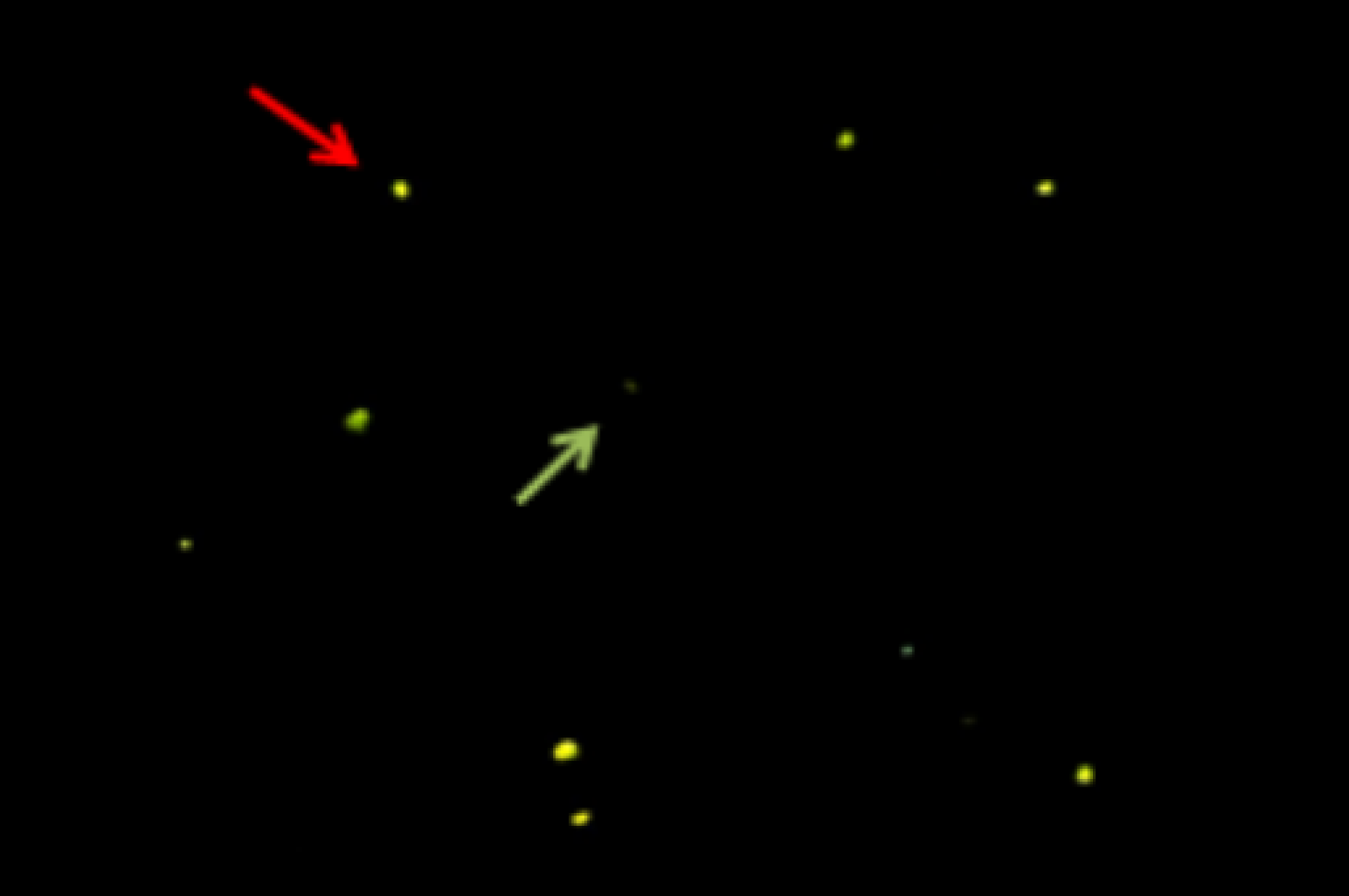
The TUNEL technique was performed using the In Situ Cell Death Detection Kit, Fluorescein (Roche®, Germany), according to the manufacturer’s instructions. After fixation, CCs were permeabilized on ice in 0.1% Triton X-100 and 0.1% sodium citrate in phosphate-buffered saline (PBS), then washed three times in PBS at room temperature. CCs were then incubated for 45 min at 37°C in 5 µl of terminal transferase (TdT) enzyme. CCs were observed under a microscope (EUROStar Germany) equipped with a reflected light fluorescence attachment in a ×20/0.40 objective (Figure 1).
DNA fragmentation detection using the TUNEL assay in cumulus cells observed under a microscope (EUROStar Germany) equipped with a reflected light fluorescence attachment in a ×20/0.40 objective. Cumulus cells with DNA fragmented show intense yellow fluorescence (red arrow), whereas normal cells appear with no colour (green arrow).

Statistical analysis
The results are expressed as the mean ± standard deviation (SD). Differences between groups were compared using the Mann–Whitney U-test (Statistical Package for the Social Sciences) software; statistical significance was defined as P ≤ 0.05.
Results
This study included 82 infertile women under ICSI treatment whose age was between 23 and 43 years. The outcomes of age, infertility length, maturity rate, embryos quality, the number of oocytes, age combined with AMH and pregnancy were divided into lower and higher groups based on the statistical analysis data with cell-free DNA (cf-DNA) and DNA fragmentation index (DFI) in oocyte microenvironment composed to FF and CCs.
As shown in Table 1, FF cf-DNA and CCs DFI levels were significantly lower in patients whose age was less than 37 years compared with those whose age was equal to or greater than 37 years (respectively P = 0.05; P = 0.02). While, in older and younger women, FF cf-DNA and CCs DFI levels when AMH rate was > 1.1 ng/ml were significantly less than in those with AMH ≤ 1.1 ng/ml.
Relationship of cell-free DNA and DNA fragmentation levels in human follicular fluid and cumulus cells with age and anti-Müllerian hormone (AMH) combined with age

Values are in mean ± standard deviation (SD).
CCs: cumulus cells; cf-DNA: cell-free DNA; DFI: DNA fragmentation index; FF: follicular fluid.
Statistical significance was defined as P < 0.05; S: significant.
According to the data presented in Table 2, we observed that cf-DNA and DFI levels were significantly higher in FF and CCs patients who had been trying to conceive for more than 5 years compared with women who had tried only for ≤ 5 years (P = 0.02 and P = 0.01, respectively). In addition, CCs and FF pools from patients with a low number of retrieved oocytes (≤6) had significantly higher levels of DFI and cf-DNA than those from women with a higher number of retrieved oocytes (>6). Indeed, we also noted a higher level of cf-DNA and DFI in the group with a maturity rate < 60% compared with the group with a maturity rate ≥ 60% (P = 0.02 and P = 0.02, respectively)
Relationship between cell-free DNA and DNA fragmentation levels in human follicular fluid and cumulus cells with infertility length, number of oocytes and ICSI outcomes

Values are in mean ± standard deviation (SD).
CCs: cumulus cells; cf-DNA: cell-free DNA, DFI: DNA fragmentation index, FF: follicular fluid.
Statistical significance was defined as P < 0.05; S: significant.
Furthermore, we noticed significantly higher cf-DNA and DFI levels in FF and CCs samples related to oocytes that generated poor-quality embryos (grades C and D) compared with those related to top embryos (grades A and B). Finally, cf-DNA and DFI levels were significantly higher in women who had not been pregnant compared with women who had been pregnant (P = 0.02 and P = 0.02, respectively; Table 2).
Discussion
The oocyte microenvironment is considered to directly affect the differential oocyte developmental capacity. Therefore, FF composition strongly influences oocyte quality, its developmental competence, and subsequent embryo quality (Baka and Malamitsi-Puchner, Reference Baka and Malamitsi-Puchner2006). Therefore, many studies have considered them potential non-invasive biomarkers for oocyte and embryo quality prediction (Baka and Malamitsi-Puchner, Reference Baka and Malamitsi-Puchner2006; Borowiecka et al., Reference Borowiecka, Wojsiat, Polac, Radwan, Radwan and Zbikowska2012). In fact, CCs play a physiological role in antral follicles contributing to metabolic support and maintaining meiotic arrest in the growing oocyte (Coticchio et al., Reference Coticchio, Dal Canto, Mignini Renzini, Guglielmo, Brambillasca, Turchi, Novara and Fadini2015; Monniaux, Reference Monniaux2016). However, there has been mounting evidence that, under certain circumstances, the higher incidence of apoptosis in CCs is associated with an increased rate of empty follicles and fewer oocytes retrieved, poor oocyte and embryo quality and low conception and pregnancy rate (Nakahara et al., Reference Nakahara, Saito, Saito, Ito, Ohta, Takahashi and Hiroi1997; Saito et al., Reference Saito, Seino, Kaneko, Nakahara, Toya and Kurachi2002).
In the follicular microenvironment, the cf-DNA levels reflect the proportion of apoptotic and necrotic cell damage (Snyder et al., Reference Snyder, Kircher, Hill, Daza and Shendure2016). Furthermore, Scalici et al., Reference Scalici, Traver, Molinari, Mullet, Monforte, Vintejoux and Hamamah2014 showed that ∼85% of FF cf-DNA is derived from cell apoptosis. Alternatively, data from Traver et al., Reference Traver, Scalici, Mullet, Molinari, Vincens, Anahory and Hamamah2015 have indicated that FF cf-DNA could be used to predict the clinical pregnancy outcome.
In the current study, we report a prospective cohort study to assess apoptosis in CCs and cf-DNA levels in FF and relate these findings to clinical parameters and pregnancy outcomes of women undergoing ICSI.
Our study has shown that cell-free DNA (cf-DNA) and DFI levels were significantly lower in FF and CCs of patients whose age was < 37 years compared with those whose age was ≥ 37 years. This finding is consistent with research that has noted increased levels of CC apoptosis with advanced age (Fujino et al., Reference Fujino, Ozaki, Yamamasu, Ito, Matsuoka, Hayashi, Nakamura, Ogita, Sato and Inoue1996; Tesarik et al., Reference Tesarik, Galán-Lázaro and Mendoza-Tesarik2021). The shortened telomeres in human oocytes are associated with reproductive age (Kalmbach et al., Reference Kalmbach, Fontes Antunes, Dracxler, Knier, Seth-Smith, Wang, Liu and Keefe2013). Several studies have been interested in evaluating CC telomere length as a function of advanced age. They have shown that the short telomeres fail to protect the chromosomal ends from being recognized as DNA double-stranded breaks, lead to genomic instability, and activate DNA repair pathways, which finally generate cellular senescence or apoptosis (Wellinger, Reference Wellinger2014). Moreover, in our previous study, we showed that 37 years of reproductive ageing was accompanied by a change in redox status imbalance in FF that impaired reactive oxygen species (ROS) scavenging efficiency (Debbarh et al., Reference Debbarh, Louanjli, Aboulmaouahib, Jamil, Ahbbas, Kaarouch, Sefrioui and Cadi2021). ROS overproduction can damage mitochondrial DNA (mtDNA). It can reduce ATP production, which increases vulnerability to apoptotic signalling (Ventura-Clapier et al., Reference Ventura-Clapier, Moulin, Piquereau, Lemaire, Mericskay, Veksler and Garnier2017), therefore causing a decrease in oocyte quality and interfering with embryonic development (Wang et al., Reference Wang, Tang, Wang, Tan, Song, Zhou and Li2021). The apoptosis of CCs with reproductive ageing may induce unfavourable environments for follicular oocyte development. This could cause the significant release of cf-DNA related to advanced age, which results in the deterioration of oocyte quality, thereby reducing fertilization rates and embryo development (Chaube et al., Reference Chaube, Shrivastav, Tiwari, Prasad, Tripathi and Pandey2014).
Age-dependent AMH production in antral follicles starts in females in the 36th week of gestation. It reaches a peak in puberty, after which there is a continuous decrease until the AMH serum level reaches undetectable levels with menopause. Therefore, AMH is a marker for predicting ovarian reserve (Gnoth et al., Reference Gnoth, Schuring, Friol, Tigges, Mallmann and Godehardt2008). Our data showed that, in young women, cf-DNA content in FF and DFI of CCs were significantly lower when AMH was > 1.1 ng/ml than when the AMH ≤ 1.1 ng/ml. These observations agreed with those of Ebner et al. (Reference Ebner, Shebl, Holzer, Oppelt, Petek, Schappacher-Tilp and Mayer2014) providing evidence that patients with lower AMH levels produce CCs of reduced quality showing strand breaks in their DNA. This result can be explained by the ovarian reserve decline via the apoptosis of the GCs responsible for the secretion of AMH, therefore promoting a significant release of the fragment-free DNA in FF (Jayaprakasan et al., Reference Jayaprakasan, Campbell, Hopkisson, Johnson and Raine-Fenning2010). Furthermore, patients with low AMH rates and diminished ovarian reserve have shown a subexpression in tropomyosin-related kinase, thought to mediate a survival signal that maintains viable CCs (Buyuk et al., Reference Buyuk, Santoro, Cohen, Charron and Jindal2011), therefore promoting apoptosis in these patients.
We also noted that cf-DNA and DFI levels were significantly higher in patients who had been trying to conceive for > 5 years compared with those who tried only for ≤ 5 years. Various studies have suggested that a long period of infertility is associated with increased psychological stress in infertile couples (Chiba et al.,Reference Chiba, Mori, Morioka, Kashiwakura, Nadaoka, Saito and Hiroi1997; Lynch et al., Reference Lynch, Sundaram, Maisog, Sweeney and Buck Louis2014), which could lead to follicular cell apoptotic events and the release of cf-DNA (Czamanski-Cohen et al., Reference Czamanski-Cohen, Sarid, Cwikel, Levitas, Lunenfeld, Douvdevani and Har-Vardi2014).
We also observed that CCs and FF pools from patients with a low number of retrieved oocytes ≤ 6 had significantly higher levels of DFI and cf-DNA than those from women with a higher number of retrieved oocytes > 6. It is largely recognized that, during the process of ovarian stimulation, the apoptotic pathway can be activated in many recruited oocytes (Tilly, Reference Tilly1997). The higher the free DNA in the FF, the greater the apoptotic cascade, resulting in either apoptotic oocytes or a limited number of oocytes (Dimopoulou et al., Reference Dimopoulou, Anifandis, Messini, Dafopoulos, Kouris, Sotiriou, Satra, Vamvakopoulos and Messinis2014).
Furthermore, in our study, a higher level of FF cf-DNA and CCs DFI was noted in the group of women with a maturity rate of < 60% compared with those with a maturity rate ≥ 60%. This observation confirms that increased levels of DNA fragmentation of CCs are associated with a higher number of immature oocytes (Ruvolo et al., Reference Ruvolo, Bosco, Pane, Morici, Cittadini and Roccheri2007; Bosco et al., Reference Bosco, Chiarelli, Roccheri, Matranga and Ruvolo2017). Therefore, we can assume that the apoptosis of CCs can reduce cAMP levels and steroid hormone biosynthesis (Chaube et al., Reference Chaube, Prasad, Khillare and Shrivastav2006), thereby generating hypoestrogenic conditions in the ovary and reducing the oocyte quality (Duffy et al., Reference Duffy, Nulsen, Maier, Engmann, Schmidt and Benadiva2005). Furthermore, the apoptotic cells release not only nuclear DNA but also mitochondrial DNA, and the oocytes with good quality have optimal mitochondrial numbers and adequate ATP levels (Van Blerkom et al., Reference Van Blerkom, Davis and Lee1995). ATP-generating capability is critical for the successful maturation of the cytoplasm and nucleus in preparation for fertilization and completion of meiosis II (St John et al., Reference St John, Facucho-Oliveira, Jiang, Kelly and Salah2010). It has been suggested that FF cf-DNA level is related to both retrieved oocyte quantity and quality, two key features for embryo production (Traver et al., Reference Traver, Scalici, Mullet, Molinari, Vincens, Anahory and Hamamah2015).
In addition, we observed that CCs DFI and FF cf-DNA levels were significantly higher in samples related to oocytes that generated poor-quality embryos (grades C and D) compared with those related to top embryos (grades A and B). This result is consistent with other research that has reported a high level of DNA fragmentation of CCs that have generated poor-quality embryos (Salehi et al., Reference Salehi, Aflatoonian, Moeini, Yamini, Asadi, Khosravizadeh, Tarzjani, Harat and Abolhassani2017; Emanuelli et al., Reference Emanuelli, Costa, Rafagnin Marinho, Seneda and Meirelles2019). Indeed, the apoptosis of the GCs reduces the communication between these CCs and the oocytes, depriving oocytes of nutrients, cell signalling molecules and survival factors (Eppig et al., Reference Eppig, Ward-Bailey, Potter and Schultz1982) which risks influencing oocyte and embryo quality. Furthermore, apoptosis is a fundamental process in releasing cell-free DNA (Aucamp et al., Reference Aucamp, Bronkhorst, Badenhorst and Pretorius2018). The origin of cell-free mtDNA might be mitochondrial dysfunction. Indeed, the mature (MII) oocytes, fertilized oocytes, and early cleavage stage embryos depend on the function of the mitochondrial pool present at ovulation (Spikings et al., Reference Spikings, Alderson and St John2006). Consequently, any adverse influence on mitochondrial function via accumulation of the mtDNA resulting from apoptosis will negatively affect the development of the preimplantation embryo. Various studies have suggested that a high level of cf-DNA generates a poor-quality embryo (Scalici et al., Reference Scalici, Traver, Molinari, Mullet, Monforte, Vintejoux and Hamamah2014; Traver et al., Reference Traver, Scalici, Mullet, Molinari, Vincens, Anahory and Hamamah2015).
We found that FF cf-DNA and CCs DFI levels were significantly higher in women who had not been pregnant compared with women who had been pregnant. These results are consistent with various reports suggesting that DNA fragments could come from massive apoptotic events that occur in the ovaries and that contribute to increasing cf-DNA levels in FF samples (Czamanski-Cohen et al., Reference Czamanski-Cohen, Sarid, Cwikel, Lunenfeld, Douvdevani, Levitas and Har-Vardi2013).
Furthermore, it has been reported that CCs are important in all processes of oocyte development, from maturation to embryo development. The CCs can prevent premature exocytosis of cortical granules and the hardening of the zona pellucida to avoid failure of sperm–oocyte recognition, allowing fertilization (Van Soom et al., Reference Van Soom, Tanghe, De Pauw, Maes and De Kruif2002).
All of the observations suggest that oocytes that develop in a cf-DNA-rich environment could have accumulated 'negative signals’ causing harmful consequences on embryo quality and development, or a lack certain ‘positive signals’ normally transmitted by viable CCs.
In conclusion, the current study showed that DFI and cell-free DNA levels in the oocyte microenvironment could have potential use in assessing and predicting oocyte and embryo quality and clinical pregnancy outcomes complementary to the morphological embryo criteria.
Financial support
This research received no specific grant from any funding agency, commercial or not-for-profit sectors.
Conflict of interest
All the authors declare that they have no conflict of interest.
Ethical standards
The authors assert that all procedures contributing to this work comply with the ethical standards of the relevant national and institutional committees on human experimentation and with the Helsinki Declaration of 1975, as revised in 2008.