Shigellae are noted for high transmission rates and may cause shigellosis with a very low infectious dose. Some strains of Shigella produce shiga toxin, similar to the verotoxin of Shiga-toxin producing Escherichia coli (STEC). STEC may cause a broad disease spectrum in humans and are often difficult to distinguish by phenotypic traits from members of Shigella (Koneman et al. Reference Koneman, Allen, Janda, Schreckenberger, Winn and Allen1997). Although O157:H7 serotype is the dominant STEC in many parts of the world, now it is recognized that STEC strains belong to a very broad range of O:H serotypes (Paton & Paton, Reference Paton and Paton1998). The ability of STEC serogroups other than O157 to cause diarrhea and hemolytic uremic syndrome has also been well documented (Gerber et al. Reference Gerber, Karch, Allerberger, Verweyen and Zimmerhackl2002; Werber et al. Reference Werber, Fruth, Buchholz, Prager, Kramer, Ammon and Tschape2003; Ethelberg et al. Reference Ethelberg, Olsen, Scheutz, Jensen, Schiellerup, Enberg, Petersen, Olesen, Gerner-Smidt and Molbak2004). More than 200 serotypes of STEC have been identified and around 160 of these have been recovered from humans with HC and HUS (Brett et al. Reference Brett, Ramachandran, Hornitzky, Bettelheim, Walker and Djordjevic2003). STEC are more prevalent in animals and as contaminants of food, therefore humans are probably more exposed to these strains, and some of them have been associated with severe illness (Boerlin et al. Reference Boerlin, McEwen, Boerlin-Petzold, Wilson, Johnson and Gyles1999; Blanco et al. Reference Blanco, Blanco, Mora, Rey, Alonso, Herrmoso, Hermoso and Alonso2003). Raw milk and beef have been confirmed to be the most likely sources of infection in outbreaks which have occurred during the last decade mainly in Canada, the USA, the UK and Japan (Paton & Paton, Reference Paton and Paton1998; Centers for Disease Control, 2007). Shigella spp. have also been implicated in outbreaks due to consumption of milk and milk products (Gracia-Fulgueiras et al. Reference Gracia-Fulgueiras, Sanchez, Guillen, Marsilla, Aladuena and Novarro2001; Zagrebneviene et al. Reference Zagrebneviene, Jasulaitiene, Morkunas, Tarbunas and Ladygaite2005; Centers for Disease Control, 2007). In India, milk is usually consumed after boiling or pasteurization. However, pathogens may survive inadequate processing and subsequently lead to food infection. Keeping in view the enormity of the problem, it becomes imperative to have rapid and specific test for the detection of Shigella and STEC in milk.
Conventional methods of pathogen detection are time-consuming and laborious, whereas antigen and antibody detection methods are limited in sensitivity in comparison with in vitro nucleic acid amplification (Versalovic & Lupski, Reference Versalovic and Lupski2002). Nucleic acid amplification by Polymerase Chain Reaction (PCR) has provided increased sensitivity, allowed for more rapid processing times, and enhanced the likelihood of detecting bacterial pathogens including Shigella and STEC (Aslam et al. Reference Aslam, Hogan and Smith2003; Aranda et al. Reference Aranda, Fagundes-Neto and Scaletsky2004; Cristancho et al. Reference Cristancho, Johnson, McEwen and Gyles2008).
In this study, a multiplex PCR assay was established for the detection of Shigella, Esch. coli and STEC for analysis of naturally contaminated milk samples.
Materials and Methods
Bacterial strains and template preparation
All strains examined by PCR were grown on Tripticase Soya Agar plates (HiMedia, India) at 37°C. DNA was extracted from bacteria by re-suspending one bacterial colony in 200 μl deionized water, boiling the suspension for 5 min, and centrifuging it for 10 000× g for 3 min. The supernatant was used as template DNA for PCR.
Specificity test
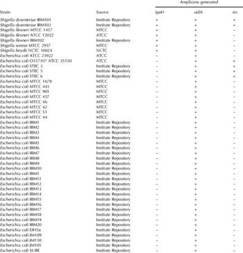
Table 1 lists the strains used for inclusivity test reactions. A total number of 46 strains of Shigella and Esch. coli were used consisting of all the four species of Shigella, four strains of STEC and several other strains of Esch. coli. The specificity of the mPCR assay was tested with 25 known cultures and 11 unidentified gram positive strains. The known cultures consisted of strains or serotypes of Salmonella, Enterobacter, Klebsiella, Pseudomonas, Citrobacter, Enterococcus, Streptococcus, Staphylococcus, Bacillus and Listeria.
Oligonucleotide primers
The targets selected for common detection of Esch. coli and Shigella, Shigella alone and STEC alone were uidA, ipaH and stx 1 genes respectively. Oligonucleotide primers were designed by Primer3 software (Rozen & Skaletsky, Reference Rozen, Skaletsky, Krawetz and Misener2000). Sequences of the primers, GenBank Acc. nos. and size of the amplicons is presented in Table 2.
Polymerase chain reaction
Polymerase chain reactions were performed using extracted genomic DNA or crude cell lysates as described by Saiki et al. (Reference Saiki and Ehrlich1989). The reaction assay of 20 μl contained 1× PCR buffer, 200 μm-dNTP mix, 1·5 mm-MgCl2, 10 pmol of each primer, 1–5 ng DNA and 1 U Taq DNA polymerase (Sigma Aldrich, USA). The PCR assays were performed in Mastercycler gradient (Eppendorf, Germany) or GeneAmp PCR System 9700 (Applied Biosystems Inc., USA). A positive and negative reaction was set up with each batch of reactions. The thermal-cycling conditions were standardized and all the four pairs of primers produced optimum results under the same conditions. It consisted of initial denaturation at 94°C for 3 min, 30 cycles of 94°C for 15 sec, 60°C for 30 sec and 72°C for 30 sec, and final extension at 72°C for 5 min.
The PCR products were run on a 1·5% agarose gel (w/v) and documented with the help of Quantity One software (Bio Rad, USA).
Sequencing of the PCR products
The amplified products were purified by Microcon-100 columns (Millipore, USA) and sequenced using ABI Prism 310 Genetic Analyzer (Perkin-Elmer, USA) following the manufacturer's instructions. All the reagents for sequencing were from Applied Biosystems Inc., USA.
Reproducibility test
The uniplex and multiplex PCR was performed more than three times with all the strains used in the inclusivity and exclusivity test reactions to determine the reproducibility of the results, whereby the DNA template of each strain was freshly prepared for each repeat.
Cultures used for inclusivity test reactions and amplicons generated with the mPCR

PCR primers used in this study

Enrichment
For template preparation from seeded or unseeded milk samples, 1 ml each sample was inoculated into 10 ml Brain Heart Infusion Broth (Himedia, India) and incubated with shaking at 37°C for 8 h. The cells were harvested from 1 ml of the culture broth and washed twice with deionized water. The cell lysates were prepared by thermal extraction in deionized water as described above.
Template preparation from milk
1 ml aliquots of milk were centrifuged at 10 000× g for 5 min, the pellet was suspended in 50 μl of 50 μg/μl lysozyme in TE (pH 8·0) and incubated at 37°C for 15 min. The pellet was dissolved in 450 μl GES buffer (5 m-guanidium thyocyanate, 100 mm-EDTA, 30% sarcosyl in MQ water), mixed thoroughly, followed by the addition of 250 μl chilled 7·5 m-ammonium acetate. After incubation on ice bath for 15 min, the samples were twice extracted with 500 μl chloroform. The DNA in the supernatant was precipitated with two volumes of absolute alcohol and pelleted at 18 000× g for 20 min. The pellet was washed with 70% ethanol, dried under vacuum and dissolved in 50 μl MQ water. 5 μl DNA was normally used in a 20 μl reaction assay. All the chemicals used for Genomic DNA extraction were obtained from Sigma-Aldrich, USA.
Estimation of sensitivity
A suspension of the culture containing 3×10⁸ cells/ml was prepared by comparing visibly the turbidity with Mac Farland Standard of 1. The cell counts were confirmed by Standard Plate Count (CFU/ml). The suspension was serial diluted and aliquots of dilutions containing approximately 1 to 106 cells were directly added to a 20 μl PCR mix.
Validation
1 ml aliquots of whole fat milk were seeded with 102 cells of Shigella flexneri, Esch. coli O157:H7, Salmonella typhimurium, Staphylococcus aureus, Listeria monocytogenes, and Bacillus cereus, separately. The samples were coded, stored at 4°C. Templates from the samples were prepared after enrichment as described above and subjected to the PCR assays.
Results
The PCR assays with primers uid1-uid2, stx1-stx2 and ipaH1-ipaH2 were standardized on templates prepared from Esch. coli ATCC 25922, Esch. coli O157:H7 ATCC 35150 and Shigella flexneri ATCC 12022, respectively using various thermal conditions and reagent concentrations. 10 pmol of each primers in a 20 μl reaction and 1·5 mmn-MgCl2 were found to generate optimum results. Each one of the PCR assays resulted in the generation of specific amplified products when target organisms were subjected to PCR even at lower annealing temperatures (tested from 55 to 62°C). The reactions generated optimum PCR products at 60°C without compromising the efficiency and specificity of the assays. The amplicons thus obtained were sequenced to confirm that the right target was amplified. The sequence of these amplicons matched perfectly with the respective targets. Templates prepared from 7 type cultures of Shigella, consisting of all the four species, 36 Esch. coli strains and 4 strains of Shiga-toxic Esch. coli were subjected to these assays to examine the selectivity and sensitivity (Table 1). Primers based on uidA gene were specific for the common detection of Esch. coli and Shigella spp., giving the same amplification for both the organisms. Each strain of Esch. coli and Shigella tested was found to produce the specific amplification with this assay. Primers based on the ipaH gene, ipaH1-ipaH2 generated specific amplicons with templates prepared from Shigella spp. only. None of the Esch. coli including the STEC strains used in this study generated any amplification with this assay. The primers based on stx 1-encoding gene produced the specific PCR product for Sh. dysenteriae, Esch. coli O157:H7 and Shiga-toxic Esch. coli used in this study. Other strains of Esch. coli and other species of Shigella – Sh. flexneri, Sh. sonnei and Sh. boydii showed negative results with this PCR assay. Furthermore, the PCR assays did not amplify any DNA fragment from the microbial strains used for exclusivity tests depicting optimum specificity.
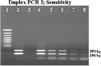
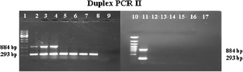
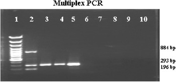
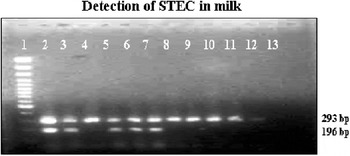
For better application, a multiplex PCR was developed for the common detection of Esch. coli and Shigella and differentiation of verotoxinogenic strains, where primers based on uidA, uid1-uid2 were used in combination with primers based on the verotoxin-encoding gene, stx1-stx2 (Fig. 1). The primers did not interfere with each other and specific amplifications were obtained. Sh. dysenteriae and STEC were found to generate both uidA and stx1 products. Another duplex PCR was developed for the detection and differentiation of Esch. coli and Shigella spp. In this PCR assay, uid1-uid2 and ipaH1-ipaH2, were used. All the species of Shigella tested generated two amplicons of 884 bp (ipaH) and 293 bp (uidA) whereas the 293 bp amplification only was obtained with the strains of Esch. coli. This assay was useful for the differentiation between Esch. coli and Shigella spp. on pure cultures. This PCR assay was found highly specific as no amplifications were generated with the DNA of the non-target organisms (Fig. 2). To combine all the three assays, the triplex PCR was standardized for the detection of Esch. coli, Shigella spp. and their verotoxinogenic strains. This PCR assay generates all three amplicons one each for ipaH, uidA and stx 1 (884 bp, 293 bp & 196 bp) for Sh. dysenteriae, two amplicons – 884 bp & 293 bp for other Shigella species, two for verotoxinogenic strains of Esch. coli (293 bp and 196 bp) and a single band for non-verotoxinogenic strains of Esch. coli (293 bp). No interference between the primers was observed and none of the assays with non-target organisms gave any DNA amplification (Fig. 3). All these PCR assays, individually or as multiplex PCR's could detect approximately a single cell in a 20 μl PCR reaction (Fig. 1).
1·5% agarose gel showing the detection of various no. of cells of Esch. coli O157:H7 by multiplex PCR with primers uid1-uid2 and stx1-stx2. Lane 1: Molecular weight marker; Lane 2: Positive control; Lane 3: Negative control; Lane 4: 104 cells; Lane 5: 103 cells; Lane 6: 102 cells; Lane 7: 10 cells; Lane 8: 1 cell.

1·5% agarose gel showing the inclusivity and exclusivity test reactions for Duplex PCR for the detection of Shigella and Escherichia coli with primers ipaH1-ipaH2 (Shigella) and uid1-uid2 (Shigella/Esch. coli). Lane 1: Molecular weight marker; Lane 2: Sh. dysenteriae; Lane 3: Sh. flexneri; Lane 4: Sh. sonnei; Lane 5–8: Esch. coli strains; Lane 9: Negative control; Lane 10: Molecular weight marker; Lane 11: Sh. dysenteriae; Lane 12: Salmonella typhi; Lane 13: Staphylococcus aureus; Lane 14: Enterbacter sp.; Lane 15: Citrobacter sp.; Lane 16: Bacillus cereus; Lane 17: Listeria monocytogenes.

1·5% agarose gel showing several inclusivity and exclusivity tests for multiplex PCR for the detection of Shigella, Escherichia coli and STEC with primers ipaH1-ipaH2 (Shigella), uid1-uid2 (Shigella/Esch. coli) and stx1-stx2 (verotoxinogenic strains) Lane 1: Molecular weight marker; Lane 2: Sh. dysenteriae; Lane 3: Esch. coli MTCC 443; Lane 4: Esch. coli MTCC 901; Lane 5: Esch. coli ATCC 25922; Lane 6: Salmonella typhi; Lane 7: Enterococcus faecalis; Lane 8: Klebsiella rhinoscleromatis; Lane 9: Pseudomonas aeruginosa; Lane 10: Streptococcus pyogenes.

During the course of validation in seeded samples of whole fat milk, our protocols detected precisely verotoxinogenic Esch. coli in 9 samples and Shigella in 2 samples.
When applied to naturally contaminated milk samples (n=100), 15 samples and 100 samples were found positive for STEC and Esch. coli, respectively (Fig. 4). DNA was purified from the samples found positive for Shiga-toxic Esch. coli and directly used as template for the mPCR. This method generated optimum results and showed agreement with enrichment based detection. Nonetheless, none of the samples of milk analysed in this study was contaminated with Shigella.
1·5% agarose gel showing the detection of Escherichia coli and its verotoxinogenic strains in milk by multiplex PCR with primers uid1-uid2 and stx1-stx2 Lane 1: Molecular weight ladder; Lane 2, 3, 5, 6, 7: presence of verotoxinogenic strains of Esch. coli in milk samples; Lane 4, 8, 9, 10, 11, 12: presence of non-verotoxinogenic strains of Esch. coli in milk samples; Lane 13: Negative control.

Discussion
Since Esch. coli and Shigella are genetically very closely related, PCR primers designed to detect members of Shigella often detect pathogenic Esch. coli as well (Olsen et al. Reference Olsen, Aabo, Hill, Notermans, Wernars, Granum, Popovic, Rasmussen and Olsvik1995). Hence, an attempt was made to establish a multiplex PCR which can not only detect but also differentiate between strains of Esch. coli, Shigella and STEC. Shigella is also noted for its high transmission rates through person-to-person contact. At present, foods are not routinely screened for the presence of human pathogenic bacteria such as Shigella, rather they are screened only after clinical and epidemiological information to identify the causative agent of an outbreak caused by the consumption of contaminated food (Lampel et al. Reference Lampel, Orlandi and Kornegay2000). Infection with Shiga toxin-producing Esch. coli (STEC) in humans has been associated with a spectrum of diseases, including diarrhoea, haemorrhagic colitis (HC), and haemolytic uremic syndrome (HUS) (Karmali, Reference Karmali1989; Griffin & Tauxe, Reference Griffin and Tauxe1991). Foods that have an animal origin have been identified as the main vehicles of these food-borne pathogens (Wani et al. Reference Wani, Pandit, Samanta, Bhat and Buchh2004). The importance of STEC transmission through food has been illustrated by many outbreaks worldwide (Bettelheim, Reference Bettelheim1997; Centers for Disease Control, 1993; Bradbury, Reference Bradbury1997). Therefore it is imperative to develop rapid methods for the detection of Esch. coli, Shigella and their verotoxinogenic strains.
In this study, three markers – uidA of Esch. coli, ipaH of Shigella and stx 1 gene of Esch. coli were targeted by PCR for the detection of Esch. coli, Shigella and their STEC strains. uid has been successfully used earlier for the common detection of Esch. coli and Shigella (Bej et al. Reference Bej, DiCesare, Haff and Atlas1991). The uidA structural gene of β-D-glucuronidase and the uidR regulatory gene, which controls uidA expression, are located at 36 min on the Esch. coli linkage map (Novel & Novel, Reference Novel and Novel1973; Jochimsen et al. Reference Jochimsen, Nygaard and Vestergaard1975). ipaH, a gene of unknown function, is present in multiple copies on the invasive plasmid and the chromosome of Shigella (Buysse et al. Reference Buysse, Stover, Oaks, Venkatesan and Kopecko1987), hence it is a stable gene to detect as loss of the plasmid will not affect the presence of the gene. The use of ipaH as a marker for the detection of Shigella has been well established. The invasive plasmid is present in all Shigella spp. and entero-invasive Esch. coli (EIEC) (Hartman et al. Reference Hartman, Venkatesan, Oaks and Buysee1990; Lampel et al. Reference Lampel, Orlandi and Kornegay2000). Hence, both uniplex and multiplex PCR with ipaH1 and ipaH2 primers will detect EIEC as well. Primers were designed in such a way so that all the three pairs have the same Tm and amplify fragments of different molecular sizes. This makes their multiplexing possible. The inclusivity and exclusivity tests indicate the high specificity and selectivity of the PCR assays. The validation studies with seeded samples of milk further emphasize the specificity of the assays developed. The multiplex PCR described here is useful for the detection of Esch. coli and Shigella and STEC. A single STEC strain can produce Stx1 or Stx2, or both. Stx1 is virtually identical to the Stx of Sh. dysenteriae type 1 (Wani et al. Reference Wani, Pandit, Samanta, Bhat and Buchh2004). For this reason, the primer pair stx1 and stx2 also produced an amplification with template DNA prepared from strains of Sh. dysenteriae. The assay will not, however, detect STEC strains possessing Stx2 alone. Multiplex PCR reduces the cost of the reaction and increases the throughput besides reducing the time of analysis. PCR assays detected a single cell of Esch. coli or Shigella depicting the high sensitivity of the assay system. Similar sensitivity has been achieved by Villalobo & Torres (Reference Villalobo and Torres1998) by using primers based on virA for the detection of Shigella. The incidence of Esch. coli (100%) and STEC (15%) in milk samples evaluated in this study was very high. Perelle et al. (Reference Perelle, Dilasser, Grout and Fach2007) have also found incidence of STEC in 21% raw milk samples in a similar study.
The multiplex PCR established in this study could be applied for detection of Esch. coli, Shigella and STEC in milk. This study also depicts high incidence of Esch. coli and STEC in raw milk and highlights the need for better hygienic conditions in dairies and appropriate processing of milk prior to consumption.
The authors are thankful to the Council of Scientific and Industrial Research, New Delhi, India for supporting this research (Grant No. 5/258/45/2005/NMITLI).