Introduction
Parasitic helminths have developed a large array of strategies to optimize transmission. For parasites with free-living stages, these strategies must include ways to maximize development and survival of eggs and larvae in the environment, and modes to avoid the large variety of constraints the host activates to fight the infection: from targeting the incoming infective stages, through distressing the established adults, to attacking their eggs on the way out of the host's body (Thomas et al., Reference Thomas, Brown, Sukhdeo and Renaud2002; Maizels et al., Reference Maizels, Balic, Gomez-Escobar, Nair, Taylor and Allen2004; Else, Reference Else2005; Pearce, Reference Pearce2005; Moxon et al., Reference Moxon, Flynn, Golden, Hamilton, Mulcahy and Brophy2010; Bourke et al., Reference Bourke, Maizels and Mutapi2011; Hewitson et al., Reference Hewitson, Harcus, Murray, van Aagtmaal, Filbey, Grainger, Bridgett, Blaxter, Ashton, Ashford, Curwen, Wilson, Dowle and Maizels2011; Viney & Cable, Reference Viney and Cable2011; Gonçalves et al., Reference Gonçalves, Rocha, Gonzaga, Gonçalves-Pires, Ueta and Costa-Cruz2012a, Reference Gonçalves, Nunes, Gonçalves-Pires, Ueta and Costa-Cruzb; Morgan & van Dijk, Reference Morgan and van Dijk2012; Viney & Diaz, Reference Viney and Diaz2012; Van Kuren et al., Reference Van Kuren, Boag, Hrubar and Cattadori2013).
Temperature and humidity are frequently reported to be critical cues for the hatchability of free-living helminth eggs. We recently showed that different temperature regimes and seasonal changes in temperature have different impacts on egg development and rate of hatching of two gastrointestinal nematodes with free-living stages spent on the herbage (Hernandez et al., Reference Hernandez, Poole and Cattadori2013). Female conditions have also been suggested to be fundamental for the quality and hatching potential of their eggs and it is generally thought that host immunity can be a severe constraint against female health and, thus, their fitness (Hein et al., Reference Hein, Pernthaner, Piedrafita and Meeusen2010; Viney & Cable, Reference Viney and Cable2011; Viney & Diaz, Reference Viney and Diaz2012). Since a large number of nematodes shed their eggs into the host gastrointestinal tract to reach the outside with the host's faeces, we can reasonably assume that host immunity, stimulated by the eggs themselves or by cross-reactive responses mounted against other parasite stages, might also prime these eggs and modulate their development and hatching in the environment. The evidence of immune responses to parasite eggs is not new. Previous studies on helminths with developmental phases spent in the host tissue have clearly shown that patency can trigger robust immune reactions targeted specifically against eggs. For example, there is a vigorous T-helper (Th)-2-like immune reaction against eggs of Schistosoma mansoni (Pearce et al., Reference Pearce, Kane, Sun, Taylor, McKee and Cervi2004; Pearce, Reference Pearce2005) and the soluble egg proteins of Fasciola hepatica stimulate a clear immunoglobulin G1 (IgG1) antibody response, which is also associated with increased interleukin (IL)-10 and transforming growth factor (TGF)-β levels during patency (Moxon et al., Reference Moxon, Flynn, Golden, Hamilton, Mulcahy and Brophy2010). Amongst nematodes, IgA and IgG responses have been shown against Strongyloides venezuelensis eggs and a diagnostic test was proposed for the detection of human strongyloidiasis based on these results (Gonçalves et al., Reference Gonçalves, Rocha, Gonzaga, Gonçalves-Pires, Ueta and Costa-Cruz2012a, Reference Gonçalves, Nunes, Gonçalves-Pires, Ueta and Costa-Cruzb). Host immunity was also suggested to negatively impact Ostertagia circumcincta but not Haemonchus contortus egg viability in the environment, supporting the hypothesis that different constraints might impact eggs from different nematode species (Jørgensen et al., Reference Jørgensen, Leathwick, Charleston, Godfrey, Vlassoff and Sutherland1998; Jørgensen, Reference Jørgensen2000; Sargison et al., Reference Sargison, Jackson and Gilleard2011). Yet, how immunity contributes to egg maturation and hatchability outside the host and how these relationships change over the course of an infection remain largely unexplored. Indeed, if immunity plays a role, eggs shed at the very beginning of an infection are expected to do better than eggs shed at a later time when the immune responses are fully developed and can influence egg conditions and/or female quality.
In the current study we examine how the hatching rate and volume of eggs collected from rabbits singly infected with the gastrointestinal nematodes Trichostrongylus retortaeformis or Graphidium strigosum are affected by the host immune response over a 15-week period. Trichostrongylus retortaeformis and G. strigosum are directly transmitted nematodes that colonize the small intestine and stomach, respectively, of the European rabbit (Oryctolagus cuniculus). We recently showed that T. retortaeformis eggs have consistently higher hatching rates than G. strigosum (Hernandez et al., Reference Hernandez, Poole and Cattadori2013). What determined this different pattern is unclear, since eggs of both parasites were exposed to identical environmental conditions in the laboratory and the field. The two species have eggs of similar shape but different size (length: T. retortaeformis ~90 μm and G. strigosum: ~100 μm) (Taylor et al., Reference Taylor, Coop and Wall2007), which might be critical for hatchability. We recently showed that rabbits mount a strong immune response to both nematodes by targeting third-stage larvae, adult worms and adult excretory/secretory compounds (Murphy et al., Reference Murphy, Nalpas, Stear and Cattadori2011, Reference Murphy, Pathak and Cattadori2013; Pathak et al., Reference Pathak, Pelensky, Boag and Cattadori2012; Cattadori et al., Reference Cattadori, Wagner, Wodzinski, Pathak, Poole and Boag2013). Eggs could also be impacted by these immune responses directly or through female conditions (Cattadori et al., Reference Cattadori, Wagner, Wodzinski, Pathak, Poole and Boag2013). This leads us to predict that the antibody response against T. retortaeformis eggs is generally lower than that to G. strigosum eggs and this will contribute to the higher and faster hatching rate of the former compared to the latter parasite. We also predict that the rate of hatching and volume of T. retortaeformis decreases with the course of the infection, as a result of the increased immune response against adult stages and their eggs, although they continue to do better than G. strigosum.
Materials and methods
Experimental design
Two New Zealand white, 60-day-old male rabbits were orally infected with a single dose of third-stage larvae of either T. retortaeformis (dose: 1500 L3 s) or G. strigosum (dose: 750 L3 s) diluted in 3 ml mineral water (Murphy et al., Reference Murphy, Nalpas, Stear and Cattadori2011). Animals were housed individually and kept on food and water ad libitum on a 12-h day–night light cycle. Given the different pre-patent periods of the two parasites (T. retortaeformis ~12 days and G. strigosum ~43 days) (Audebert et al., Reference Audebert, Hoste and Durette-Desset2002; Massoni et al., Reference Massoni, Cassone, Durette-Desset and Audebert2011), we planned the G. strigosum infections such that both rabbits would start shedding parasite eggs in the same week. This first week of eggs shed in host's faeces was referred to as week 0. Cage trays were cleaned every Monday and faeces collected the following morning for each week of the 15-week trial; this allowed us to have fresh eggs ( < 24 h) at the very beginning of their development.
It is important to highlight that we were interested primarily in detecting an immune response to these helminth eggs and then to examine whether this response influenced the egg hatching rate over time. To address our aims, we infected a single rabbit for each species and followed the hatching success of eggs produced by a cohort of parasites of the same age exposed to an immune response that changed over the course of the trial. We were not interested in comparing the hatching rate with the immune response from different rabbits but to examine the hatching profile of generations of eggs shed by a single population of worms inhabiting a single study site, namely, a rabbit. While there is some variability in the immune response between rabbits (Murphy et al., Reference Murphy, Nalpas, Stear and Cattadori2011, Reference Murphy, Pathak and Cattadori2013; Cattadori et al., Reference Cattadori, Wagner, Wodzinski, Pathak, Poole and Boag2013), the effect of this variability on egg hatchability will be addressed in a different study.
Parasite egg collection and hatching rates
Faeces were soaked in water for 15 min, homogenized in a commercial blender and filtered through a 212 μm sieve. The filtrate was evenly distributed across 50-ml tubes and centrifuged for 10 min at 1500 × g at 4°C. For each tube, the supernatant was removed, the pellet vortexed and mixed with ~0.02 g of kaolin clay powder and 20 ml of saturated sodium chloride solution (~36% dry weight/volume, w/v). The resulting supernatant was then filtered through a 36-μm sieve and kept under running water for 30 min to clean the residual salt and clay off of the eggs. Finally, the sieve content was collected into a 50-ml tube, adjusted to 30 ml with tap water and the total number of eggs counted.
To estimate the rate of hatching, eggs collected weekly were placed into a climatic chamber (VWR International, Radnor, Pennsylvania, USA) with programmable temperatures (Series 982 microprocessor, Watlow Controls, Pittsburgh, Pennsylvania, USA). Based on our previous work, we selected a day–night temperature cycle between 15°C and 7°C, where the temperature cycles by steadily decreasing or increasing between the two temperatures every 12 h; this regime was run for 8 days (additional details in Hernandez et al., Reference Hernandez, Poole and Cattadori2013). About 100 eggs of T. retortaeformis or G. strigosum, purified as described above, were dispensed on to 45 Petri dishes (6 cm), each previously layered with a 10 ml solution of 1% agarose (w/v) in tap water. Plates were then transferred to the climatic chamber and the cycle started at 12.00 hours (noon) and 15°C on the first day of the experiment. For each parasite, five plates were collected on days 1, 2, 3, 4, 5, 7 and 8, and eggs and hatched larvae counted to estimate changes in the daily hatching rate of both parasites over the 15-week experiment. An additional group of five plates was counted at day 0. Data were expressed as hatching rate, which is the percentage of eggs hatched over the total number of larvae and eggs counted on each plate.
Antibody responses to eggs and estimation of egg volume
To quantify the host immune response to parasite eggs, two different approaches were used. The first approach quantified the systemic antibody response by using indirect enzyme-linked immunosorbent assays (ELISAs) to measure anti-egg IgA and IgG responses from rabbit serum challenged with whole egg homogenates as antigen. The second approach estimated the local immune responses by measuring the amount of antibodies bound to the shells of eggs purified from faeces, using immuno-fluorescence and laser scanning confocal microscopy. While the approaches were complementary, in that they allowed us to compare systemic and local immune responses, they differed fundamentally in that the serum ELISA measures the immunoglobulins that react with egg homogenate, and thus can include possible cross-reactive antibodies to other parasite developmental stages, whereas the immuno-fluorescence approach measured the amount of antibodies naturally bound to the egg shell. Therefore the immuno-fluorescence data give us a true representation of the antibody response at the host–egg interface.
Unless stated otherwise, all reagents and consumables were purchased from VWR International.
For the serum antibodies, blood was collected once a week from the rabbits, centrifuged at 3000 × g for 3 min at room temperature and serum supernatants stored at − 20°C prior to use in ELISAs. For the egg homogenates, 10,000 eggs were washed extensively with phosphate-buffered saline, pH 7.4 (PBS) supplemented with a protease inhibitor cocktail (1 tablet/50 ml PBS, Roche Applied Science, Indianapolis, Indiana, USA) prior to storage in 50-ml tubes at − 80°C in the same buffer. Egg homogenates were prepared by freeze–thawing the tubes seven times and finally grinding the eggs with 40 strokes in a ground-glass homogenizer (Kimble-Chase, Vineland, New Jersey, USA). Homogenization efficiency was confirmed under a stereo-microscope. Homogenates were centrifuged at 5200 × g for 15 min to remove large debris and resulting supernatants were used to coat 96-well ELISA plates (Greiner Bio-One, Monroe, North Carolina, USA). Plates coated with either T. retortaeformis or G. strigosum egg homogenates were blocked with 5% non-fat milk (w/v) in PBS supplemented with 0.05% (volume/volume, v/v) Tween-20 (PBS-T) at 37°C for 1 h and then washed twice with PBS-T, prior to incubation with sera from infected rabbits. All sera were diluted in blocking buffer (v/v in 5% non-fat milk in PBS-T) and incubated for 1.5 h at 37°C. For G. strigosum, sera were diluted to 1:5 and 1:15 for detecting IgA and IgG, respectively, while for T. retortaeformis, sera dilutions of 1:10 and 1:40 were used for IgA and IgG, respectively. Plates were washed four times with PBS-T and then incubated with horseradish peroxidase-conjugated goat anti-rabbit IgA (Abcam plc, Cambridge, Massachusetts, USA) or IgG (SouthernBiotech, Birmingham, Alabama, USA) diluted to 1:2500 in blocking buffer (v/v) for 1 h at 37°C. After washing the plates four times with PBS-T, reactions were developed with 2,2′-azino-bis(3-ethylbenzothiazoline-6-sulphonic acid) (ABTS) as colorimetric reagent for 15 min and colour intensities recorded on a spectrophotometric plate reader (Bio-Tek Instruments, Winooski, Vermont, USA). Each plate incorporated positive and negative control sera which were pooled from single infected and naive animals, respectively, as part of a separate study (Murphy et al., Reference Murphy, Nalpas, Stear and Cattadori2011). Naive sera were included to account for background reactivity resulting from any contaminants that may have co-purified with the eggs prior to the preparation of egg homogenates. All dilutions and reaction conditions outlined here were optimized before the experiment in a checkerboard format as described previously (Murphy et al., Reference Murphy, Nalpas, Stear and Cattadori2011). Raw data were transformed into optical density (OD) indices for the subsequent analysis (Murphy et al., Reference Murphy, Nalpas, Stear and Cattadori2011).
For the detection of antibodies bound to eggshells, 1500 eggs of each parasite, purified as described previously for the hatching studies, were dispensed into three 1.5-ml tubes each and fixed overnight (~16 h) at 4°C in 1.5 ml of 10% (w/v) neutral buffered formalin. Optimal antibody dilutions were established previously from the inflection points obtained by incubating eggs with two-fold incremental dilutions of sera and corresponding fluorophore conjugates (Cy3-conjugated goat anti-rabbit IgG Heavy+Light chain (H+L), Jackson Immunoresearch, West Grove, Pennsylvania, USA) in a tube-based modified checkerboard format adapted from Pathak et al. (Reference Pathak, Creppage, Werner and Cattadori2010). Specificity of these fluorophore conjugates for rabbit antibodies were confirmed in a preliminary test where no binding was observed to eggs stained with the corresponding isotype controls of non-specific goat IgG antibodies conjugated to Cy3 fluorophore (Jackson Immunoresearch). Formalin-fixed eggs were washed by centrifuging the tubes at 17,000 × g for 1 min, removing the supernatant and re-suspending the pellet in 1.5 ml PBS. Eggs were washed a further two times in the same manner prior to blocking them in 1 ml of 10% goat serum (Jackson Immunoresearch) prepared in PBS-T for 30 min. All incubations were performed on a rocking platform shaker at room temperature. Eggs in two of the three tubes (control samples) were washed twice with PBS-T and re-suspended in 1 ml of PBS-T supplemented with positive or background control sera diluted 1:100 (v/v) and incubated for an additional 60 min at room temperature. Eggs in the third tube (experimental samples) were left undisturbed in the blocking solution for this 60-min period. Following incubations, all control and experimental samples were washed three times with 1.5 ml PBS-T and stained with 1 ml of PBS-T containing 1:400 diluted (v/v) goat anti-rabbit IgG (H+L) conjugated to Cy3 fluorophore (Jackson Immunoresearch) and incubated for an additional 60 min at room temperature. Eggs were finally washed three times with 1.5 ml PBS-T and once with 1.5 ml PBS prior to microscopy. From each tube (controls and experimental), 50–150 stained eggs were visualized under a confocal laser scanning microscope and fluorescence intensity recorded at the equatorial perimeter of the ellipsoid egg. Images were obtained with a 10 × objective (Olympus Fluoview® FV 1000, Olympus America Inc., Center Valley, Pennsylvania, USA) pre-calibrated at identical lamp intensities (485 V) and acquisition parameters (28%) throughout the study, to allow weekly comparisons of staining intensities. Level of binding (i.e. intensity of staining) was quantified with the iVision software package (BioVision Technologies, West Exton, Pennsylvania, USA) and expressed as mean fluorescent intensity (MFI) using only positive eggs (i.e. fluorescent eggs). Overall, from the shedding of faeces on to the cage tray to the fluorescence measurements, eggs were cycled between 4°C and room temperature for 36–48 h, depending on the procedures implemented. While our approach was unable to stop egg development, we did standardize our procedures to maintain the processing time within 48 h to allow data comparison.
To examine whether egg volume – as indicative of egg quality – changed with the course of the infection and affected egg hatchability, a random sample of 50 eggs, digitalized during immuno-fluorescence microscopy, was measured every week for each parasite species. Egg size (length and width) was quantified using a digitalized system and the software Image J (version1.45, http://rsbweb.nih.gov/ij) (Chylinski et al., Reference Chylinski, Boag, Stear and Cattadori2009). Egg volume was then estimated assuming their shape to be a prolate spheroid (4/3πa 2b, where a represents the minor axis or egg width and b describes the major axis or egg length).
Data analysis
To highlight significant changes in the egg hatching rate over the course of the infection (weeks) or the amount of time in the chamber (days), linear mixed-effect models (LME-REML, package NLME in R (Pinheiro & Bates, Reference Pinheiro and Bates2000)) were applied. This approach was also used to investigate the relationship between the rate of hatching and the antibody responses, both serum and egg immuno-fluoresence data, as well as temperature changes (maximum and minimum) in the chamber. To take into account variability between the Petri dishes, the dish ID was initially included as a random effect; however, since very low between-dish variability was observed, the ‘dish effect’ was not considered in subsequent analyses. Based on the combination of variables examined, the experimental week, or sampling day, was included as a random factor. To consider the effect of pseudo-replication, i.e. sampling the same rabbit over the course of the infection, an autoregressive function of order 1 (AR-1) was included, which took into account the correlation of the hatching rate, or egg volume, between sampling weeks. To examine changes in egg volume by week of sampling, generalized linear models (GLM with normal error distribution) were used, while LME-REML was used to investigate the relationship between egg volume and immune responses. Egg hatching rates were arc sin transformed.
Results
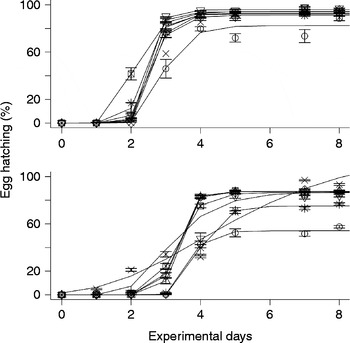
Overall and between the two parasites, the rate of weekly hatching was significantly higher for T. retortaeformis than G. strigosum (coeff. ± SE: − 0.17 ± 0.06, df = 1124, P< 0.01) and the second parasite showed a tendency to be more variable than the first parasite (fig. 1). Similarly, and within weeks, daily hatching was higher for T. retortaeformis than G. strigosum (two-way interaction species–sampling day, coeff. ± SE: − 0.03 ± 0.005, df = 1140, P< 0.0001) (fig. 1).
Mean percentage egg hatching (with SE bars) and predicted Gompertz curves (lines) by experimental day for T. retortaeformis (top panel) and G. strigosum (bottom panel). The following alternate weeks are shown as week: 1 (○), 3 (▲), 5 (+), 7 (x), 9 (◇), 11 (▾), 13 ( ) and 15 (*).

For T. retortaeformis, egg hatching rate did not change significantly among the experimental weeks, from when rabbits started shedding eggs to 15 weeks into the trial (fig. 1 top panel). For all the sampled weeks, hatching rate increased with experimental day: the longer the eggs were kept in the thermal chamber, the higher the rate of eggs that hatched (fig. 1 top panel, coeff. ± SE: 0.18 ± 0.004, df = 544, P< 0.0001).
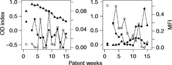
No significant association was found between the mean weekly rate of hatching and serum IgG level, which decreased with the course of infection (fig. 2). Both the egg-bound antibodies (MFI) and the species-specific serum IgA OD index exhibited background levels throughout the infection, hence, their effect on hatching rate or egg volume was not examined.
Optical density (OD) indices of serum IgA (●) and serum IgG (▲), and mean fluorescent intensities (MFI) of egg-bound antibodies (○) by sampling week for T. retortaeformis (left panel) and G. strigosum (right panel). Lines connect continuous points.

In the climatic chamber, T. retortaeformis hatching rate was positively associated with minimum temperature (interaction min. temperature–sampling day, coeff. ± SE: 0.05 ± 0.005, df = 337, P< 0.0001) and negatively related to maximum temperature (two-way interaction max. temperature–sampling day, coeff. ± SE: − 0.01 ± 0.002, df = 337, P< 0.0001), suggesting that warmer temperatures stimulate egg development and hatching, while temperatures that are too high have the opposite effect, indicative of an optimal thermal range for the development of this helminth.
The phenology of hatching, i.e. the first day within each week when eggs had hatched in at least one Petri dish, was about 2 days (mean ± SE: 1.86 ± 0.14). No significant relationship was observed between the first day of hatching and antibody responses, which is not surprising considering the narrow variability in the phenology of hatching observed during the 15 weeks of infection.
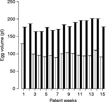
The average volume of T. retortaeformis eggs (mean (picolitre) pl ± SE: 98.86 ± 0.06) was consistently smaller than that of G. strigosum eggs (mean pl ± SE: 181.29 ± 0.93) (t-test, df = 1481.596, P< 0.0001). Trichostrongylus retortaeformis egg volume slowly decreased with the progression of the infection, although some variation was observed between the weeks (fig. 3, coeff. ± SE: − 0.08e − 5 ± 0.02e − 5, df = 716, P< 0.001). No significant relationships were found between the weekly egg volume and serum IgG response. Egg hatchability was not associated with egg volume.
Mean egg volume (with SE bars) of T. retortaeformis (white bars) and G. strigosum (black bars) by sampling week. Note the very narrow SE.

For G. strigosum, the hatching rate of eggs was more variable than for T. retortaeformis but did not significantly differ amongst the experimental weeks. Consistent with T. retortaeformis, hatching rate increased with the number of days spent in the thermal chamber (fig. 1 bottom panel, coeff. ± SE: 0.15 ± 0.003, df = 583, P< 0.0001).
No significant associations were found between weekly mean rate of hatching and serum or egg-bound antibodies. MFI of antibodies bound to eggs and IgA OD indices decreased throughout the infection (MFI and IgA versus weeks, respectively, coeff. ± SE: − 0.03 ± 0.01, P< 0.0001 and − 0.02 ± 0.01, P< 0.05; for both: df = 13), while IgG OD levels increased (coeff. ± SE: 0.07 ± 0.01, df = 13, P< 0.0001) (fig. 2). MFIs for this helminth were 20-fold higher than for T. retortaeformis.
In line with T. retortaeformis, hatching rate increased with minimum temperature (interaction min. temperature–sampling day coeff. ± SE: 0.03 ± 0.003, df = 371, P< 0.0001) and was negatively associated with maximum temperature (two-way interaction max. temperature–sampling day coeff. ± SE: − 0.01 ± 0.001, df = 371, P< 0.0001).
Across the experimental weeks, the mean first day of hatching was 1.73 ( ± 0.26). No relationship was found between first day of hatching and antibody responses.
Graphidium strigosum egg volume positively increased with sampling week (fig. 3, coeff. ± SE: 0.02e − 4 ± 0.03e − 5, df = 833, P< 0.0001), although some variability was observed between weeks. Egg volume was negatively related to MFI (coeff. ± SE: − 0.04e − 3 ± 0.02e − 3, df = 13, P< 0.05) but positively associated with IgG (coeff. ± SE: 0.03e − 3 0.06e − 4, df = 12, P< 0.001). The hatchability of G. strigosum eggs was not related to their volume.
Discussion
In the current study, eggs of T. retortaeformis developed faster than G. strigosum eggs and this pattern was consistent both within and between weeks over the course of the experiment. These contrasting trends were comparable with our previous findings using this system and the same temperature regime (Hernandez et al., Reference Hernandez, Poole and Cattadori2013). The volume of G. strigosum eggs was significantly greater than those of T. retortaeformis, and increased for the first but decreased for the second parasite throughout the trial. The hypothesis that parasite eggs stimulated an immune response and that this accounted for the temporal patterns observed was only partially confirmed. We found that eggs shed directly into the lumen by helminths inhabiting the gastrointestinal tract do stimulate an antibody response, and eggs of G. strigosum elicited a stronger reaction than those of T. retortaeformis. However, while this could contribute to the relatively lower hatching rate of G. strigosum, changes in antibody levels could not explain the dynamics of hatching nor the temporal variation in egg volume for both helminths. These general findings suggest that either the eggs can tolerate the host immune responses, or these attacks are fundamentally too frail to cause any clear change in development and hatchability. Other factors, such as genetic differences, contrasting life history strategies or intrinsic differences in female worms and egg qualities, are probably the primary drivers of the dissimilarities observed.
Rabbits infected with a single dose of T. retortaeformis or G. strigosum regularly shed viable parasite eggs, and the rate of hatching did not change significantly with time. Within each week, hatching rate showed a characteristic logistic growth, with the majority of eggs hatching between the second and third day under a cyclic 15°C–7°C daily temperature regime. Contrary to our previous studies, phenology of hatching seemed to favour G. strigosum, with a slightly earlier first day of hatching than T. retortaeformis, although this difference was not significant and might have been caused by the way the variable was calculated (Hernandez et al., Reference Hernandez, Poole and Cattadori2013).
In general, the volume of G. strigosum eggs was twice that of T. retortaeformis, in agreement with previous reports on the size of these eggs (Taylor et al., Reference Taylor, Coop and Wall2007). Egg volume can be an important factor for egg hatchability. The development of G. strigosum eggs might take longer and require more energetic investment simply because of their larger size compared to those of T. retortaeformis. Yet, it is worth noting that while the egg volume changed over the course of the infection, the hatching rate was comparable between weeks for both helminths, and no significant relationship was found between these two variables. This suggests that such changes are probably not sufficient to alter hatchability, or egg volume is not a critical constraint for hatching success. The experimental design prevented us from gaining any information on female quality and specific immune responses, although previous studies have repeatedly advocated a critical role of helminth female conditions on offspring performance (Viney & Cable, Reference Viney and Cable2011; Viney & Diaz, Reference Viney and Diaz2012). We recently found a positive correlation between female length and number of eggs in the uterus both for T. retortaeformis and G. strigosum (Cattadori et al., Reference Cattadori, Wagner, Wodzinski, Pathak, Poole and Boag2013); however, it is still unclear if this relationship translates into eggs of higher quality.
Based on the antibody levels in the serum, we found that IgG, and partly IgA, trends were reasonably consistent with our previous work describing the antibody profiles against somatic antigen from third-stage infective larvae (L3) and adult worms, as well as the adult excretory/secretory products (Murphy et al., Reference Murphy, Nalpas, Stear and Cattadori2011, Reference Murphy, Pathak and Cattadori2013). In these studies we found clear evidence of cross-reactivity between L3 and adult somatic compounds and we do not exclude that cross-reactivity can also occur between these stages and the egg somatic products. However, in the current work the level of serum IgA to T. retortaeformis eggs was negligible, suggesting that cross-reactivity might not be as strong and widespread as expected, at least for this helminth. Similarly, the egg immuno-fluorescence signal was notably lower and rarely above background values for T. retortaeformis, also indicative of an overall low antibody signal to the eggs of this species. In contrast, the immuno-fluorescence levels were 20-fold higher against G. strigosum, which might partly explain the lower rate of hatching, although no relationship was found between antibodies and hatching. Given the similar trend between MFI and serum IgA, it is also possible that IgA contributes the most to the antibodies bound to the egg. Altogether, these findings show that T. retortaeformis eggs are fundamentally less immunogenic than G. strigosum eggs, and stress the importance of examining infection processes at the host–parasite interface.
In conclusion, eggs shed by rabbits singly infected with either T. retortaeformis or G. strigosum showed contrasting rates of hatching, which appeared to be only partially affected by the host antibody responses. The eggs of these two helminths were partly immunogenic and their hatching efficiency remained consistently high over the course of the infection, despite changes in the antibody responses, indicating that other factors could be more important in modulating hatching over time. Eggs shed in the environment are a crucial component for the transmission and long-term persistence for a large number of parasitic helminths. More work is needed to understand the role of the host in affecting egg development and hatchability, and how this varies across parasite species. This is fundamental information required to appreciate the factors driving risk of infection and, more broadly, species distribution and dynamics.
Acknowledgements
The authors would like to thank Graham Thomas, Alexander D. Hernandez and the Microscopy and Cytometry Core Facility at PSU for technical support.
Financial support
K.A.L. was supported by National Science Foundation-Research Experience for Undergraduates supplemental funds to I.M.C. This study was funded by the National Science Foundation (DEB-1145697 and DEB-0716885).
Conflict of interest
None.
Ethical standards
The authors assert that all procedures contributing to this work comply with the ethical standards of the United States Department of Agriculture–Animal and Plant Health Inspection Service and have been approved by the Institutional Animal Care and Use Committee of The Pennsylvania State University (USA).