INTRODUCTION
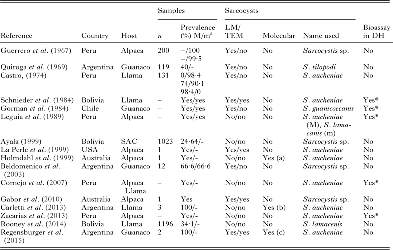
There is considerable confusion concerning the species of Sarcocystis in camelids (Dubey et al. Reference Dubey, Calero-Bernal, Rosenthal, Speer and Fayer2015a ). Historically, a macroscopic sarcocyst observed in a llama (Lama glama) was named Sarcocystis aucheniae by Brumpt (Reference Brumpt1913) in his book but no other detail was provided. Sarcocysts were found in a guanaco (Lama guanicoe) and named Sarcocystis tilopodi by Quiroga et al. (Reference Quiroga, Lombardero and Zorrilla1969), based on finding it in a different species of Lama. In Chile, Gorman et al. (Reference Gorman, Alcaino, Munoz and Cunazza1984) found macroscopic and microscopic sarcocysts in L. guanicoe and the authors suggested that the parasite in L. glama should be named Sarcocystis lamacanis and the parasite in the species L. guanicoe should be called S. guanicoecanis. In a review paper, Leguía recognized 2 species and called the parasite, S. aucheniae (macroscopic cysts) and S. lamacenis (microscopic cysts); the author probably misspelled S. lamacanis as lamacenis and this name was copied by Taylor et al. (Reference Taylor, Coop and Wall2007) and by Rooney et al. (Reference Rooney, Limon, Vides, Cortez and Guitian2014). Microscopic cysts detected in heart muscle from llamas and alpacas were named as S. lamacanis (Leguía and Casas, Reference Leguía and Casas1999). In Germany, Schnieder et al. (Reference Schnieder, Kaup, Drommer, Thiel and Rommel1984) found both microscopic and macroscopic sarcocysts in L. glama from Bolivia; dogs but not cats fed both types of sarcocysts excreted sporocysts. They thought that microscopic sarcocysts could either be developmental stages of S. auchenie or sarcocysts of a separate species. A history of sarcocysts detection in South American camelids (SAC) is summarized in Table 1.
Detection of Sarcocystis spp. sarcocysts in SAC
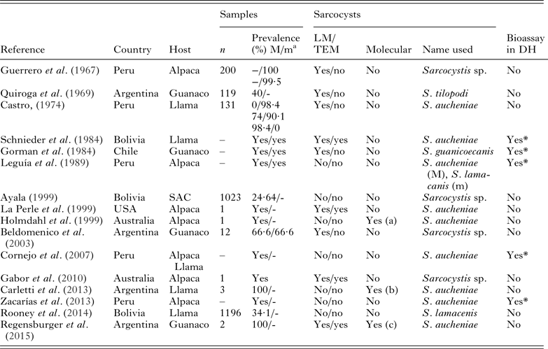
a M, Macroscopic cysts; m, microscopic cysts; (-), not informed. 18S rRNA gene sequence reported in the GenBank as (a) = AF017123; (b) = JX110660- JX110668; (c) = KT382799. DH: definitive hosts/ (*), dogs shed sporocysts after experimental infection.
A critical examination of the taxonomy of Sarcocystis species (Dubey et al. Reference Dubey, Calero-Bernal, Rosenthal, Speer and Fayer2015a ), led to the conclusion that there is only 1 valid name, S. aucheniae, and that there is no valid reason separating species infecting llamas and alpacas (Vicugna pacos). Therefore, the names S. guanicoecanis, S. lamacanis, S. lamacenis should be nomen nudum/dubia; as well as the names with hyphen since taxonomically are not permitted. Because the sarcocyst that Brumpt (Reference Brumpt1913) first recognized was macroscopic it seems reasonable to designate macroscopic species as S. aucheniae (Dubey et al. Reference Dubey, Calero-Bernal, Rosenthal, Speer and Fayer2015a ).
In the present paper, we redescribe the macroscopic sarcocyst forming S. aucheniae and describe and name the microscopic sarcocyst forming species, as S. masoni, after Dr Eugene Mason who first recognized sarcocystosis in a camel.
MATERIALS AND METHODS
Samples collection and microscopy
Samples (50 g) of muscles from the neck and lumbar region were collected from different camelids from Argentina and Peru (Table 2). Muscles were transported refrigerated at 4 °C and processed immediately by the direct observation and fresh microscopic examination.
Details of camelids muscle samples and sarcocysts examined by LM and TEM

The muscle samples were cut with a scalpel and examined for the presence of macroscopic sarcocysts. Sarcocysts were separated from muscle and individual cysts samples were observed by light microscopy (LM), fixed in 2% glutaraldehyde for transmission electron microscopy (TEM) and conserved at −20 °C for molecular studies. The muscles were further processed by homogenization and examined by LM essentially as previously described (Moré et al. Reference Moré, Abrahamovich, Jurado, Bacigalupe, Marin, Rambeaud, Venturini and Venturini2011). Microscopic cysts observed were collected with a micropipette and a sterile tip and processed as for macroscopic cysts. TEM studies were conducted as previously for sarcocysts obtained from cattle samples (Moré et al. Reference Moré, Abrahamovich, Jurado, Bacigalupe, Marin, Rambeaud, Venturini and Venturini2011). Additionally, glutaraldehyde-fixed macroscopic cysts from a llama sample were processed for scanning electron microscopy (SEM).
All the morphological descriptions were performed with the average of at least 20 measurements.
Molecular analysis
DNA was extracted from intact or broken cysts with commercial kit (Wizard genomics, Promega, USA) according to manufacturer instructions. Samples of DNA were amplified by PCR using primers SarcoFext and SarcoRext proceeding as previously (Moré et al. Reference Moré, Schares, Maksimov, Conraths, Venturini and Schares2013). Amplicons obtained which reach a gel-estimated concentration of about 40 ng µL−1, were purified using a commercial kit (Wizard SV clean up system, Promega) according to manufacturer instructions, and submitted for sequencing to the Genomic Unit, Biotechnology Institute CICVyA – CNIA – INTA, Argentina. Samples of DNA from microscopic cysts were amplified with primers ERIB1 – PrimerB and purified as previously, and submitted for sequencing with primers 3H, 4H, S3 and S5 (Moré et al. Reference Moré, Schares, Maksimov, Conraths, Venturini and Schares2013). Additionally, DNA from a macroscopic cyst from Argentinean llama was amplified to reach the full length of the 18S rRNA gene, purified, cloned into plasmids and sequenced as previously (Moré et al. Reference Moré, Schares, Maksimov, Conraths, Venturini and Schares2013). Sequences obtained were aligned and analysed with the GENEIOUS software (7·1 version available at http://www.geneious.com), and analysed and compared by BLASTn function from NCBI (http://blast.ncbi.nlm.nih.gov/).
Consensus sequences obtained were phylogeneticaly analysed based on a ClustalW multi-alignment using 18S rRNA gene sequences from several Sarcocystis spp. with the 18S rRNA gene sequence of Toxoplasma gondii (GenBank M97703) as an out-group (tree function of the GENEIOUS program). A neighbour-joining method was applied with a Tamura–Nei-model genetic distance calculation and with 1000 bootstrap replicates using 50% of support threshold.
RESULTS
Description of S. masoni, n. sp.
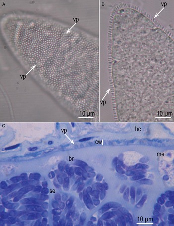
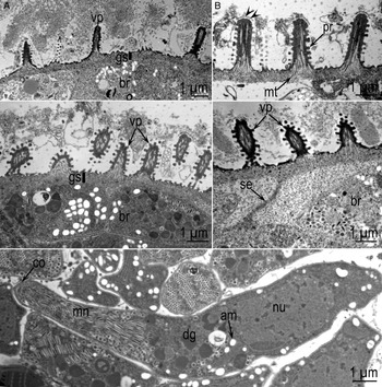
Sarcocysts were up to 800 µm long and 35–95 µm wide. The sarcocyst wall in isolated unstained cysts appeared striated with radial projections which were not always visible (Fig. 1A, B). In Toluidine blue stained sections the sarcocyst wall was 2·5–3·5 µm thick, and had small protrusions (Fig. 1C). By TEM, the sarcocyst wall had conical to cylindrical villar protrusions (vp), 2–2·8 µm long and 0·5–0·7 µm wide (Fig. 2A–D). Some vp appeared wider at the base (Fig. 2C, D). They were lined by a 25–45 nm thick electron dense parasitophorous vacuolar membrane (pvm). The vp were at irregular distances and the host myocyte was often degenerated along the vp giving the impression that vp were apart (Fig. 2A). Each vp had several microtubules (mt) from the tip of the villus to the middle of the ground substance (gs) layer. The mt were smooth, without granules. Several rows (11 or more) of knob-like protrusions (pr) out-pocketed from pvm and they appeared to be interconnected. Evenly distributed hair-like electron dense, structures were seen on vp tips (Fig. 2B). The gs layer was 0·5–1·0 µm thick (Fig. 2A). The deeper part (juxtaposed with bradyzoites) of the gs was smooth and more electron dense than the outer part towards the vp (Fig. 2C), but this distinction was not always apparent. The total thickness of the sarcocyst wall (from the base of gs to vp tip) was 2·5–3·5 µm. Bradyzoites were 11–14 × 2–3·5 µm in size. Bradyzoites contained numerous micronemes located in the anterior half and amylopectin granules located in the posterior half (Fig. 2E).
Sarcocystis masoni sarcocysts from Lama glama from Argentina. (A) Surface view showing tips of vp. Unstained. (B) Flattened sarcocyst showing striations on the cyst wall. Unstained. (C) Section (1 µm) stained with Toluidine blue. Note relatively thin cyst wall (cw), conical vp and elongated bradyzoites (br).

TEM of Sarcocystis masoni sarcocysts. Note villar protrusions (vp), ground substance layer (gs), microtubules (mt), bradyzoites (br), septum (se). (A–D) Cyst wall of 4 sarcocysts. Note variability of vp shapes and sizes. A from Lama glama, Argentina, B from Lama guanicoe, Argentina, C from L. guanicoe, Argentina, D from L. glama, Peru. (E) Longitudinally cut bradyzoite. Note conoid (co), amylopectin granules (am), dense granules (dg) and nucleus (nu). From L. glama, Peru.

Full-length 18S rRNA gene amplicons evidenced sequencing results only with primers 3H, 4H, S3 and S5 and consensus sequences of 1125 and 1134 bp from an Argentinean llama and guanaco sample were obtained, respectively (GenBank accession numbers KU527109 and KU527111). For the remaining 5 amplicons (partial sequences), consensus sequences obtained ranged from 755 to 805 bp and correspond to cyst samples from Argentinean guanacoes (n = 2) and llama (n = 1), and from Peruvian alpacas (n = 2) (GenBank KU527107, KU527108, KU527110, KU527112 and KU527113). The 7 sequences obtained showed a sequence identity ranging 98·6–99·8% among them. BLAST analysis revealed the higher sequence identity (only 95–96%) with other Sarcocystis spp. sequences such as S. silva (JN226124) and S. tarandi (GQ250971 and GQ250968). All sequences obtained from microscopic cysts from different SAC species positioned together in a unique branch closely related with other sequences of Sarcocystis spp. which used canids as definitive hosts (Fig. 3).
Phylogenetic tree. Neighbour-Joining consensus tree from a multiple Sarcocystis spp. 18S rRNA gene sequence alignment. A Toxoplasma gondii sequence (GenBank M97703) was used as an out-group. Branch support is represented as percentage from 1000 bootstraps. Sequences obtained from South American Camelids are in bold and the sequences obtained in the present study include the host name and country (Arg = Argentina).

Taxonomic summary of S. masoni, n. sp. (Figs 1, 2, 7A)
Diagnosis
Sarcocysts microscopic (up to 800 µm long and 35–95 µm wide). The sarcocyst wall was 2·5–3·5 µm had conical to cylindrical vp (2–2·8 µm long and 0·5–0·7 µm wide) with mt and knob-like projections.
Type intermediate host
Llama (L. glama).
Other intermediate hosts
Guanaco (L. guanicoe), Alpaca (V. pacos).
Distribution
South America. Probably follow the intermediate host distribution.
Definitive host
Unknown (dog and other canids are the most likely DH).
Etymology
Species named after Dr Eugene Mason who was the first to describe sarcocysts in camelids in a report in 1910.
Specimens deposited
Sections of Syntypes of toluidine blue stained cyst sections deposited in the United States National Parasite Collection in the Division of Invertebrate Zoology and National Museum of Natural History, Smithsonian Institution, Washington, D.C.
Sequences were deposited in NCBI GenBank (accession numbers KU527107-KU527113).
Redescription of S. aucheniae
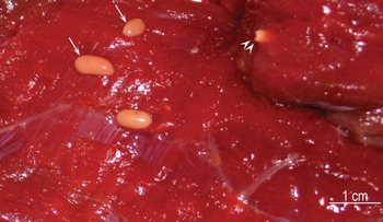
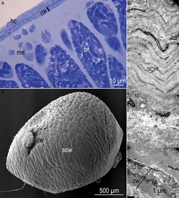
Sarcocysts were macroscopic up to 1·2 cm long; most cysts were 2–7 mm (Fig. 4). They were pale yellow in colour. Some cysts were embedded deep in muscles while some were superficial. Cysts were frequently surrounded by a dense and laminar secondary cyst wall (scw), approximately 50 µm thick (Figs 5B, C). In Toluidine blue stained sections, the sarcocyst wall was 8–10 µm thick (Fig. 5A), and the centre of the cysts contained fluid material, often without viable bradyzoites. By TEM, the cyst wall was up to 10 µm thick, including the gs layer and the scw consisted of degenerated host cells, often appearing laminated structures (Fig. 5C). The proper sarcocyst wall had branched vp, appearing like cauliflower. The vp were difficult to measure because of branching; they were approximately 3–4·5 µm high by 2·5–3·5 µm wide. The gs was 3–5 µm and appeared homogenous without granules. In some sections, the vp had a conical cap (Figs 6B and 7B) with criss-crossing mt, without granules. Hair-like structures were visible on the tip of the villar cap. Oval to elongated bradyzoites were packed in sacs separated by septa (Fig. 6C). By TEM, the bradyzoites were 13–18 µm × 3–5 µm in size. Each bradyzoite contained numerous micronemes in conoidal third of the bradyzoite, several dense granules, 2 rhoptries and numerous amylopectin granules (Fig. 6D).
Macroscopic sarcocysts of Sarcocystis aucheniae from Lama glama, Argentina. Unstained. Note sarcocysts on the surface (arrow) and embedded (arrow heads).

Sarcocyst of Sarcocystis aucheniae. (A) Toluidine blue stained section (1 µm) from Lama glama, Peru. Note host cell (hc), cyst wall (cw) and bradyzoites (br). (B) SEM of a sarcocyst enclosed in capsule from Lama glama, Argentina. Part of the secondary cyst wall (SCW) is broken and the cw proper is visible. (C) TEM of sarcocyst from L. glama, Argentina. The cw is relatively thin compared with thickness of the secondary cyst wall.

TEM and SEM of Sarcocystis aucheniae sarcocyst from Lama glama, Argentina. (A) Low magnification of the cyst wall showing branched protrusions (vp). (B). Higher magnification of vp showing villar tip containing microtubules (mt) and hair-like projections (arrow heads). (C) SEM of cyst showing bradyzoites (br) and septa (se). (D) TEM of bradyzoites. Note amylopectin granules (am), dense granules (dg), a conoid (co) and nucleus (nu).

Comparison of villar protrusions (vp) of Sarcocystis masoni and Sarcocystis aucheniae. Note smooth microtubules (mt). (A) Cylindrical vp of S. masoni. Arrow points to hair-like projections on villar tip. (B) Conical vp of S. aucheniae.

Full-length 18S rRNA gene sequence of 1879 bp was obtained after cloning in plasmids from an Argentinean llama cyst sample (accession number KU527117). A total of 2 clones were sequenced and consensus sequences were identical. The sequence revealed the highest identity (⩾99%) with sequences of S. aucheniae reported in GenBank (KF383266–KF3832668 and KT382799). A total of 13 amplicons from macroscopic cysts samples were submitted for sequencing and in 3 samples the results were inconclusive. From the remaining 10 samples, consensus sequences ranging from 511 to 794 bp were obtained from Argentinean llamas (n = 2) and guanacos (n = 4) and from alpacas (n = 3) and llamas (n = 1) from Peru. All sequences were registered on the GenBank (accession numbers KU527114-KU527124) and evidenced a 98–99% of sequence homology with S. aucheniae sequences (KT382799, KF383266, KF3832667, KF3832668 and AF017123) by the BLASTn analysis. Comparison of the 11 sequences obtained from macroscopic cysts evidenced a sequence identity ranging 99·4–100% among them.
The phylogenetic tree performed positioned all the obtained S. aucheniae sequences in the same branch with other S. aucheniae previously reported sequences. The S. aucheniae branch positioned separated from other sequences of Sarcocystis spp. which used canids as definitive host (Fig. 3).
Taxonomic summary of S. aucheniae (Brumpt, Reference Brumpt1913)
Diagnosis: Sarcocysts macroscopic (up to 1·2 cm). The sarcocyst wall is 10 µm thick and frequently surrounded by a 50 µm thick secondary cyst wall. The branched vp with ‘cauliflower-like’ shape measured 3–4·5 µm high by 2·5–3·5 µm width, mt in vp are smooth. Mature sarcocysts evidenced a solid package of bradyzoites within septa in the peripheral area and fluid material without viable bradyzoites in the centre. Bradyzoites measured 13–18 × 3–5 µm.
Intermediate host
South American Camelids (SAC).
Distribution
South America. Probably follow the intermediate host distribution.
Definitive host
Dog (according experimental infection results).
DISCUSSION
The sarcocysts found in the present study were clearly distinguished as 2 species based on morphological and molecular data. Until now, sarcocysts in camelids were recognized as microscopic and macroscopic, sometimes regarded the same species. This is the first full morphological description of the microscopic cyst.
The structure of sarcocyst wall is a useful criterion to distinguish Sarcocystis spp. within a given host. A recent review indicated that there were more than 200 Sarcocystis species with at least 42 types and several subtypes of sarcocyst wall (Dubey et al. Reference Dubey, Calero-Bernal, Rosenthal, Speer and Fayer2015a ). Among all species of Sarcocystis, S. cameli from the domestic camel has a distinctive cyst wall, classified as ‘type 9j’. Sarcocyst wall of S. masoni, described here is similar to cyst wall of S. cameli (Dubey et al. Reference Dubey, Hilali, Van Wilpe, Calero-Bernal, Verma and Abbas2015b ).
Although the macroscopic S. aucheniae sarcocysts were recognized for decades, this is the first complete description of the sarcocyst. Schnieder et al. (Reference Schnieder, Kaup, Drommer, Thiel and Rommel1984) described the ultraestructure of S. aucheniae cysts; however, their focus was on transmission of the parasite. They found that of dogs and cats fed individual macroscopic sarcocysts, only the dog shed sporocysts. This is an unusual finding because most macroscopic sarcocysts of other animals are transmitted by cats and not dogs (Dubey et al. Reference Dubey, Calero-Bernal, Rosenthal, Speer and Fayer2015a ). The structure of macroscopic S. aucheniae sarcocysts in the present study is essentially similar to sarcocysts of Sarcocystis gigantea of sheep described in detail from the oesophagus of sheep (Mehlhorn and Scholtyseck, Reference Mehlhorn and Scholtyseck1973). In the present study, we provided the description of the scw of S. aucheniae, including the surface view by SEM. The proper cyst wall had vp that were highly branched, like a cauliflower. Other species producing macroscopic cysts like S. gigantea of sheep, Sarcocystis fusiformis of the water buffalo (Dubey et al. Reference Dubey, Hilali, Van Wilpe, Verma, Calero-Bernal and Abdel-Wahab2015c ), Sarcocystis cafferi of the African buffalo (Dubey et al. Reference Dubey, Lane, van Wilpe, Suleman, Reininghaus, Verma, Rosenthal and Mtshali2014) and Sarcocystis rileyi of ducks (Dubey et al. Reference Dubey, Cawthorn, Speer and Wobeser2003) have been described to have cauliflower-like branching of vp. However, these sarcocysts are structurally distinct. For example, the vp in S. fusiformis have dumbbell-like protrusions that were not seen in S. aucheniae. Also, the conical villar tips with prominent mt found in the present study were not reported previously for other macroscopic cysts. Whether some of these differences are related to techniques and fixatives used needs further investigation.
Sarcocystis spp. mixed infections are frequently observed in herbivore muscles (Moré et al. Reference Moré, Pantchev, Skuballa, Langenmayer, Maksimov, Conraths, Venturini and Schares2014a ; Dubey et al. Reference Dubey, Calero-Bernal, Rosenthal, Speer and Fayer2015a ). In the present study, most of the samples contained sarcocysts from 2 species. Because in some transmission studies, dogs that excreted sporocysts had been fed naturally infected camelid muscles (Gorman et al. Reference Gorman, Alcaino, Munoz and Cunazza1984; Leguía and Casas, Reference Leguía and Casas1999), the definitive host for Sarcocystis masoni is uncertain. Therefore, there is a need to perform experimental studies using individual sarcocysts as we did for the present molecular studies.
From the literature it appears that sarcocysts in domestic camel are different than from sarcocysts in SAC. Camels are native to Asia and the Middle Eastern countries and not present in South America. The macroscopic sarcocyst of S. aucheniae has not been recorded in domestic camel and Sarcocystis ippeni of camel has not been found in the SAC. Additionally, S. ippeni has characteristic sarcocyst wall structure, ‘type 32’ not found in other species of Sarcocystis (Dubey et al. Reference Dubey, Hilali, Van Wilpe, Calero-Bernal, Verma and Abbas2015b ).
In the present study, we provided additional molecular data on Sarcocystis species in camelids and supporting morphological findings. While reviewing literature on Sarcocystis infections in SAC, we encountered that several publications were conference proceedings or in local journals (Table 1), a fact which complicate proper comparisons.
The 18S rRNA gene fragments sequences from S. masoni were almost identical among different sampled SAC, but showed only 96% of sequence identity with S. aucheniae and other Sarcocystis spp. gene sequences reported on the GenBank. Previous reported sequences of S. lamacanis DQ100056 and AY840990 (Medrano et al. Reference Medrano, Hung and Rubio2006) as well as EF640651 and EF640652 (Arias and Hung unpublished) did not show higher homology and/or did not showed a high coverage with our sequences. Probably all previous short 18S rRNA gene sequences (ranging 280–343 bp) were obtained from a different gene region as we amplified, which did not include hyper-variable regions. In addition, if these previously reported sequences (for example DQ100056 and EF640652) are analysed by BLASTn, they have 99% identity with many other Sarcocystis spp. sequences (including S. cruzi, S. tenella, S. gracilis, S. silva, S. hjorti, etc.). Moreover, any of the previous studies have performed a morphological characterization of the Sarcocystis sp. prior amplification and sequencing. The present sequences from the microscopic S. masoni sarcocysts should serve as reference for future molecular studies. Phylogenetical analysis positioned S. masoni sequences in the same branch as other Sarcocystis spp. using canids as definitive hosts. Therefore, based on this molecular finding and the results of dog infection with heart muscles containing ‘microscopic’ sarcocysts (Leguía et al. Reference Leguía, Guerrero, Sam and Chavez1989), it is possible to suggest that S. masoni used canids as definitive hosts.
With respect to molecular characterization and comparison of different 18S rRNA gene fragment sequences from macroscopic cysts a high-sequence identity (⩾99%) was observed among the obtained from different SAC samples as well as with the sequences reported on the GenBank as S. aucheniae from llamas and guanaco from Argentina (KF383266, KF383267, KF3832668 and KT382799) and an alpaca in Australia (AF017123) (Holmdahl et al. Reference Holmdahl, Morrison, Ellis and Huong1999; Carletti et al. Reference Carletti, Martin, Romero, Morrison, Marcoppido, Florin-Christensen and Schnittger2013; Regensburger et al. Reference Regensburger, Gos, Ctibor and Moré2015). Interestingly, when analysed phylogenetically, S. aucheniae sequences aligned in a separated branch from other Sarcocystis spp. which used canids as definitive host. A similar phylogenetical construction and observation was previously achieved (Holmdahl et al. Reference Holmdahl, Morrison, Ellis and Huong1999; Carletti et al. Reference Carletti, Martin, Romero, Morrison, Marcoppido, Florin-Christensen and Schnittger2013; Moré et al. Reference Moré, Pantchev, Herrmann, Vrhovec, Ofner, Conraths and Schares2014b ).
In conclusion, 2 morphologically and molecularly different Sarcocystis species, S. masoni (microscopic cysts) and S. aucheniae (macroscopic cysts), were identified affecting different SAC from Argentina and Peru. Further studies are needed in order to identify natural infected definitive hosts for S. masoni and S. aucheniae.
ACKNOWLEDGEMENTS
We would like to thank Isidoro Ercoli, Marco Quispe Huacho and Roxana Peralta for their excellent technical assistance.
FINANCIAL SUPPORT
Financial support was partially obtained through Agencia Nacional de Promoción Científica y Tecnológica (PICT2012-0506), Argentina (GM); and by a researcher exchange program PE13-05 MINCYT-CONCYTEC (Argentina-Peru, GM and MESM).
CONFLICT OF INTEREST
The authors declare that they have no conflict of interest.