Implication
Improvement in intestinal development is the first step to rescue the intrauterine growth-restricted (IUGR) individual, and it is evident that intestinal development can be improved at a higher nutrient intake level through increasing the levels of glucagon-like peptide-2 (GLP-2) in pigs with normal birth weight (NBW). Therefore, we wanted to test the hypothesis that increased nutrient intake levels could also be effective in stimulating similar beneficial effects in IUGR pigs via the action of GLP-2. Our results show that intestinal development of IUGR piglets was improved in a nutrient-intake-level-dependent fashion, and the depressed intestinal development in pigs fed the low nutrient intake levels could be restored by exogenous GLP-2 injection. These results imply that the nutrient-intake-level-dependent regulation of intestinal development could also be effective in IUGR individuals via GLP-2 administration, which provides a strategy to improve the digestive and absorptive function in newborn individuals with low birth weight.
Introduction
Neonates born with IUGR have increased risk of postnatal morbidity and mortality, and IUGR is considered to be a major health concern worldwide for both humans and commercially raised domestic animals (Bateson et al., Reference Bateson, Barker, Clutton-Brock, Deb, D’Udine, Foley, Gluckman, Godfrey, Kirkwood, Lahr, McNamara, Metcalfe, Monaghan, Spencer and Sultan2004; Wu et al., Reference Wu, Bazer, Wallace and Spencer2006). IUGR has been primarily attributed to placental insufficiency during the intrauterine phase (Wu et al., Reference Wu, Bazer, Wallace and Spencer2006), and can be regarded as the failure of a fetus to reach its genetic growth potential. The small intestine, which is the portal for absorption of virtually all nutrients into the blood stream, can be dramatically altered in IUGR neonates (D’Inca et al., Reference Hansen, Strathe, Kebreab, France and Theil2010). This, along with compromised intestinal growth and development, can lead to atrophy of the intestine and impairment of nutrient absorption and utilization (Weaver et al., Reference Weaver, Austin and Cole1991; Baserga et al., Reference Baserga, Bertolotto, Maclennan, Hsu, Pham, Laksana and Lane2004; Bjornvad et al., Reference Burrin, Stoll, Guan, Cui, Chang and Holst2005). However, it remains unclear whether this impairment in intestinal development during the intrauterine growth phase will persist through the postnatal phase.
The newborn has a high cellular turnover rate with renewal of the entire intestinal epithelium every 2 to 3 days (Stoll et al., Reference Stoll, Chang, Fan, Reeds and Burrin2000). This high turnover rate is only possible via increased cell proliferation in the face of apoptotic processes. It is known that the homeostasis of the intestinal apoptotic system is sensitive to enteral nutrient intake (Stoll et al., Reference Stoll, Chang, Fan, Reeds and Burrin2000). Thus, the apoptotic process can be quickly initiated if enteral nutrient intake is insufficient, which can in turn lead to intestinal atrophy (Niinikoski et al., Reference Niinikoski, Stoll, Guan, Kansagra, Lambert, Stephens, Hartmann, Holst and Burrin2004; Ito et al., Reference Ito, Uchida, Yokote, Ohtake and Kobayashi2010; Dodge et al., Reference D’Inca, Kloareg, Gras-Le Guen and Le Huërou-Luron2012). However, it remains unclear whether such nutrient-intake-level-dependent regulation of the development of the small intestine exists in IUGR neonates. If this is indeed the case, then the rescue of the IUGR individual could be initiated by the nutritional regulation of intestinal development.
Improvements in the intestinal development of neonatal piglets by increased enteral nutrient intake levels might be controlled by specific intestinotrophic factors. GLP-2 is produced by the intestinal endocrine L-cell and by various neurons in the central nervous system, and has been shown to increase intestinal blood flow which in turn stimulates intestinal cell proliferation (Guan et al., Reference Gabler, Spencer, Webel and Spurlock2003). It has been demonstrated that GLP-2 is secreted in a nutrition-dependent manner from the entero-endocrine L-cells, and that GLP-2 receptor-mediated-signaling pathways can accelerate crypt cell proliferation and nutrient absorption, inhibit enterocyte apoptosis and decrease mucosal permeability (Andreassen et al., Reference Andreassen, Paerregaard, Schmiegelow, Rechnitzer, Heilman, Hartmann, Holst and Michaelsen2005; Brinkman et al., Reference Burrin, Stoll, Jiang, Chang, Hartmann, Holst, Greeley and Reeds2012). However, whether the trophic effects of GLP-2 are effective in improving intestinal development of IUGR individuals needs further investigation.
Therefore, two nutrient intake levels were administered to piglets with NBW or IUGR, to test the hypothesis that the intestinal development of IUGR neonates can be improved by nutritional levels. To further elucidate the role of GLP-2 in this process, GLP-2 was administered to IUGR piglets via subcutaneous injection to examine whether GLP-2 could benefit the intestinal development of IUGR piglets when the nutrient intake level was restricted.
Material and methods
All experimental procedures were approved by the Animal Care and Use Committee of the Chinese Academy of Agricultural Sciences, and followed the current laws of animal welfare.
Experimental design, diets and animals
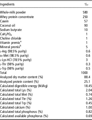
In experiment 1, newborn IUGR and NBW male piglets were obtained from first-parity Large White×Landrace sows bred by Duroc boars. The sows were of similar body condition, genetic background and age, which facilitated the selection of the target piglets. The criteria for the selection of IUGR and NBW piglets was as previously described (Liu et al., Reference Liu, He, Yang, Yu, Mao, Yu and Chen2014a). Briefly, an IUGR piglet was defined as being 2 SD below the average birth weight within a litter of 9 to 13 piglets, and a NBW piglet was defined as having a birth weight closest to the average value within a litter. A total of 16 IUGR piglets and 16 NBW piglets were selected from 16 sows, with each sow providing one IUGR and one NBW piglet. The average birth weight of NBW and IUGR piglets were 1.45±0.03 and 0.99±0.04 kg, respectively. From delivery to day 7 postnatal, these piglets were allowed to suckle their dams freely. To achieve this for the IUGR neonates, which have reduced ability to compete for milk, IUGR piglets were given assistance in finding the mammary gland. On day 8 postnatal, the 32 piglets were allocated to treatments of either consuming normal nutrient intake (NNI) or restricted nutrient intake (RNI). The nutrient intake level of RNI piglets was maintained at 50% of that of piglets in NNI feeding regimes. The estimated nutrient allowance level of NNI was determined according to the normal milk yield of a sow as previously reported (Hansen et al., Reference Guan, Stoll, Lu, Tappenden, Holst, Hartmann and Burrin2012). Piglets in the NNI regimen were fed 150 and 210 g/day dry matter from day 8 to day 15 and day 16 to day 28, respectively. The milk replacer was formulated as previously described (Han et al., Reference Han, Hu, Xuan, Ding, Luo, Bai, He, Zhang and Che2013), with minor modifications and is presented in Table 1. The diet was provided to piglets after mixing with water at a ratio of 1 : 4. Finally, four experimental groups (birth weight/nutrient level) were produced, including NBW/NNI, NBW/RNI, IUGR/NNI and IUGR/RNI (n=8/group). Piglets were fed every 3 h from 0600 to 1200 h using bottles. If a piglet refused to suckle the milk replacer, a syringe was used to gavage the remaining formula. In our previous experience, the ad libitum nutrient intake level, if calculated on a relative BW basis, was higher in IUGR pigs compared with NBW pigs (Liu et al., Reference Liu, He, Yu, Mao, Zheng, Huang, Yu and Chen2014b), indicating that the IUGR pigs developed the adaptive response of consuming higher amounts of nutrients to compensate for their inadequate intrauterine growth. This motivated us to examine whether IUGR piglets that consumed similar amounts of nutrients as NBW piglets would demonstrate improved intestinal development.
Composition of the experimental diet (as-fed)

a Supplied (per kg diet): vitamin A, 12 800 IU; vitamin E, 44 IU; vitamin D, 2600 IU; vitamin K, 4 mg; vitamin B1, 2.4 mg; riboflavin, 8.8 mg; niacin, 32 mg; pantothenic acid, 4 mg; biotin, 0.5 mg; folic acid, 2 mg; and vitamin B12, 0.05 mg.
b Supplied (per kg diet): Zn, 90 mg; Mn, 4.0 mg; Fe, 90 mg; Cu, 6.0 mg; I, 0.2 mg; Se, 0.3 mg.
In experiment 2, a total of 18 IUGR piglets were chosen from 18 litters where each sow provided one piglet. Six IUGR piglets fed the RNI regimen were injected subcutaneously with GLP-2 (h[Gly2]-GLP-2, a human GLP-2 analogue; Phoenix Pharmaceuticals Inc., Belmont, CA, USA) twice daily at 0800 and 1600 h at 40 μg/kg BW per day from day 8 to day 15 postnatal as previously described (Sigalet et al., Reference Sigalet, de Heuvel, Wallace, Bulloch, Turner, Wales, Nation, Wizzard, Hartmann, Assad and Holst2014). This form of GLP-2 has a longer half-life than the natural peptide due to the altered sequence and increased resistance to the proteolytic action of enzyme dipeptidyl peptidase IV. An additional 12 IUGR piglets that were fed on either the NNI (n=6) or RNI (n=6) regimen were injected with saline as the physiological control. This resulted in a total of three treatment groups, which were NNI/saline, RNI/saline and RNI/GLP-2. Diet composition and management were the same as that in experiment 1.
Sample collection and small intestinal morphology
At 0600 h on the day of slaughter, all piglets were given one meal providing 14% of the total amount of feed on that day. At 0700 h, which is about 15 to 30 min after their first meal and 15 h after last GLP-2 injection in experiment 2, 10 ml blood samples were collected from the jugular vein and were centrifuged at 2400×g for 15 min at 4°C to collect the serum which was then stored at −20°C for analysis of GLP-2 concentrations. Piglets were then killed after being anaesthetized with an intravenous injection of pentobarbital sodium (15 mg/kg BW) on day 28 (experiment 1) or on day 15 (experiment 2). Immediately after opening the abdominal cavity, the length of the small intestine was recorded. Jejunum samples for real-time quantitative PCR were snap-frozen in liquid nitrogen and stored at −80°C. The middle section (5 cm) of jejunum samples were fixed with a solid tissue paper to avoid crispation, and were then embedded in paraffin, and stained with haematoxylin–eosin for morphological analysis. Villous heights and crypt depths were measured using an image processing and analysis system (Optimus software version 6.5; Media Cybergenetics, Silver Spring, MD, USA). The intestinal protein content was determined by the Commassie method, and DNA content was determined as has been previously described (Labarca and Paigen, Reference Labarca and Paigen1980).
Ussing chamber
Ussing chambers were used to evaluate the nutrient transport capacity of the jejunum as previously described, with minor modifications (Gabler et al., Reference Gabler, Radcliffe, Spencer, Webel and Spurlock2009). In brief, a 20 cm segment of the proximal jejunum was removed and placed in chilled Krebs–Henseleit buffer (pH 7.4) consisting of 25 mM NaHCO3, 120 mM NaCl, 1 mM MgSO4, 6.3 mM KCl, 2 mM CaCl and 0.32 mM NaH2PO4 (21). Tissue samples were mounted in the Ussing chambers (Beijing Kingtech Technology Co. Ltd, Beijing, China) and were washed on the mucosal and serosal surfaces (opening area, 1.0 cm2) with 10 ml of Krebs solution and oxygenated with a 95% O2–5% CO2 mixture. Tissues were stabilized in the Ussing chambers for 20 min and were then challenged with 10 mmol/l d-glucose added to the serosal buffer. d-glucose was added on the mucosal side for absorption studies. Because the short-circuit current was delivered by a voltage-clamp apparatus, the potential difference across the tissue was measured for 30 min after each challenge by open-circuit conditions every 10 s. The change in maximal current was recorded, and tissue conductance was calculated from the short-circuit current and potential difference using Ohm’s law.
Enzyme activities
The activity of brush border enzymes was determined in the intestinal mucosa. In brief, 0.5 to 1.3 g of mucosal samples were homogenized in ice-cold ethylenediaminetetraacetic acid-saline phosphate buffer (pH 7.4) and centrifuged at 3000 r.p.m. for 10 min at 4°C. Total protein content was determined according to the Bradford method. The activities of lactase, maltase, sucrase and caspase-3 were measured by a commercial kit supplied by Nanjing Jiancheng Bioengineering Ltd (Jiangshu, China).
Serum glucagon-like peptide-2 concentrations
The serum concentration of GLP-2 was determined by radioimmunoassay with a commercial kit (Phoenix Pharmaceuticals Inc.). GLP-2 from human, bovine, rat and mouse are all 33 amino acid peptides, porcine GLP-2 is a 35 amino acid peptide with Ser and Leu at the C-terminal end. The assay used in the present study allowed the determination of circulating concentrations of active form of GLP-2 using the antibody targeting N-terminus and cross-reacting 100% with porcine GLP-2.
Gene expressions
Real-time quantitative PCR was used to detect the transcriptional abundance of mRNA in the jejunum section. Detailed procedures were followed as previously described (Liu et al., Reference Liu, He, Yu, Mao, Zheng, Huang, Yu and Chen2014b). Primers used for target genes are p53 (Genebank no. AF098067), sense primer CACTGGATGGCGAGTATTTCAC and antisense primer CTTAGACTTCAGGTGGCTGGA, Bcl-2 (Genebank no. NM214285), sense primer CTGGTGGTTGACTTTCTCTCCT and antisense primer TAGGGGTTTCCGCTTCTGAT, Bax (Genebank no. AJ606301.1), sense primer GGTCGCGCTTTTCTACTTTG and antisense primer CGATCTCGAAGGAAGTCCAG, Na+-dependent glucose transporter 1 (SGLT1) (Genebank no. NM_001164021.1), sense primer CCACTTTCCCTATAAAACCTCAC and antisense primer CTCCATCAAACTTCCATCCTCAG, glucose transporter 2 (GLUT2) (Genebank no. NM_001097417.1), sense primer CCTGCTTGGTCTATCTGCTGTG and antisense primer TTGATGCTTCTTCCCTTTCTTT, 18S RNA (Genebank no. JF903663.1), sense primer TCCGACTTTCGTTCTTGATTAATG and antisense primer TGGACCGGCGCAAGAC. The primers were synthesized commercially by TaKaRa Biotechnology (Dalian, China). Real-time quantitative PCR was conducted in a 96-well CFX-96 Real-Time PCR detection System (Bio-Rad, Hercules, CA, USA). The 18S RNA was used as house-keeping gene and the relative gene expression levels were expressed as fold changes relative to average mRNA levels of genes in the NBW/NNI group (Pfaffl, Reference Pfaffl2001).
Statistical analysis
In experiment 1, data were analyzed using the MIXED procedure of SAS (SAS Institute, Cary, NC, USA). Birth weight (NBW or IUGR) and nutritional level (NNI or RNI) were included as fixed effects, and the interactions of birth weight×nutritional level were also considered as fixed effects. In experiment 2, data were analyzed by a one-way ANOVA to determine statistical differences between treatment groups. The individual pig was included as a random effect in each experiment. Means were separated using the Tukey procedure and differences with probabilities of P<0.05 were considered as statistically significant.
Results
Experiment 1
Growth traits
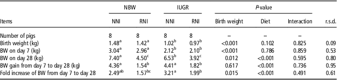
As might be expected, the initial BW on day 7 was greater for NBW compared with IUGR piglets (P<0.01), and BW on day 28 was significantly elevated by the NNI feeding regimen compared with the RNI feeding regimen (P<0.01) (Table 2). BW gain was greater in piglets fed the NNI regimen compared with the RNI regimen (P<0.01), and was not affected by birth weight. However, the fold increase of BW was higher in IUGR piglets (P<0.05), and was improved in piglets fed the NNI regimen compared with piglets fed the RNI regimen (P<0.01).
Growth traits in piglets fed either the normal nutrient intake (NNI) or the restricted nutrient intake (RNI) regimen

NBW=normal birth weight; IUGR=intrauterine growth restriction; r.s.d.=root mean square of the error.
a,b,cData with different superscripts denotes P<0.05.
Intestinal development
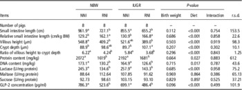
The intestinal development traits of piglets are shown in Table 3. The length of the small intestine was significantly enhanced by the NNI regimen compared with the RNI regimen (P<0.01), regardless of birth weight. The length of the small intestine relative to BW was greater in RNI piglets than that in NNI piglets (P<0.01) and was independent of birth weight (P>0.05). Piglets fed the NNI regimen had greater villous heights and villous-height to crypt-depth ratios (P<0.01), but lower crypt depths compared with piglets fed the RNI regimen (P<0.01). The villous height in IUGR piglets fed the NNI regimen was similar to that of NBW piglets fed the NNI and RNI regimens. The amount of DNA and protein in jejunum samples was greater in NNI piglets than in RNI piglets (P<0.05), but were not influenced by birth weight (P>0.05).
Small intestinal development of piglets fed either the normal nutrient intake (NNI) or the restricted nutrient intake (RNI) regimen

NBW=normal birth weight; IUGR=intrauterine growth restriction; GLP-2=glucagon-like peptide-2; r.s.d.=root mean square of the error.
a,bData with different superscripts denotes differences at P<0.05.
Digestive enzyme activities
Lactase activity was not affected by birth weight (P>0.05), but was elevated in piglets fed the NNI regimen compared with piglets fed the RNI regimen (P<0.01) (Table 3). Maltase and sucrase activities were not affected by birth weight or diet (P>0.05).
Absorptive capacity
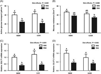
As shown in Figure 1A, active glucose transport in the jejunal section of piglets fed the NNI regimen was greater than that of piglets fed the RNI regimen (P<0.01). Mucosa resistance (Figure 1B), reflecting the intestinal permeability, was greater in piglets fed NNI than that of piglets fed RNI (P<0.01). NBW piglets fed NNI had a greater mucosa resistance than IUGR piglets fed RNI (P<0.05). Paralleling changes in active glucose transport, the mRNA transcriptional abundance of the two main intestinal glucose transporters, SGLT1 and GLUT2, were upregulated in the jejunum of the piglets fed the NNI regimen compared with piglets fed the RNI regimen (P<0.01; Figure 1C and D).
Influence of birth weight and diet on the absorptive capacity of the jejunum in piglets (n=8/treatment group). (A) active glucose transport as measured by the Ussing chamber method; (B) mucosa resistance; (C) relative messenger RNA (mRNA) expression of SGLT1; (D) relative mRNA expression of GLUT2. a,bData with different superscripts denote differences at P<0.05. NBW=normal birth weight; IUGR=intrauterine growth restriction; NNI=normal nutrient intake; RNI=restricted nutrient intake; SGLT1=Na+-dependent glucose transporter 1; GLUT2=glucose transporter 2.

Apoptotic process
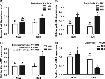
The parameters relating to the apoptotic process are presented in Figure 2. Caspase-3 activity, which is a sign of an activated apoptotic cell, was found to be greater in piglets fed RNI than that in piglets fed NNI (P<0.05). The RNI-fed IUGR piglets had a greater caspase-3 activity than pigs fed the NNI regimen (P<0.05, Figure 2A). The transcriptional abundance of p53, Bax and Bcl-2, which are apoptosis-related genes, was significantly different among treatment groups. The mRNA transcriptional abundance of p53 was greater in piglets fed the RNI regimen compared with that in piglets fed the NNI regimen (P<0.01), and were not affected by birth weight (Figure 2B). Bax mRNA expression was affected by diet (P<0.05), birth weight (P<0.05) and the interaction of birth weight with diet (P<0.05), and was significantly greater (P<0.05) in IUGR piglets fed the RNI regimen than that of other groups (Figure 2C). Bcl-2 mRNA expression was lower (P<0.05) in piglets fed the RNI regimen compared with the other piglets (Figure 2D).
Influence of birth weight and diet on parameters relating to apoptotic processes of the jejunum in piglets (n=8/treatment group). (A) caspase-3 activity; (B) relative messenger RNA (mRNA) expression of p53; (C) relative mRNA expression of Bax; (D) relative mRNA expression of Bcl-2. a,bData with different superscripts denote differences at P<0.05. NBW=normal birth weight; IUGR=intrauterine growth restriction; NNI=normal nutrient intake; RNI=restricted nutrient intake; Bcl-2=B-cell lymphoma-2; Bax=bcl-2-like protein 4.

Glucagon-like peptide-2 concentration
As shown in Table 3, the GLP-2 concentration was significantly elevated in NNI-fed piglets compared with RNI-fed piglets (P<0.01), but was not affected by birth weight (P>0.05).
Experiment 2
Glucagon-like peptide-2 concentrations
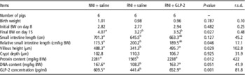
GLP-2 injection elevated circulating GLP-2 concentrations (Table 4) in IUGR piglets fed the RNI regimen compared with the saline-injected piglets (P<0.05), and resulted in similar levels of GLP-2 as in IUGR piglets fed the NNI regimen.
Intestinal development in intrauterine growth restriction (IUGR) piglets fed the restricted nutrient intake (RNI) regimen and injected with glucagon-like peptide-2 (GLP-2)

NNI=normal nutrient intake; r.s.d.=root mean square of the error.
a,bData with different superscripts denotes P<0.05.
Intestinal development
As shown in Table 4, final BW at the end of the experiment was greater in piglets fed the NNI regimen as compared with piglets fed the RNI regimen (P<0.05). There was no effect of GLP-2 injection on final BW when piglets were fed the RNI (P>0.05). Intestinal length and villous height were lower in RNI-fed piglets compared with NNI-fed piglets (P<0.05). Relative to BW, the small intestinal length of piglets fed RNI was greater than that of piglets fed NNI (P<0.05). The villous height of piglets fed the RNI regimen was elevated to similar levels as NNI-fed piglets by injection with GLP-2 (P<0.05). The protein and DNA contents of the intestine were lower in piglets fed the RNI regimen than that of NNI-fed piglets (P<0.05), but were elevated to similar levels as the NNI-fed piglets when RNI-fed piglets were treated with GLP-2.
Absorptive processes
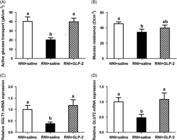
The gene expression of SGLT1 (P<0.05) and GLUT2 (P<0.05), as well as active transport of glucose (P<0.01) and mucosa resistance (P<0.05), were lower in RNI-fed piglets compared with NNI-fed piglets and RNI-fed piglets treated with GLP-2 injection (Figure 3) as determined by the Ussing chamber method.
Influence of GLP-2 injection on absorptive capacity of the jejunum in intrauterine growth restriction piglets (n=6/treatment group). (A) active glucose transport as measured by the Ussing chamber method; (B) mucosa resistance; (C) relative messenger RNA (mRNA) expression of SGLT1; (D) relative mRNA expression of GLUT2. a,bData with different superscripts denote differences at P<0.05. NNI=normal nutrient intake; RNI=restricted nutrient intake; GLP-2=glucagon-like peptide-2; SGLT1=Na+-dependent glucose transporter 1; GLUT2=glucose transporter 2.

Parameters relating to apoptotic processes
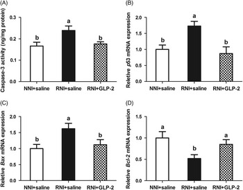
The p53, Bax and Bcl-2 mRNA transcriptional abundance are shown in Figure 4. Caspase-3 activity was greater in piglets fed the RNI regimen than that of piglets on either the NNI regimen or the RNI+GLP-2 group (P<0.05, Figure 4A). In RNI-fed piglets compared with NNI-fed piglets the gene expression levels of pro-apoptotic factors p53 and Bax (P<0.05) were upregulated, whereas the anti-apoptotic factor Bcl-2 (P<0.01) was downregulated. These factors were at similar levels as the NNI-fed piglets when GLP-2 was injected (Figure 4B–D).
Influence of GLP-2 injection on parameters relating to apoptotic processes of the jejunum in intrauterine growth restriction piglets (n=8/treatment group). (A) caspase-3 activity; (B) relative messenger RNA (mRNA) expression of p53; (C) relative mRNA expression of Bax; (D) relative mRNA expression of Bcl-2. a,bData with different superscripts denotes P<0.05. GLP-2=glucagon-like peptide-2; NNI=normal nutrient intake; RNI=restricted nutrient intake; Bcl-2=B-cell lymphoma-2; Bax=bcl-2-like protein 4.

Discussion
In commercial swine farms, low birth weight piglets have increased risks of postnatal morbidity and mortality, as well as long-term compromised growth performance (Wu et al., Reference Wu, Bazer, Wallace and Spencer2006). The gastrointestinal tract is the portal for digestion and absorption of virtually all nutrients into the blood, and its health plays a vital role in the survival of the newborn pig. The transfer of nutrients in neonates switches from the placenta to the gastrointestinal tract at birth which presents an additional challenge for the already compromised IUGR piglet. Our data indicate that when IUGR piglets were fed with NNI, which is a similar level of nutrient intake as experienced by normal suckling piglets, the fold increase of bodyweight was higher (3.21) compared with NBW piglets fed NNI (2.49). As a result, the relative BW of IUGR to NBW piglets reached a final average percentage of 87% compared with an initial 70%. This demonstrates that the compensatory growth of IUGR/NNI piglets is the result of an adequate nutrient intake level, indicating that the genetic growth potential of IUGR piglets could be met under certain postnatal nutritional conditions. However, it remains unclear whether this kind of catch-up in BW would have any undesirable implications in later phases of growth, as this mismatch of pre and postnatal nutrition can lead to an increased risk of developing metabolic disorders in adulthood (Liu et al., Reference Liu, Chen, Yao, Yu, Mao, He, Huang and Zheng2012).
Growth of the intestine was improved by NNI compared with the RNI regimen, regardless of birth weight. The gastrointestinal tissues consume 15% to 35% of whole-body oxygen use and have a significantly higher protein turnover rate despite representing only 5% of total BW (Edelstone and Holzman, Reference Ebner, Schoknecht, Reeds and Burrin1981; Ebner et al., Reference Dodge, Bertolo and Brunton1994). Thus, the high metabolic demand of the intestinal tissue requires sufficient nutrient intake to avoid subsequent villous atrophy and suppression of protein synthesis (Niinikoski et al., Reference Niinikoski, Stoll, Guan, Kansagra, Lambert, Stephens, Hartmann, Holst and Burrin2004). This may explain why intestinal development did not appear to be compromised when IUGR piglets were fed a NNI level, and does not support the hypothesis that IUGR piglets are destined to have impaired intestinal development (Wang et al., Reference Wang, Lin, Liu, Feng, Zhou, Wang, Li, Wu and Wang2014). On the contrary, intestinal development was also compromised in NBW piglets fed the RNI regimen. Therefore, these results demonstrate that the intestinal growth of newborn IUGR piglets has a similar response as NBW piglets to the nutrient intake level during the neonatal phase.
The absorptive function was improved in piglets by the NNI regimen. The main nutrient transporters mediating the intestinal uptake of glucose in newborn piglets are SGLT1 and GLUT2 (Gabler et al., Reference Edelstone and Holzman2007). As measured by gene expression, the abundance of SGLT1 and GLUT2 were significantly increased in IUGR piglets by the NNI feeding regimen. Similar to the increased transcriptional abundance of SGLT1 and GLUT2, active glucose transport was greater in piglets fed at normal levels of nutrient intake as evidenced by the Ussing chamber method. To date, very few researches have reported the expression pattern of nutrient transporters in newborn IUGR piglets. Nevertheless, the increase in nutrient transport capacity seen here indicates a higher absorptive capacity for piglets fed a normal level of nutrients.
Lactase is an enzyme responsible for hydrolysis of the major milk carbohydrate, lactose. In the present study, lactase activity was elevated by the NNI feeding regimen. This was consistent with a previous study which indicated that lactase activity in the intestine was suppressed in piglets fed a low enteral nutrient intake, but was stimulated by increasing enteral nutrient intake (Burrin et al., Reference Bjornvad, Schmidt, Petersen, Jensen, Offenberg, Elnif and Sangild2000). However, the activity of maltase, an enzyme located in the brush border of the small intestine that breaks down the disaccharide maltose, and sucrase, an enzyme located in the brush border of the small intestine that catalyze the hydrolysis of sucrose to fructose and glucose, were not influenced by birth weight and nutrient intake levels. The explanation for this observation warrants further investigation. These results demonstrate that the digestive and absorptive function of both NBW and IUGR piglets can be improved by a NNI.
Recent clues from transcriptomic profiles of the intestine have revealed molecular evidence of IUGR-induced impairment in intestinal growth that may result from changes in the cell proliferation–apoptosis balance (D’Inca et al., Reference Hansen, Strathe, Kebreab, France and Theil2010). For the control of cell apoptosis, p53 has received significant attention (Miyashita and Reed, Reference Miyashita and Reed1995). Up-regulated p53 mRNA expression was associated with increased cell apoptosis under chronic and acute stress (Cummings, Reference Cummings1996). Earlier findings revealed that altered gene (p53, Bax and Bcl-2) expressions was related to abnormal apoptotic homeostasis in IUGR rats (Baserga et al., Reference Baserga, Bertolotto, Maclennan, Hsu, Pham, Laksana and Lane2004). The anti-apoptotic (Bcl-2) or pro-apoptotic (Bax) proteins were downstream molecules regulated by p53 to play a role in small intestine damage surveillance. The present study observed decreased Bcl-2 and increased Bax mRNA expression in IUGR piglets as compared with NBW piglets, but their transcriptional abundance was reversed to a similar level as in NBW/NNI piglets when IUGR piglets received the NNI regimen. These results are consistent with previous research that showed that the enteral nutrient intake level could benefit intestinal growth through improved apoptotic homeostasis in a nutrient-level-dependent manner (Burrin et al., Reference Bjornvad, Schmidt, Petersen, Jensen, Offenberg, Elnif and Sangild2000; Stoll et al., Reference Stoll, Chang, Fan, Reeds and Burrin2000). Furthermore, the altered gene expression levels in IUGR neonates fed NNI might be the cause of the greater villous height in these piglets.
GLP-2 is produced by the intestinal endocrine L-cell and by various neurons in the central nervous system, and has been shown to act in an endocrine fashion to link intestinal growth and metabolism with nutrient intake (Guan et al., Reference Gabler, Spencer, Webel and Spurlock2003). Introduction of increased enteral nutrient intake can increase plasma GLP-2 concentration (Petersen et al., Reference Petersen, Hartmann, Holst, Le Huerou-Luron, Bjørnvad and Sangild2003), and facilitate the intestinal development of newborn piglets through improved intestinal cell survival and proliferation (Burrin et al., Reference Brinkman, Murali, Hitt, Solverson, Holst and Ney2005). Our data show that piglets fed the NNI regimen had greater GLP-2 concentrations, regardless of birth weight. Coupled with the enhanced expression levels of genes related to the anti-apoptotic process, the elevated GLP-2 concentration caused by higher nutrient intake could very well be responsible for the improvement of intestinal development through regulation of the apoptotic process, as previously described (Burrin et al., Reference Brinkman, Murali, Hitt, Solverson, Holst and Ney2005). To further confirm the physiological role of GLP-2 in the nutrient-level-dependent regulation of intestinal development, exogenous GLP-2 was administered to the IUGR piglets fed the RNI regimen to examine its role in intestinal growth. The IUGR piglets fed the RNI regimen showed suppressed intestinal development, characterized by impaired active glucose transport and villous development. However, injection of GLP-2 returned the absorptive function to similar levels as that of NNI-fed piglets, and this improvement was associated with the improved anti-apoptotic process induced by GLP-2. In a preterm pig model of intestinal resection, GLP-2 administration induced structural and functional adaptation, resulting in higher relative absorption of fluid and macronutrients (Vegge et al., Reference Vegge, Thymann, Lund, Stoll, Bering, Hartmann, Jelsing, Qvist, Burrin, Jeppesen, Holst and Sangild2013). These results demonstrate that the intestine in IUGR neonates responded positively to GLP-2 stimulation when the piglets had limited nutrient intake.
For our results, however, we could not exclude the possibility that a higher level of nutrient intake than fed here would induce additional potential risk, as previous research demonstrated that enteral feeding of formula for preterm IUGR pigs induced mucosal structural changes that may predispose them to later development of necrotizing enterocolitis (Oste et al., Reference Oste, Van Haver, Thymann, Sangild, Weyns and Van Ginneken2010). In our previous research, IUGR did not predispose piglets to necrotizing enterocolitis, nor did we detect any compromised postnatal intestinal adaptation in IUGR pigs fed a high-fat diet (Liu et al., Reference Liu, He, Yu, Mao, Zheng, Huang, Yu and Chen2014b). This inconsistency might be related to the digestibility of nutrients for the premature pig. It has been reported that the less-digestible maltodextrin-based formula induced higher incidences of necrotizing enterocolitis compared with the highly digestible lactose-based formula in preterm pigs (Thymann et al., Reference Thymann, Møller, Stoll, Støy, Buddington, Bering, Jensen, Olutoye, Siggers, Mølbak, Sangild and Burrin2009). This raises the question of how the composition of the formula could impact the intestinal development of IUGR neonates.
In summary, our results suggest that the intestinal growth of IUGR piglets responded to the level of nutrient intake in a similar way to that of NBW piglets. In addition, based on the trophic effect of GLP-2 seen here, GLP-2 administration could also be considered an effective nutritional strategy to improve the intestinal development of both human and porcine IUGR individuals.
Acknowledgements
The authors gratefully acknowledge Shanchuan Cao and Ruqing Zhong for their varied roles in the contributions to this study.
Financial Support
This work was supported by grants from the State Key Laboratory of Animal Nutrition of China (2004DA125184F1424), the National Science and Technology Support Program (2012BAD39B01), and the Agricultural Science and Technology Innovation Program (ASTIP-IAS07) in China. The funding agencies had no role in the design of this study, the analysis of data or the writing of this article.