Introduction
Until recently, data regarding the digenean fauna of mugilid fish species from the southern Russian Far East were limited to the description of two species of flukes in the redlip mullet Liza haematocheila (Temminck and Schlegel), Skrjabinolecithum spasskii Belous, Reference Belous1954 and Parasaccocoelium mugili Zhukov, Reference Zhukov1971 (Belous, Reference Belous1954; Skrjabin, Reference Skrjabin and Skrjabin1956; Zhukov, Reference Zhukov1971). Overstreet & Curran (Reference Overstreet, Curran, Jones, Bray and Gibson2005) synonymized Parasaccocoelium Zhukov, Reference Zhukov1971 with Pseudohapladena Yamaguti, Reference Yamaguti1952, which also included Ps. scatophagi Yamaguti, Reference Yamaguti1952 (type species), Ps. pearsoni (Martin, Reference Martin1973), Ps. martini (Madhavi, Reference Madhavi1979), Ps. huidongensis (Lu, 1993), Ps. megaorchis Liu et Yang, 2002 and Ps. lizae Liu et Yang, 2002, which are parasites of mugilid and scatophagid fish species in the Indo-West Pacific Region (Overstreet & Curran, Reference Overstreet, Curran, Jones, Bray and Gibson2005). Parasaccocoelium was synonymized with Pseudohapladena based on a longer prepharynx than pharynx and relatively short sac-like caeca ending near the mid-body, and other signs that are characteristic of Pseudohapladena.
While investigating the parasite fauna of the redlip mullet L. haematocheila and the striped mullet Mugil cephalus L., we found several species of haploporids. Based on newly obtained material from type hosts near the type locality, we added new data to the description of the type species of Parasaccocoelium and restored the classification of this genus. We have included Pa. mugili and two new species in Parasaccocoelium.
Materials and methods
Collection of trematodes
Trematodes were recovered from two striped mullet from Kievka River, one from Razdolnaya River basin and two from Karasyk River, and five redlip mullet from each of three river sites. Fresh specimens of fish were dissected and the intestines removed for examination and identification of trematodes.
Worms from the fish species were rinsed in distilled water, identified to species, killed in boiling distilled water and preserved in 70% ethanol. Whole-mounts for adult descriptions were made by staining the specimens with alum carmine, dehydrating the worms in a graded ethanol series and clearing in xylene, followed by mounting in Canada balsam under a coverslip on a slide. To establish the time of development of the miracidia, the eggs, which were collected from mature worms, were placed in distilled water with the temperature at approximately 20°C. All measurements are given in micrometres (μm). Mean and standard deviation of coefficient of variation (CV) were calculated according to Steel & Torrier (Reference Steel and Torrier1980).
Descriptions were based on 100 whole-mounts of Pa. mugili; 12 of Parasaccocoelium haematocheilum n. sp. and 34 of Parasaccocoelium polyovum n. sp. were studied.
Molecular and phylogenetic analyses
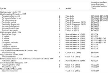
Adult Parasaccocoelium specimens (n= 14), including Pa. mugili (n= 4), Pa. haematocheilum n. sp. (n= 7) and Pa. polyovum n. sp. (n= 5), from the Kievka River, Primorsky Region, were used for molecular analysis (table 1). Total DNA was extracted from flukes, which were fixed in 96% ethanol, using a ‘hot shot’ technique (Truett, Reference Truett and Kieleczawa2006).
List of taxa incorporated in the molecular analysis of subfamilies of Haploporoidea Nicoll, 1914.

Nuclear 28S rDNA was successfully amplified using polymerase chain reaction (PCR) with the following primers: DIG12 (5′- AAG CAT ATC ACT AAG CGG -3′) and 1500R (5′- GCT ATC CTG AGG GAA ACT TCG-3′) (Tkach et al., Reference Tkach, Littlewood, Olson, Kinsella and Swiderski2003). The initial PCR reaction was performed in a total volume of 20 μl containing 0.25 mm of each primer pair, 1 μl of DNA in water, 1 × Taq buffer, 1.25 mm deoxynucleoside triphosphates (dNTPs), 1.5 mm magnesium and 1 unit of Taq polymerase. The amplification of a 1200-bp fragment of 28S rDNA was performed in a GeneAmp 9700 (Applied Biosystems, Grand Island, New York, USA), with a 3-min denaturation at 94°C, 40 cycles of 30 s at 94°C, 30 s at 52°C and 2 min at 72°C, and a 7-min extension at 72°C. Negative and positive controls using both primers were used. The PCR products were directly sequenced using an ABI Big Dye Terminator v. 3.1 Cycle Sequencing Kit (Applied Biosystems), as recommended by the manufacturer, with the internal sequencing primers of 300F, ECD2, 900F and 1200R (Tkach et al., Reference Tkach, Littlewood, Olson, Kinsella and Swiderski2003). The PCR products were analysed using an ABI 3130 genetic analyser at the Institute of Biology and Soil Sciences, Far Eastern Branch, Russian Academy of Sciences. The sequences have been submitted to the European Nucleotide Archive (ENA), with the following accession numbers: HF548461–HF548478 (table 1). The ribosomal DNA sequences were assembled with SeqScape v. 2.6 software and aligned using MEGA 5.22 (Tamura et al., Reference Tamura, Peterson, Peterson, Stecher, Nei and Kumar2011) alignment explorer with the default options. The regions that could not be unambiguously aligned were excluded from the analyses. Calculation of a number of variable and parsimony-informative sites and ancestral sequence test were performed using MEGA v. 5.22. Genetic divergence was estimated using genetic p-distance values, which were calculated including all substitution types. The phylogenetic analysis of the nucleotide sequences was performed using Bayesian algorithms with MrBayes v. 3.1.2 software (Huelsenbeck, Reference Huelsenbeck2000). The best nucleotide substitution model, general time reversible with estimates of invariant sites and gamma-distributed among-site variation (GTR+I+G), was estimated with Modeltest v. 3.7 software (Posada & Crandall, Reference Posada and Crandall1998). Bayesian analysis was performed using 10,000,000 generations and with four independent runs. Burn-in (sump and sumt) values were 10,000. The significance of the phylogenetic relationship was estimated by posterior probabilities (Huelsenbeck et al., Reference Huelsenbeck, Ronquist, Nielsen and Bollback2001). The test on ancestral sequence inferring was performed on the basis of an MP phylogenetic tree using a maximum parsimony algorithm with MEGA v. 5.22 software. The phylogenetic relationships among the Parasaccocoelium species were inferred from our data and the nucleotide sequences of 28S rDNA from other trematode specimens obtained from the National Center for Biotechnology Information (NCBI) GenBank database (Tkach et al., Reference Tkach, Pawlowski, Mariaux, Swiderski, Littlewood and Bray2001; Olson et al., Reference Olson, Cribb, Tkach, Bray and Littlewood2003; Curran et al., Reference Curran, Tkach and Overstreet2006; Blasco-Costa et al., Reference Blasco-Costa, Balbuena, Kostadinova and Olson2009; Pulis & Overstreet, Reference Pulis and Overstreet2013; Pulis et al., Reference Pulis, Fayton, Curran and Overstreet2013). The other sequences were from the Haploporidae and Atractotrematidae families. Monorchis monorchis, family Monorchidae, was used as an outgroup (table 1).
Family Haploporidae Nicoll, 1914; Subfamily Waretrematinae Srivastava, 1937; Genus Parasaccocoelium Zhukov, Reference Zhukov1971
Diagnosis of the genus
Body oval, fusiform or retort-shaped; anterior end retractable or not, posterior retractable. Eye-spot pigment dispersed. Oral sucker subterminal. Ventral sucker larger than oral sucker placed on border of anterior and middle third of body, or in middle third of body. Prepharynx and the oesophagus variable in length. Pharynx transversally oval or spherical. Caeca short, sac-shaped. Testis single, variable in form: longitudinal, longitudinally oval, spherical, transversally oval, irregular, bilobed or V-shaped. Ovary and testis in end of middle or in posterior third of body. Hermaphroditic sac sacciform or oval. Posterior margin of sac does not cross level of posterior margin of ventral sucker. External seminal vesicle sac-shaped, club-shaped or tubular. Genital pore anterior to ventral sucker. Genital atrium absent. Ovary from large cells, pretesticular, medial. Uterus short, extending between posterior end of hermaphroditic sac and middle of testis. Eggs operculate, light yellow, embryonated when laid. Developed miracidium with eye spot. Vitellarium in lateral fields, from large compact follicles of irregular form, extends between anterior margin of ventral sucker or pharynx and posterior margin of testis. Vitelline follicles absent in post-testicular region. Excretory bladder Y-shaped. In mugilid fish species. Type species: Parasaccocoelium mugili Zhukov, Reference Zhukov1971.
Parasaccocoelium mugili Zhukov, Reference Zhukov1971
Species description
Synonyms. Pseudohapladena mugili (Zhukov, Reference Zhukov1971) Overstreet et Curran, 2005; Pseudohapladena lizae Liu et Yang, 2002.
Type host. Redlip mullet Liza haematocheila.
Other host. Striped mullet Mugil cephalus.
Site. Intestine.
Intensity of infection. In redlip mullets, from 1 to 254; in striped mullets, up to 10 worms per fish.
Localities. Razdolnaya River basin (43°20′N, 131°47′E), Karasyk River (42°32′N, 130°38′E), Kievka River (42°52′N, 133°39′E). Flukes were found in both fish species in each river.
Material examined. 100 specimens.
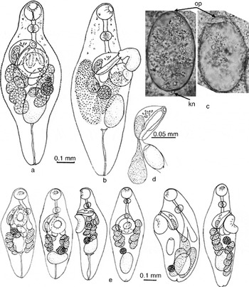
Adult worm (fig. 1). Body pyriform when posterior end retracted, as typical in life, or fusiform. Small spines on tegument, except in last sixth of body length, can be lost during fixation and staining. Eye-spot pigment dispersed in anterior third of body. Ventral sucker larger than oral sucker, about one-third body length from anterior end. Prepharynx usually conspicuous, sometimes almost overlapping anterior edge of ventral sucker, or else very short. Pharynx spherical or transversally oval. Oesophagus short. Caeca sac-shaped, terminating near or slightly posterior to posterior margin of ventral sucker. Testis single, oval, spherical or irregular in shape. Hermaphroditic sac (fig. 1d) dorsal to ventral sucker and not extending beyond posterior margin of sucker, oval in dorsal or ventral view, but sac-shaped in lateral view, with an internal seminal vesicle, the duct of which extends into the proximal part of the hermaphroditic canal and prostate cells occupy the length of this canal. Hermaphroditic canal with muscular walls. External seminal vesicle sac-shaped, somewhat variable in volume and position. Genital pore with muscular sphincter, median, immediately anterior to ventral sucker. Ovary round or oval, somewhat variable in position but usually overlapping testis, median or submedian. Uterus short, between hermaphroditic sac and anterior margin of testis, contains single large, light-yellow, operculate egg with curved knob at aboperculate pole (fig. 1c) and with 1–2 embryonic and vitelline cells. Uterine seminal receptacle present. Metraterm short, with fine walls. Mehlis' gland consists of 6–8 cells. Vitellarium between anterior margin of ventral sucker and posterior margin of testis, or anterior margin of testis in some cases, from compact groups of six large, variably shaped follicles on each side of body, sometimes coalescing in larger worms and often appearing as homogeneous masses in whole mounts (fig. 1b). Lateral groups of vitelline cells never confluent. Excretory bladder Y-shaped.
Parasaccocoelium mugili Zhukov, Reference Zhukov1971: (a) ventral view; (b) lateral view; (c) eggs with operculum (op) and knob (kn); (d) hermaphroditic sac; (e) variations in the position of organs.

Miracidium. Hatches after 14 days in distilled water and possesses an eye spot.
28S rRNA gene sequence data
Ribosomal 28S rRNA gene fragment of approximately 1200 bp was obtained for species of Parasaccocoelium. Six specimens of Pa. mugili were analysed. Partial 28S rDNA fragments of 1143 bp were identical from each of six P. mugili specimens.
Remarks
Zhukov (Reference Zhukov1971) described a new digenean species from the family Haploporidae (Waretrematinae) isolated from the intestines of red mullets that were caught in the Sea of Japan and the Yellow Sea, and erected the new genus Parasaccocoelium with a single species, Pa. mugili.
Overstreet & Curran (Reference Overstreet, Curran, Jones, Bray and Gibson2005) revised the family Haploporidae and transferred Parasaccocoelium to a junior synonym of the genus Pseudohapladena. In this genus, in addition to Ps. mugili, they included Saccocoelioides martini Madchavi, 1980, S. pearsoni Martin, Reference Martin1973 and S. huidongensis Lu, 1993. These species were transferred to Pseudohapladena because they have a prepharynx that is longer than the pharynx, short caeca, an ovary and testis that are close to each other and lie near the ends of the caeca, and elongated vitelline fields.
As proposed by Overstreet & Curran (Reference Overstreet, Curran, Jones, Bray and Gibson2005), the genus Pseudohapladena includes seven species: the type species Ps. scatophagi Yamaguti, Reference Yamaguti1952 and Ps. mugili (Zhukov, Reference Zhukov1971), Ps. pearsoni (Martin, Reference Martin1973), Ps. martini (Madhavi, 1979), Ps. huidongensis (Lu, 1993), Ps. megaorchis Liu et Yang, 2002 and Ps. lizae Liu et Yang, 2002.
Overstreet & Curran (Reference Overstreet, Curran, Jones, Bray and Gibson2005) changed the diagnostic features of this genus to include the following characteristics: (1) body fusiform to elongate (previously body small, elongate, covered with spines); (2) eye-spot pigment dispersed (previously eye spot absent); (3) oral sucker terminal or subterminal (previously oral sucker terminal, with subterminal aperture); (4) testis subspherical to irregular to elongate, near the middle or posterior portion of the hindbody (previously testis single, median, near posterior extremity); (5) seminal receptacle canalicular (previously seminal receptacle absent); and (6) vitellarium with elongate groups of follicles that usually extend at least one-half of a body length, extending to the posterior end of the body in some and to the posterior margin of the testis in others (previously vitellarium tubular, divided into two groups). Yamaguti (Reference Yamaguti1952) described a genital atrium in the genus Pseudohapladena, but Overstreet & Curran (Reference Overstreet, Curran, Jones, Bray and Gibson2005) did not mention this characteristic. However, Overstreet & Curran (Reference Overstreet, Curran, Jones, Bray and Gibson2005) did indicate that eggs are operculate, at least in some species, and that miracidia lack eye spots.
We believe that a revision of the Pseudohapladena complex, which includes genera Pseudohapladena, Skrjabinolecithum and Platydidymus, is necessary. In many cases, to define the systematic status of a genus or species, Overstreet & Curran (Reference Overstreet, Curran, Jones, Bray and Gibson2005) used variable features, such as the length of the prepharynx, the ratio of the prepharynx to pharynx, forms of the caeca and the hermaphroditic sac, insignificant differences in the length of the caeca, and the position of the ovary and testis relative to the end of the caeca. We noted great variation in the features of Pa. mugili (fig. 1e) and in the other species described herein.
The most stable features of Pa. mugili and other species include the lateral position of the vitellaria in the mid and posterior third of the body, with no vitelline connection behind the testis and the presence of a short uterus containing a small number of eggs. The type species of Pseudohapladena, Ps. scatophagi, and other members of this genus, such as Ps. martini and Ps. megaorchis, possess vitelline fields that join behind the testis and extend to the posterior end of the body (Zhukov, Reference Zhukov1971; Martin, 1973; Madhavi, Reference Madhavi1979; Liu & Yang, Reference Liu and Yang2002). Thus, this species and Pa. mugili cannot be combined within a single genus. Additionally, Ps. scatophagi possesses a long uterus with many eggs. Pseudohapladena pearsoni, described by Martin (Reference Martin1973), is atypical of the genus in that vitellaria, apart from containing only a few follicles, extend from the hermaphroditic sac to the posterior end of the testis, whereas the uterus is short with only a small number of eggs. However, because of differences in the position of the vitellaria, Ps. pearsoni has not been transferred to the genus Parasaccocoelium.
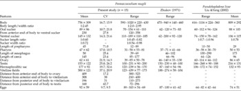
It is difficult to distinguish Ps. lizae, described by Liu & Yang (Reference Liu and Yang2002) from Liza carinatus (Cuvier et Valenciennes) from Pa. mugili. Specimens of both species are similar in size and morphology, including body shape, structure and position of hermaphroditic sac, uterus, vitellarium, testis and ovary (table 2). Therefore, we consider Ps. lizae to be a junior synonym of Pa. mugili.
Comparative morphometrics (μm) of Parasaccocoelium mugili Zhukov, Reference Zhukov1971 with Pseudohapladena lizae Liu & Yang, Reference Liu and Yang2002, to show ranges, means and coefficient of variation (CV), with first values for length and second values for width; n=number of specimens examined.

We consider that Parasaccocoelium and Pseudohapladena sensu Yamaguti (Reference Yamaguti1952) differ in having (1) dispersed or absent eye-spot pigment; (2) a subterminal or terminal oral sucker; (3) absent or present genital atrium; and (4) a vitellarium in the form of two clusters of large, compact vitelline masses or a tubular vitellarium that is divided into two groups. In addition to these criteria, Yamaguti (Reference Yamaguti1952) noted in a description of the type species Ps. scatophagi that vitelline follicles occupy almost the entire post-testicular region; but vitelline follicles do not occupy this zone in specimens of Parasaccocoelium. From this diagnosis of Pseudohapladena, Overstreet & Curran (Reference Overstreet, Curran, Jones, Bray and Gibson2005) accordingly proposed that the genus Parasaccocoelium differs in these features: (1) a single testis that is longitudinal, longitudinally oval, spherical, transversally oval, irregular, bilobed or V-shaped, or a subspherical testis that is irregular to elongate; (2) vitellarium in lateral fields formed from large compact follicles with an irregular form that extend between the anterior margin of the ventral sucker or the pharynx and posterior margin of testis, and possesses vitelline follicles that do not occupy the post-testicular region; or a vitellarium that consists of elongate groups of follicles that usually extend at least one-half of a body length to the posterior end of the body in some and to the posterior margin of the testis in others; (3) eggs operculate, unembryonated, developed when laid, or eggs which, in some cases, are only operculate; and (4) a developed miracidium with or without an eye spot.
In the light of these differences, we restore the genus Parasaccocoelium with the type species Pa. mugili Zhukov, Reference Zhukov1971.
Parasaccocoelium haematocheilum n. sp.
Species description
Type host. Redlip mullet Liza haematocheila.
Site. Intestine.
Intensity of infection. 1–10 worms per fish.
Type locality. Razdolnaya River basin, 43°20′N, 131°47′E.
Type deposition. Holotype No. 51-Tr, paratypes No. 52-Tr. 63-Tr. This material is held in the collection of the Zoological Museum, Institute of Biology and Soil Sciences, Far East Branch of the Russian Academy of Sciences, Vladivostok, Russia (e-mail: petrova@ibss.dvo.ru). Deposited 29 July 2010.
Etymology. The specific name refers to the specific name of its definitive host.
Material examined. 12 specimens.
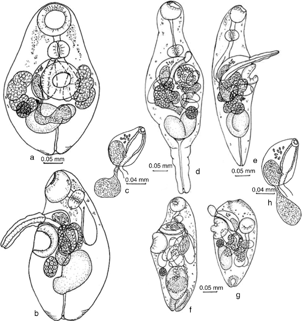
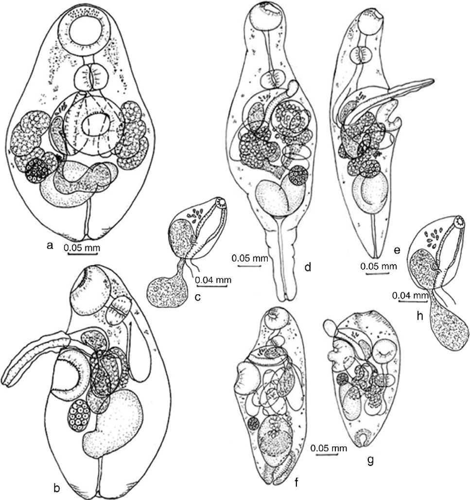
Adult worm (fig. 2a–c). Body oval. Tegumental spines only before opening of oral sucker and around opening of ventral sucker. Pigmented eye spots mainly in anterior end of body and small pigmented spots occur in the middle third of body. Oral sucker subterminal. Ventral sucker larger than oral sucker, about one-third body length of anterior end or in middle third of body. Prepharynx short. Pharynx longitudinally oval. Oesophagus short. Caeca sac-shaped, terminating near posterior margin of ventral sucker. Ovary, testis and vitellarium usually between middle and beginning posterior third of body. Testis longitudinally or transversally elongate, from two equal or unequal lobes (holotype with lobes 69 × 77 and 31 × 39). Hermaphroditic sac (fig. 2c) oval in dorsal or ventral view but sac-shaped in lateral view, dorsally from ventral sucker. Posterior margin of hermaphroditic sac does not reach level of posterior margin of ventral sucker. Hermaphroditic sac contains internal seminal vesicle (duct of vesicle occupies the beginning of hermaphroditic canal) and some prostatic cells along hermaphroditic canal. Hermaphroditic canal with muscular walls. External seminal vesicle sac-shaped, reaches testis or most often lies on level of ovary and testis. Genital pore with muscular sphincter, on midline of body immediately anterior to ventral sucker. Ovary round or oval, joined to testis or some distance from it on midline of body or to right on level of testis. Uterus short, bounded by hermaphroditic sac and anterior border of testis, contains 1–4 oval, light yellow, operculate eggs with curved knob at aboperculate pole, and with 1–2 embryonic and vitelline cells. Uterine seminal receptacle present. Metraterm short, with fine walls. Mehlis' gland present. Vitellarium in two lateral fields between anterior margin of ventral sucker and posterior margin of testis, and partly covers ovary and testis, forming 4–5 compact follicles each. Lateral groups of vitelline cells never confluent. Excretory bladder Y-shaped.
Parasaccocoelium haematocheilum n. sp.: (a) ventral view; (b) lateral view; (c) hermaphroditic sac. Parasaccocoelium polyovum n. sp.: (d) ventral view; (e) lateral view; (f) posterior extremity retracted; (g) anterior extremity retracted; (h) hermaphroditic sac.
Miracidium. Hatches after 14 days in distilled water and possesses an eye spot.
28S rRNA gene sequence data
Partial 28S rDNA fragments of 1143 bp were identical from each of seven specimens of Pa. haematocheilum n. sp.
Remarks
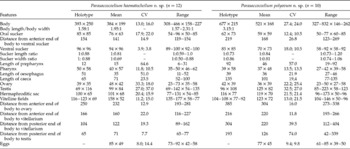
Morphological features of this species are found in accordance with the generic diagnosis. Compared with the type species Pa. mugili, they are smaller in almost all dimensions (tables 2, 3), such as the bilobed testis and eggs in the uterus (1–4 versus only 1 in Pa. mugili). Molecular data also confirm an independence of described species. The 1143-bp 28S rDNA fragment of Pa. haematocheilum n. sp. and Pa. mugili contained eight variable sites. Genetic differentiation between 28S rDNA partial sequences of Pa. haematocheilum n. sp. and Pa. mugili was 0.73%.
Comparative morphometrics (μm) of Parasaccocoelium haematocheilum and Pa. polyovum, to show ranges, means and coefficient of variation (CV), with first values for length and second values for width; n=number of specimens examined.

Parasaccocoelium polyovum n. sp.
Species description
Type host. Redlip mullet Liza haematocheila.
Other host. Striped mullet Mugil cephalus.
Site. Intestine.
Intensity of infection. 21–104 worms per redlip mullet, 18 in striped mullet.
Type locality. Razdolnaya River Basin, 43°20′N, 131°47′E (only redlip mullet was infected).
Other locality. Kievka River, 42°52′N, 133°39′E (both fish species were infected).
Type deposition. Holotype No. 71-Tr, paratypes No. 72-Tr.–79-Tr. This material is held in the collection of the Zoological Museum, Institute of Biology and Soil Sciences, Far East Branch of the Russian Academy of Sciences, Vladivostok, Russia (e-mail: petrova@ibss.dvo.ru). Deposited: 29 July 2010.
Etymology. The specific name refers to the fact that these worms have the greatest number of eggs in the uterus among all described species of this genus.
Material examined. 34 specimens.
Adult worm (fig. 2d–h). Body fusiform or oval with retractable anterior and posterior ends, spined only on anterior end of body. Eye-spot pigment dispersed over entire body surface. Oral sucker subterminal. Prepharynx usually well developed. Pharynx transversally oval, oesophagus short, sac-shaped caeca terminate near posterior margin of ventral sucker. Ventral sucker equal to or larger than oral sucker and lies on border of anterior and middle third of body, or in beginning of middle third. Hermaphroditic sac (fig. 2h) sac-shaped, dorsally from ventral sucker. Ovary, testis and vitellarium between middle and beginning posterior third of body and the testis is V-shaped. Posterior margin of hermaphroditic sac does not cross level of posterior margin of ventral sucker. Hermaphroditic sac contains internal seminal vesicle (duct of vesicle occupies the beginning of hermaphroditic canal) and some prostatic cells along hermaphroditic canal. Hermaphroditic canal with muscular walls. External seminal vesicle, depending on number of genital products, can extended to the ovary and testis and can be tubular or sac-shaped. Genital pore with muscular sphincter, immediately anterior to ventral sucker. Ovary round or oval and level with, or situated to the left of, the anterior margin of the testis. Uterus short and, when containing up to 7 eggs, is located from hermaphroditic sac up to anterior margin of the testis or, with up to 12 eggs, to the mid-region of the testis. Uterine seminal receptacle present. Metraterm short, with fine walls. Eggs light yellow, oval, operculate, with curved knob at abopercular pole, at various stages of embryogeny. Vitellarium in two lateral fields formed from compact follicles of various forms, extending between anterior margin of ventral sucker and middle of testis, and can partly cover ovary and testis. Lateral groups of vitelline cells never confluent. Excretory bladder Y-shaped.
Miracidium. Hatches after 5 days in distilled water and possesses an eye spot.
28S rRNA gene sequence data
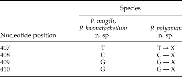
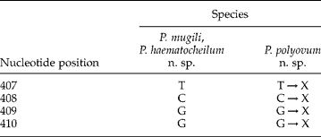
Partial 28S rDNA fragments of 1139 bp were identical from each of five specimens of P. polyovum n. sp. The shorter length of the investigated DNA fragment was due to a four-nucleotide deletion in the 28S rDNA sequences of Pa. polyovum n. sp. between base-pair positions 407 and 410 in comparison with the sequences of Pa. mugili and Pa. haematocheilum n. sp.
Remarks
The described species belongs to the genus Parasaccocoelium but differs from others by a suite of features. Compared with Pa. mugili, specimens of Pa. polyovum n. sp. differ by the smaller maximum and mean dimensions of the body and most organs (suckers, pharynx, ovary and testis), and by the smaller eggs (tables 2 and 3). Their dimensions more closely resemble those of Pa. haematocheilum n. sp. than Pa. mugili. Adult Pa. polyovum n. sp. have greater maximum and mean sizes of the length of the body and hermaphroditic sac, smaller maximum and mean sizes of the oral sucker, and a vitelline field that is shorter and wider than that of Pa. haematocheilum. In addition, Pa. polyovum n. sp. differs from these species by the V-shaped testis, the presence in the uterus of up to 12 eggs and the presence of pigment spots over the entire surface of the body. Molecular data support the validity of P. polyovum n. sp. The 1139 bp 28S rDNA fragment of Pa. polyovum n. sp. and 1143 bp fragment of Pa. mugili contained 34 variable sites, which were also parsimony-informative. 28S rDNA fragments of Pa. polyovum n. sp. and Pa. haematocheilum n. sp. contained 37 variable and parsimony-informative sites. Genetic differentiation between 28S rDNA partial sequences of Pa. polyovum n. sp. and Pa. mugili was 2.8%, and between sequences of Pa. polyovum n. sp. and Pa. haematocheilum n. sp. it was 3.1%.
Discussion
We attribute three species – Pa. mugili Zhukov, Reference Zhukov1971 and two newly described species, Pa. haematocheilum n. sp. and Pa. polyovum n. sp. – to the genus Parasaccocoelium. Studies of the morphology of mature worms of this genus showed that certain parameters, such as the length of the prepharynx and oesophagus, the ratios of prepharynx to pharynx lengths and oesophagus to pharynx lengths, the form and size of the caeca, the seminal vesicles, the ovary, the testis, the relative position of the ovary and testis, and the relative position of the testis and ventral sucker, are very variable. The most stable characteristics of this genus are the structure of the vitellarium, which includes large compact and irregular follicles, the position of the vitellarium in the posterior part of the body up to the level of the posterior margin of the testes and no connection between the vitelline fields behind the testes, the position of the posterior margin of the hermaphroditic sac not far from the posterior margin of the ventral sucker, and a short uterus between the posterior margin of the hermaphroditic sac and the middle of the testis. Eggs are operculate and possess a curved knob. Miracidia, each with an eye spot, develop only in water and not in utero.
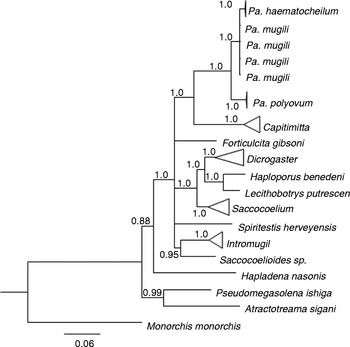
Our molecular results supported the species designation for the specimens under investigation. The minimum p-distance values were obtained between Pa. mugili and Pa. haematocheilum n. sp., which were also considerably differentiated from each other by morphometrical data. Based on the interspecific variation of 28S rDNA sequences for haploporid trematodes, earlier estimates ranged from 0.9 to 4.6% for the genus Saccocoelium (Blasco-Costa et al., Reference Blasco-Costa, Balbuena, Kostadinova and Olson2009) and from 2.7 to 2.8% for the genera Capitimitta (Pulis et al., Reference Pulis, Fayton, Curran and Overstreet2013) and Intromugil (Pulis & Overstreet, Reference Pulis and Overstreet2013). Genetic differentiation between species of the genus Parasaccocoelium from our study ranged from 0.73 to 3.11%. These species have considerable morphological differences. We argue that such interspecific ranges of genetic differences in the genus Parasaccocoelium can be accepted, whereas intrageneric molecular data are lacking for the family Haploporidae. The 28S rDNA sequences of Pa. polyovum species are characterized by a four-nucleotide deletion between the positions 407 and 410, which we considered to be possible apomorphy (table 4). This conclusion is based on the results of the inferred ancestral sequences test using a maximum parsimony algorithm to re-construct a phylogenetic tree for the species of Parasaccocoelium. The results were statistically significant (probability of inference >90%) and showed that the four nucleotides between the 407 and 410 positions of the 28S rDNA fragment were deleted (table 4). This species, therefore, can be considered to be the younger species. Bayesian analysis of partial 28S rDNA sequences showed that species of Parasaccocoelium formed a monophyletic cluster (fig. 3). Parasaccocoelium haematocheilum n. sp. was closely related to Pa. mugili, and Pa. polyovum n. sp. appeared as a sister species to the cluster containing Pa. mugili and Pa. haematocheilum with 3.2% sequence differentiation. Species of the genus Parasaccocoelium clustered with species of genus Capitimitta (subfamily Waretrematinae). The results presented here support the taxonomical status of the genus Parasaccocoelium and the inclusion of this genus in the subfamily Waretrematinae.
Ancestral sequences for species of the genus Parasaccocoelium with the probability of inference>90%.
Phylogenetic tree of different genera of the family Haploporidae based on the analysis of partial sequences of 28S rDNA using the Bayesian algorithm. Nodal numbers indicate posterior probabilities.

Financial support
Partial funding support for this research by grants No. 09-1P23-02 from the FEB RAS Presidium and No. MK-394.2014.4 from the President of the Russian Federation is gratefully acknowledged.
Conflict of interest
None.