Introduction
Correctly diagnosing dementia type is increasingly important in an era when potentially disease-modifying agents are soon likely to be marketed. Alzheimer's disease (AD) affects a range of cognitive domains including memory, executive functions, language and visuospatial function. Affected individuals show a range of impairment profiles across these functions, and these impairments may progress differently in each domain. Characteristically memory is affected early in AD (Grady et al., 1988; Welsh et al., Reference Welsh, Butters, Hughes, Mohs and Heyman1991; Greene et al., Reference Greene, Patterson, Xuereb and Hodges1996) and in severe AD all domains are affected (Price et al., Reference Price, Gurvit, Weintraub, Geula, Leimkuhler and Mesulam1993). Atypical presentations of AD are, however, recognized and relate closely to the pattern of pathology at autopsy (Kanne et al., Reference Kanne, Balota, Storandt, McKeel and Morris1998; Galton et al., Reference Galton, Patterson, Xuereb and Hodges2000). Impairments of executive and language functions may be early prominent features of AD (Becker et al., 1988; Binetti et al., Reference Binetti, Magri, Padovani, Cappa, Bianchetti and Trabucchi1996; Gorno-Tempini et al., Reference Gorno-Tempini2008) while behavioral abnormalities may also manifest early and have prognostic value (Stern et al., Reference Stern, Mayeux, Sano, Hauser and Bush1987; Mega et al., Reference Mega1996). A frontal variant of AD with a neuropathological correlate of greater numbers of neurofibrillary tangles within the frontal lobes than is seen in other AD patients is reported (Johnson et al., Reference Johnson, Head, Kim, Stark, Arnold and Cotman1999).
It can be difficult to distinguish AD patients with disproportionate frontal features from patients with frontotemporal dementia (FTD) – now known as frontotemporal lobar degeneration (FTLD) (Lebert et al., Reference Lebert, Pasquier, Souliez and Petit1998; Storey et al., Reference Storey, Slavin and Kinsella2002). The NINCDS-ADRDA diagnostic criteria for AD have low specificity when comparing AD with FTLD patients, where most FTLD patients can fulfill NINCDS-ADRDA criteria for AD (Varma et al., Reference Varma, Snowden, Lloyd, Talbot, Mann and Neary1999). Additionally, the Lund-Manchester clinical criteria for FTLD frequently misdiagnose AD, with 34% of clinically diagnosed AD cases in a community sample of 185 dementia cases fulfilling these non-AD criteria (Ikeda et al., Reference Ikeda, Ishikawa and Tanabe2004). This clinical difficulty in diagnosis may reflect the underlying pathology, with 7–32% of patients clinically diagnosed as having FTLD found to have AD pathology alone or in combination with other pathology at autopsy (Kertesz et al., Reference Kertesz, McMonagle, Blair, Davidson and Munoz2005; Knopmann et al., Reference Knopmann2005; Forman et al., Reference Forman2005; Knibb et al., Reference Knibb, Xuereb, Patterson and Hodges2006). An autopsy study of 45 well-characterized subjects with dementia had eight cases with both AD and FTLD pathology (Woodward et al., Reference Woodward, Hsiung, Feldman, Jacova and Mackenzie2010a) and more recently the Alzheimer's Disease Neuroimaging Initiative reported that one of the first nine cases to come to autopsy had coexistent AD and FTLD (TDP-43) pathology (Cairns et al., Reference Cairns, Taylor-Reinwald and Morris2010). Patients with FTLD may have prominent early memory loss, further blurring the distinction between FTLD and AD (Hodges et al., Reference Hodges2004). Indeed, it has been proposed that AD and FTLD are opposite ends of a spectrum with much overlap clinically, pathologically and genetically (Liscic et al., Reference Liscic, Storandt, Cairns and Morris2007; van der Zee et al., Reference van der Zee, Sleegers and Van Broeckhoven2008).
Several scales assess the cognitive profile of frontal executive function and neuropsychiatric symptoms referable to frontal structures. The Frontal Assessment Battery (FAB) (Dubois et al., Reference Dubois, Slachevsky, Litvan and Pillon2000; Slachevsky et al., Reference Slachevsky, Villalpando, Sarazin, Hahn-Barma, Pillon and Dubois2004) and the EXIT-25 (Royall et al., Reference Royall, Mahurin and Cornell1994) assess cognitive functions, particularly dysexecutive features, while the Frontal Behavioral Inventory (FBI) (Kertesz et al., Reference Kertesz, Nadkarni, Davidson and Thomas2000; Marczinski et al., Reference Marczinski, Davidson and Kertesz2004) and the Frontotemporal Behavior Scale (Lebert et al., Reference Lebert, Pasquier, Souliez and Petit1998) assess neuropsychiatric (“behavioral”) symptoms. It has previously been demonstrated that AD patients with greater frontal neuropsychiatric features as measured by higher FBI scores differ from AD cases with lower FBI scores across a range of features including cognition, function and behavior and more closely resemble subjects with FTLD in several of these features (Woodward et al., Reference Woodward, Jacova, Black, Kertesz, Mackenzie and Feldman2010b). It was proposed in that study that these “high FBI AD” patients had frontal variant AD (FvAD), an AD syndrome variant based on clinical features.
We hypothesized that this proposed frontal variant of AD could also be clinically identified by lower scores on the FAB (indicating poorer executive function) and that these subjects would resemble FTLD subjects and both would differ from other AD subjects on a range of features including the degree of cognitive, behavioral and functional impairment, use of psychotropic medications, the degree of caregiver burden and the amount of change over 12 months.
Methods
PRIME (Prospective Research In MEmory clinics, NCT00297271) is an ongoing non-prescriptive, longitudinal convenience cohort study. The primary purpose of PRIME is to provide a cohort of patients that can be examined to quantify complex relationships between a number of interrelated predictor and outcome variables. The study population is representative of Australian dementia patients treated by specialists, working in memory clinics and experienced in dementia management. A description of Australian memory clinics has been recently published (Woodward and Woodward, Reference Woodward and Woodward2009).
Patients were eligible for inclusion if they had been diagnosed with dementia, by a clinician at a specialized centre of excellence, using the Diagnostic and Statistical Manual of Mental Disorders, 4th edition (DSM-IV; American Psychiatric Association, 2000) criteria or mild cognitive impairment under the Petersen criteria (Petersen et al., Reference Petersen, Smith, Waring, Ivnik, Tangalos and Kokmen1999); were living in the community with fewer than 40 hours/week nursing care; had a caregiver willing to provide consent for required components of the study; were fluent in English; and could provide informed consent, or provision of written informed consent by a legal guardian/proxy was obtained. Clinicians utilized all available information to establish a diagnosis, including neuroimaging and neuropsychological assessments where available, and only initial diagnoses were utilized in this study. Patients were excluded if they had any concomitant life-threatening illness which was considered likely to interfere with the patient's ability to complete the study or if they were concurrently participating in a clinical trial of an investigational drug (phase I, II or III).
The following data were prospectively collected: baseline demographics and baseline, 6- and 12-monthly ratings on the Clinical Dementia Rating (CDR; Morris, Reference Morris1993); Mini-mental State Examination (MMSE; Folstein et al., Reference Folstein, Folstein and McHugh1975); Alzheimer's Disease Assessment Scale - Cognitive (ADAS-Cog; Rosen et al., Reference Rosen, Mohs and Davis1984); Functional Autonomy Measurement System (SMAF; Hebert et al., 1988); Neuropsychiatric Inventory (NPI; Cummings et al., Reference Cummings, Mega, Gray, Rosenberg-Thompson, Carusi and Gorbein1994); Clock Drawing Test (Sunderland et al., Reference Sunderland1989); Frontal Assessment Battery (FAB), and Zarit Caregiver Burden Interview (ZBI) (Bédard et al., Reference Bédard, Molloy, Squire, Dubois, Lever and O'Donnell2001). Instruments were recommended, but not required, to be completed at all visits. Only patients who had been enrolled for 12 months or more were included in this analysis.
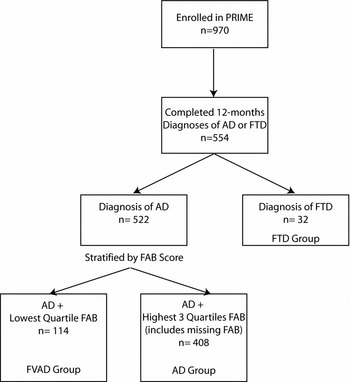
For this analysis, patients were grouped by diagnosis. The first group of patients consisted of those with a diagnosis of FTLD as assessed by the clinician, using the Lund-Manchester criteria (Lund and Manchester Groups, 1994). Patients with a diagnosis of AD were stratified by Frontal Assessment Battery (FAB) scores into two groups, with the quartile with the lowest FAB scores (i.e. reflecting greater executive dysfunction) nominated as the frontal variant Alzheimer's disease (FvAD) group, with the remainder called the AD group (Figure 1). Patients without a valid recorded baseline FAB score were included in the AD group. The data were also analyzed excluding those AD patients with missing FAB scores to determine the sensitivity of the findings to these AD cases with missing FAB scores.
Flowchart of patient selection, whole group, unmatched for severity.
PRIME = Prospective Research In MEmory clinics; AD = Alzheimer's disease; FTD = frontotemporal dementia; FAB = Frontal Assessment Battery; FVAD = frontal variant Alzheimer's disease.

To assess whether apparent differences between groups were reflecting differences in baseline severity, an additional analysis was conducted with the SMAF used as a marker of severity. Each FTLD subject was matched with four AD cases with the closest SMAF score, and these four cases were then removed from the pool of AD cases before the next FTLD case was matched. The AD cases were then arrayed on the basis of their FAB score, with the lowest quartile again nominated as the FvAD group. AD cases with missing FAB scores were excluded in this analysis.
Statistical analyses
All analyses were made pairwise between FTLD and FvAD, FvAD and AD and FTLD and AD groups respectively. Baseline demographics and assessments, six-month assessments and 12-month assessments, and the number of patients institutionalized at 12 months were all examined with Wilcoxon two sample tests or t-tests for continuous data and Fisher's exact test or χ2 for categorical data. Wilcoxon two sample tests were performed upon the individual items of FAB at baseline for the FTLD and FvAD groups. Continuous variables are presented as mean (± standard deviation) and categorical variables are presented as (n(%)) unless otherwise stated. No a priori adjustments were made for multiplicity and significance was defined at the level of 0.05.
Results
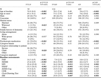
At the time of this analysis, 523 patients with a diagnosis of AD and 31 patients with FTLD had been enrolled in this study for at least 12 months. One subject with FTLD had no baseline SMAF and as the subject was excluded from the matched-severity analysis the subject was also excluded from the initial analyses. The baseline demographics and assessments for the three groups, unmatched for severity, are presented in Table 1 along with the results of the pairwise comparisons between groups. In stratifying AD patients by baseline FAB scores, the 25th percentile fell between scores of 10 and 11. All patients with scores of 10 or less were included in the FvAD group.
Baseline demographics and assessments

FTLD = Frontotemporal Lobar Degeneration; FvAD = Frontal Variant Alzheimer's disease; AD = Alzheimer's disease; FAB = Frontal Assessment Battery; MMSE = Mini-mental state Examination; NPI = Neuropsychiatric Inventory; SMAF = Functional Autonomy Measurement System; ADAS-Cog = Alzheimer's disease Assessment Scale, Cognitive subscale; CDR = Clinical Dementia Rating scale; ZBI = Zarit Burden Inventory.
The FvAD group had a similar age at both onset and baseline to the AD group but significantly differed from this group across all assessment scales, showing greater cognitive, functional, neuropsychiatric and global impairment and greater caregiver burden. The FTLD group more closely resembled the AD group than the FvAD group across all scales, differing significantly from the AD group in only the NPI and caregiver burden, where the mean scores of the FTLD group were in the direction of the FvAD group and away from that of the AD group.
Predictably, the FTLD patients were younger than both AD groups, and had a younger age at symptom onset. They did not resemble the FvAD group across most of the assessment scales, showing fewer frontal features (FAB), less cognitive and global impairment (MMSE, ADAS-Cog, Clock Drawing, CDR) and less functional impairment (SMAF). They did not significantly differ from the FvAD group in neuropsychiatric symptoms (NPI) but subjects with FTLD were associated with greater caregiver burden (Zarit) than subjects with FvAD and were more likely to be receiving psychotropic medication.
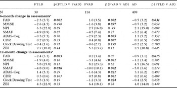
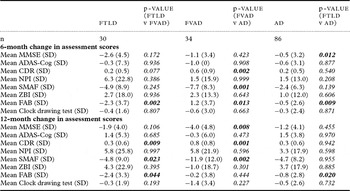
The changes in assessment scores and pairwise significance tests at six and 12 months are shown in Table 2 for each patient group. At six and 12 months, subjects with FTLD developed a significantly greater increase in frontal dysexecutive features than both the FvAD and AD groups, whose FAB scores remained relatively stable. There were also differences between the FTLD and FvAD groups at 12 months in the changes in the SMAF, where there was deterioration in both groups, but more so in the FvAD group. In all other scales the FTLD and FvAD groups showed similar changes at both six and 12 months. The FvAD group were found to deteriorate significantly more at six and 12 months than the AD group on most scales.
Change in assessment scores over 6 and 12 months from baseline

*Means and standard deviations calculated from available data, missing data ignored.
FTLD = Frontotemporal Lobar Degeneration; FvAD = Frontal variant Alzheimer's disease; AD = Alzheimer's disease; FAB = Frontal Assessment Battery; MMSE = Mini-mental state Examination; NPI = Neuropsychiatric Inventory; SMAF = Functional Autonomy Measurement System; ADAS-Cog = Alzheimer's disease Assessment Scale, Cognitive subscale; CDR = Clinical Dementia Rating scale; ZBI = Zarit Burden Inventory.
Apart from the FTLD group having a significantly greater decline in executive function at six and 12 months than the AD group, there were insignificant differences between these two groups at 12 months in the changes in the other scales.
Only a small number of patients were institutionalized and the proportions were similar in both the FTLD and FVAD patient groups at both six months (two patients, 7.4% versus 11 patients, 12.0%, p = 0.73) and 12 months (cumulatively three patients, 11.5% versus 13 patients, 16.9%, p = 0.75). Statistically more FvAD patients were institutionalized at both six (11 patients, 12.0% versus 19 patients, 5.2% p = 0.032) and 12 months (13 patients, 16.9% versus 24 patients, 7.3% p = 0.014) compared with the AD group.
The reanalysis of the data excluding those AD cases with missing FAB scores showed little change from the results when these were included. Very few p values for differences between the groups moved over the 0.05 boundary, suggesting the results were not sensitive to the inclusion or exclusion of these patients. The major effect was in the changes over 6 and 12 months (shown in Table 2) where the significant differences between the FvAD and AD groups were attenuated, with the FvAD group no longer deteriorating significantly more than the AD group in MMSE and CDR at 6 months and ADAS-Cog and Clock drawing at 12 months.
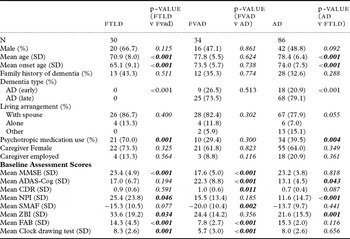
When we analyzed the data matching for baseline dementia functional severity the FvAD group differed substantially from the AD group across most assessment scales, with the exception of the NPI and the Zarit, and was now much more similar to FTLD on all assessment scales except the MMSE and clock drawing (Tables 3 and 4). As in the analysis not controlled for severity, the FAB score was lower for the FvAD group than for the FTLD group. The FvAD subjects differed from the FTLD group in being older and using fewer psychotropics but were similar to both the other groups on other baseline measures.
Baseline demographics and assessments, matched for severity

FTLD = Frontotemporal Lobar Degeneration; FvAD = Frontal variant Alzheimer's disease; AD = Alzheimer's disease; FAB = Frontal Assessment Battery; MMSE = Mini-mental state Examination; NPI = Neuropsychiatric Inventory; SMAF = Functional Autonomy Measurement System; ADAS-Cog = Alzheimer's disease Assessment Scale, Cognitive subscale; CDR = Clinical Dementia Rating scale; ZBI = Zarit Burden Inventory.
Change from baseline in assessment scores at 6 and 12 months, matched for severity at baseline

FTLD = Frontotemporal Lobar Degeneration; FvAD = Frontal variant Alzheimer's disease; AD = Alzheimer's disease; FAB = Frontal Assessment Battery; MMSE = Mini-mental state Examination; NPI = Neuropsychiatric Inventory; SMAF = Functional Autonomy Measurement System; ADAS-Cog = Alzheimer's disease Assessment Scale, Cognitive subscale; CDR = Clinical Dementia Rating scale; ZBI = Zarit Burden Inventory.
Over six and 12 months the matched FvAD group changed to a similar degree in most parameters as the other two groups except that these FvAD cases deteriorated more functionally and globally; they also deteriorated more cognitively than the AD group on the MMSE but not on the ADAS-Cog. The FTLD group was again found to deteriorate more on the FAB than the other two groups.
Discussion
Consistent with our hypothesis, we confirmed that a subset of AD patients with greater dysexecutive features differed substantially across a wide range of features from the rest of the subjects with AD. These subjects were more cognitively and functionally impaired, exhibited more behavioral symptoms and showed a greater decline on several measures over six and 12 months. Their greater neuropsychiatric morbidity was associated with greater caregiver burden. These differences were present despite the groups not differing significantly with respect to age, gender, marital status, family history of dementia, living arrangement, caregiver characteristics or use of psychotropic medication. The greater cognitive, functional and behavioral impairment was associated with a greater rate of institutionalization.
These FvAD subjects also differed substantially from the FTLD group in most domains, with the exception of neuropsychiatric symptoms. The FTLD group more closely resembled the AD group than those with FvAD in all domains except age of onset, neuropsychiatric symptoms, use of psychotropics and caregiver burden. This is a surprising finding of this study as it was hypothesized that the frontal features of the FvAD group would make them similar to the FTLD group. Rather than define a discrete non-frontal AD group that differs from both the FvAD and FTLD groups, we have separated out an FvAD group that differs substantially from both the other groups. What is perhaps most remarkable is that in using just one measure of impairment, a lower score on the FAB, we have separated off a group of AD patients who significantly differ from the rest of AD patients in all other domains that were measured by an impairment scale (cognitive, functional, behavioral/neuropsychiatric and global).
This finding is at variance with the similar Canadian study (Woodward et al., Reference Woodward, Jacova, Black, Kertesz, Mackenzie and Feldman2010b) that used a different frontal assessment scale – the FBI. In both studies the clinical features at enrolment were measured and utilized in the analyses. In the Canadian study the FTLD and the proposed FvAD groups were similar across several domains including function and neuropsychiatric symptoms but, as in this study they differed with respect to age of onset (younger for the FTLD group) and cognitive impairment (greater in the proposed FvAD group). On nearly all individual FBI items, the FTLD group and the FvAD group were very similar. In essence, the Canadian study showed that the most dissimilar of the three groups was the (non-frontal) AD group whereas we have demonstrated in this study that the most dissimilar group was the FvAD group.
A possible explanation for the varying findings from these two studies is that the FBI identifies frontal behavioral impairments whereas the FAB mostly identifies the cognitive dysexecutive features. As FTLD is frequently characterized by early behavioral changes, an FvAD subgroup defined by a scale measuring these behavioral changes could be expected to resemble the FTLD group. Our FTLD group was indeed more behaviorally disturbed than the other groups, as reflected in the higher NPI score, greater use of pychotropics and higher caregiver burden. Dysexecutive features are an early feature of AD but in FTLD these features often present later so an FvAD group defined by executive impairment could be expected to differ from the FTLD cases in earlier stages. In support of this, on the FAB our FTLD group declined far more at six and 12 months than the FvAD group and this greater degree of change was statistically significant at both time points. In essence, the FTLD group, which began with an almost identical FAB score to the (non frontal) AD group and indeed the whole AD group, was “catching up” with the FvAD group in their degree of executive dysfunction over the subsequent 12 months and leaving behind the AD group which was deteriorating far less in their FAB score. Thus, the concept that an FvAD group exists may not be disproven by our finding that this group did not initially resemble the FTLD group when FvAD is defined solely by executive dysfunction.
It does seem likely however that the FvAD groups defined by these two different approaches may not be similar groups. Further supporting this is the finding that the mean NPI scores of our FvAD group were lower than those of the FTLD group, suggesting less initial behavioral disturbance. In the Canadian study, the mean NPI scores of both the proposed FvAD group and the FTLD group were almost identical (29.8 and 29.0 respectively), and differed substantially from that of the other subjects with AD (8.3). Indeed, the mean NPI scores of the FTLD and FvAD groups in that study were higher than those of the FTLD subjects in our study (25.4) suggesting considerable behavioral disturbance in both groups in that study. Others have found that, for the same degree of dementia severity, subjects with FTLD have a greater degree of behavioral disturbance than those with AD (Mendez et al., Reference Mendez, Perryman, Miller and Cummings1998).
Is there any clinical value in separating off this proposed FvAD group using a measure of executive dysfunction, as we have done? These patients would appear to differ enough, at least initially, from the FTLD subjects not to pose difficulties in the differential diagnosis between AD and FTLD. They were more impaired across most measures, older and less likely to be on psychotropic medications. Indeed, they seem far easier to differentiate from subjects with FTLD than do those AD cases with prominent early behavioral features (Woodward et al., Reference Woodward, Jacova, Black, Kertesz, Mackenzie and Feldman2010b). Our findings suggest that where the differential diagnosis of AD and FTLD is being considered, prominent executive dysfunction in the absence of prominent behavioral impairments supports a diagnosis of AD, not FTLD. In essence, the early features of some cases of AD overlap with features of subjects with FTLD more in the behavioral domain than in the dysexecutive domain.
Our operationalization of FvAD may simply have led to the selection of subjects with more advanced AD. As AD progresses the characteristic neuropathology becomes more widespread, and can involve the frontal lobes (Braak and Braak Reference Braak and Braak1991). This could be expected to lead to more apparent frontal symptomatology. Our FvAD group had more impairment than the (remaining) AD group on all assessment scales but were not older at either entry into the study or age at onset of symptoms.
Our further analyses, where the AD and FTLD cases were matched for baseline severity, as measured by the SMAF, suggest that baseline severity may have affected the other characteristics of the two AD groups. Both groups are now marginally less affected on almost all parameters, suggesting the matching process eliminated mainly more severely affected cases which had been included before the matching. The biggest effect of this matching process however is that it created an FvAD group that look more like the FTLD than the AD group, differing less from the FTLD in most baseline assessment scale scores – insignificantly on many parameters when, post hoc, the p-values were adjusted for multiple comparisons. The FTLD and FvAD groups do differ in the degree of executive dysfunction and in two of the cognitive scales, the MMSE and the Clock-drawing score, the latter also reflecting executive function, but they did not differ on the ADAS-Cog. The FvAD group are still significantly more functionally impaired than the AD group. Their greater cognitive and functional impairment than both the other groups is interesting and suggests that cognitive, functional and executive impairment in the AD group may be linked. Thus, selecting a group that is more impaired on any of the scales will likely lead to the selection of a more severely impaired group of AD patients even when most severely impaired AD cases are excluded from the selection pool. The 6- and 12-month change data still show the FvAD group deteriorating more functionally and globally than the AD group, and the FTLD group deteriorating more on the FAB than both other groups- again supporting a “catch up” in the degree of executive dysfunction in a group that initially and surprisingly showed only as much executive dysfunction as the AD group.
On balance, our findings seem to indicate a greater disease burden in the FvAD group but they are also consistent with the FvAD group having a distinct clinical presentation characterized by prominent executive dysfunction but no difference in disease duration or age of onset to the other AD cases. The further severity-matched analysis has not changed this essential difference between the FvAD and the AD cases, or the similarity between the AD and FTLD groups, but has created an FvAD group that now looks more like FTLD and, as in the other study (Woodward et al., Reference Woodward, Jacova, Black, Kertesz, Mackenzie and Feldman2010b) still raises the possibility that there may be a frontal variant of AD that looks more like FTLD than AD.
We concede that there are limitations to our findings. There could have been selection bias in the administration of the FAB within the study as the protocol did not require all subjects to have all scales administered at all visits. We did identify 139 subjects who did not have the FAB administered at baseline. There is also clearly circularity in selecting FvAD cases according to a frontal dysexecutive score and then characterizing FvAD thus defined on this and other clinical measures. There is, however, justification for examining subjects at the statistical margins of any disease, rather than just the “average” patient. The circularity, however, and a limited availability of information around the presenting complaints of our FvAD cases and their longitudinal change prior to entry into our study limit the clinical validity of our observations. Additionally, we relied on clinical diagnosis to assign diagnostic categories, which may well have been incorrect in some cases. To somewhat offset this risk, the clinicians were working in centers of excellence with access to neuroimaging and other assessments, which have been shown to produce a diagnostic accuracy, when correlated with autopsy data, of around 90% (Lopez et al., Reference Lopez2000). Penultimately, even matching the AD cases to the FTLD cases for baseline functional severity and then differentiating a subgroup with the lowest FAB scores made it difficult to differentiate the effects of severity from type as we again separated the most severely dementing AD cases. Lastly, there was no neuropathological confirmation of the clinical diagnoses presented in this study.
Notwithstanding these limitations, our data are consistent with the existence of a subgroup of AD patients with greater executive dysfunction who are probably a more severely affected group of those with AD and who do not initially have a strong clinical overlap with the subjects with FTLD, who had a greater degree of behavioral disturbance. It is only when baseline severity is matched that this FvAD group resembles the FTLD group, and such matching is impractical in the clinical setting. The study does encourage future prospective clinical studies aimed at the further characterization of FvAD, however defined, and investigations into the neuropathological basis of this proposed variant of AD. Clinicians should be cautious when assigning patients with dementia and behavioral or executive dysfunction to a diagnosis of either FTLD or AD. Clinical differentiation can be difficult and the future may lie in combining clinical assessment with other investigations such as biomarkers.
Conflict of interest declaration
The first four authors have all been members of advisory boards funded by the companies marketing galantamine, donepezil and rivastigmine. They have been members of the PRIME database Scientific Advisory Committee (SAC), funded by Janssen-Cilag Pty Ltd Australia, and were funded for this activity. Greg Blanch is an employee of Janssen-Cilag Pty Ltd Australia and Robert Balshaw is an employee of Syreon Corporation, Canada, which processed the data and was paid by Janssen-Cilag Pty Ltd Australia to do this.
Because Professor Ames is also Editor-in-Chief of International Psychogeriatrics, the peer review process was handled entirely by one of the Journal's Deputy Editors.
Description of authors' roles
Michael Woodward designed the concept, directed the statistical analyses and wrote the majority of the paper. Henry Brodaty, Karyn Boundy and David Ames contributed to the interpretation of the data and the final version of the paper. Greg Blanch contributed to the final draft of the paper but did not direct content. Robert Balshaw and Syreon Corporation assisted with statistical analyses, as directed by the Scientific Advisory Committee.
Acknowledgments
We thank Andrea Malcolm (Janssen-Cilag Australia Pty Ltd) for assistance during the preparation of this paper and Craig Batty for project management assistance with the PRIME data base (through Janssen-Cilag Australia Pty Ltd).