Absent pulmonary valve syndrome is a rare cardiac malformation, described for the first time by Chevers in 1847, characterised by a dysplastic or rudimentary pulmonary valve associated with varying degrees of hypoplasia of the pulmonary annulus.Reference Chevers 1 – Reference Rao and Lawrie 3 Owing to the concomitant occurrence of valve stenosis and regurgitation, absent pulmonary valve syndrome is almost always associated with massive enlargement of the pulmonary trunk and its branches.Reference Emmanoulides and Gutgesell 4 , Reference Jacobs 5 Ductus arteriosus agenesis is a common but not consistent feature of the syndrome.Reference Momma, Kondo and Matsuoka 6 Absent pulmonary valve syndrome can be isolated but is usually associated with tetralogy of Fallot.Reference Razavi, Sharland and Simpson 7 Even though tetralogy of Fallot is the most frequent cyanotic heart defect (0.26–0.48/1000 live births), tetralogy of Fallot with absent pulmonary valve syndrome only represents 2.5% of all patients with tetralogy of Fallot.Reference Zucker, Rozin, Levitas and Zalzstein 8
The perinatal mortality rate of tetralogy of Fallot with absent pulmonary valve syndrome is above 60%, closely related to the existence of severe heart failure, respiratory distress, and/or associated malformations.Reference Razavi, Sharland and Simpson 7 , Reference Becker, Schmitz, Guschmann, Wegner, Stiemer and Entezami 9 Even though fetal echocardiography is the best diagnostic tool for diagnosing cardiac malformations and for quantifying pulmonary artery dilatation and vascular compromise, this technique is still not able to adequately predict the postnatal outcome of the affected fetuses and particularly to specify the degree of respiratory compromise.Reference Wertaschnigg, Jaeggi and Chitayat 10 To the best of our knowledge, abnormal fetal lung appearance has only been reported twice in fetuses with tetralogy of Fallot with absent pulmonary valve.Reference Wertaschnigg, Jaeggi and Chitayat 10 , Reference Chelliah, Berger, Blask and Donofrio 11 In both cases, the fetus presented with an enlarged lung displacing the mediastinum, associated with a small contralateral lung. Dilated pulmonary arteries were presumed to compress the main bronchi leading to fetal lung trapping and lung overexpansion.Reference Wertaschnigg, Jaeggi and Chitayat 10 , Reference Chelliah, Berger, Blask and Donofrio 11 The aims of our study were to analyse the prenatal echocardiographic lung appearance in tetralogy of Fallot with absent pulmonary valve syndrome, to correlate the prenatal and postnatal cardiopulmonary findings, and to assess the outcome.
Materials and methods
The present study was approved by the ethics committee of our institution.
Patient population
This was a retrospective study performed at a tertiary referral centre for prenatal diagnosis of fetal pathology between July, 2002 and January, 2014. A search in the database of the paediatric cardiology department showed nine cases of tetralogy of Fallot with absent pulmonary valve syndrome diagnosed prenatally. The prenatal diagnosis was made on the basis of rudimentary valve leaflets in combination with the concurrent occurrence of pulmonary stenosis and regurgitation, ventricular septal defect, overriding ascending aorta, and enlargement of central pulmonary arteries.
All patients with tetralogy of Fallot with absent pulmonary valve syndrome having at least one prenatal ultrasound performed in the medical imaging department and another one in the cardiology department of our institution were included.
Data analysis
Prenatal data
The following variables were assessed: indication for referral, gestational age at diagnosis (weeks of gestation), associated cardiac, extracardiac, or chromosomal abnormalities, pregnancy, and fetal/neonatal outcome.
Fetal echographic evaluations were performed in the medical imaging department on Toshiba Aplio XG or Toshiba Aplio 500 (Toshiba America Medical Systems, Tustin, California, United States of America). The following parameters were evaluated: gestational age, abnormalities of lung parenchymal echotexture and location, visualisation of pulmonary inferior lobar artery in the four-chamber view, presence of intrauterine growth restriction, and amniotic fluid volume disorder. As normal fetal lungs appear homogeneous at ultrasound, lungs echogenicity was considered abnormal if definitely heterogeneous without any possible confounding artefacts. The pulmonary inferior lobar artery should normally not be seen in the fetus in the four-chamber view, and this parameter is considered abnormal if visualised. The echographic examinations were reviewed independently by two radiologists with over 10 years of experience and one fellow.
Cardiac fetal ultrasound examinations were performed at the Paediatric Cardiology Echocardiography Unit with a Philips Sonons 5500 (Philips Healthcare, Andover, Massachusetts, United States of America) with a 4- or 6-MHz transducer. Fetal echocardiographic evaluation included the cardiac axis, cardiothoracic circumference ratio, side of the aortic arch, diameter of the main pulmonary artery, and the ascending aorta, measured just above the valve annulus in mm, diameter of the pulmonary arterial branches, measured immediately after their origin in mm, presence or absence of the ductus arteriosus, and calculation of main pulmonary artery/ascending aorta ratio. Vascular dimensions were measured from two-dimensional recordings with an insonation perpendicular to the long axis of the vessel and following the trailing-to-leading edge method. The cardiac axis was defined as abnormal if the angle was >60° for leftward deviation and <20° for rightward deviation. Cardiomegaly was defined as cardiothoracic circumference ratio more than 50%, and was considered as mild if the cardiothoracic ratio was between 50% and 70% and moderate if the cardiothoracic ratio was more than 70%. All the measurements were compared with established normal sizes (z-score). All echocardiographic parameters were reviewed and analysed by a paediatric cardiologist with over 10 years of experience.
Postnatal data
Postnatally, the following clinical variables were assessed: APGAR score at 1, 5, and 10 minutes, gestational age at delivery, weight at delivery, length of perinatal hospitalisation, need for preoperative ventilatory support, age at surgical correction, preoperative thoracic CT imaging, and outcome. Postnatal spiral chest CTs were performed on a GE Light Speed VCT CT (GE Healthcare, Milwaukee, Wisconsin, United States of America) after administration of intravenous contrast agent at a dose of 2 ml/kg of body weight. Parameters used were set at 80 kV and 90–200 mAs. Multiplanar reconstructions were obtained. We looked for air trapping and/or atelectasis and their location, abnormal vascular pulmonary branching, and for tracheo-bronchial compression. According to Rabinovitch et alReference Rabinovitch, Grady and David 12 and Milanesi et al,Reference Milanesi, Talenti, Pellegrino and Thiene 13 abnormal pulmonary branching was defined as tufts of vessels emerging at the segmental artery level. Significant tracheo-bronchial compression was defined as a reduction in the diameter of the airway of more than 50%. The main pulmonary artery/ascending aorta ratio was also measured with a similar method to fetal echocardiography. Cardiothoracic circumference ratio was calculated for comparison with fetal data.
All the data were retrieved from the clinical files of the patients and/or from recorded videotapes and stored images. When discordance was noted, a consensus reading was obtained.
Statistical analysis
No statistical analysis was performed due to the small size of the population studied.
Results
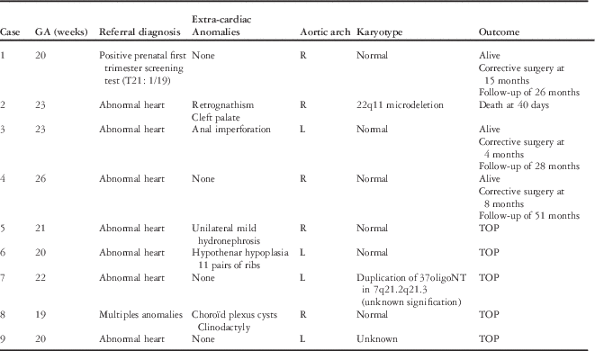
A total of nine patients who met inclusion criteria were included in the present study (Table 1).
Study subjects with tetralogy of Fallot and absent pulmonary valve.

GA=gestational age; L=left; R=right; TOP=termination of pregnancy
All fetal cardiac malformations were detected between 19 and 26 weeks of gestation. Seven cases were referred to our institution for abnormal second trimester ultrasound screening, one case for positive prenatal first trimester screening, and one case after detection of multiple extracardiac anomalies. No fetus presented with intrauterine growth restriction and/or amniotic fluid disorder. Among those nine cases, five pregnancies had been terminated, and four pregnancies were full term (Table 1). Karyotype analysis was performed in eight patients. Results were normal in six cases, and two patients had chromosomal anomalies, including one proven 22q microdeletion.
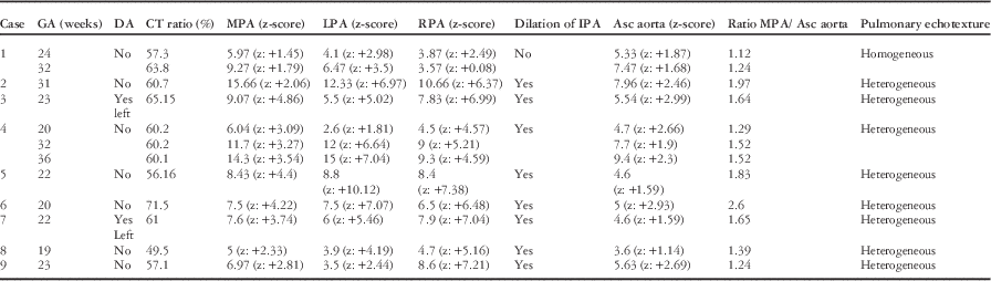
The prenatal cardiothoracic data for the nine subjects are summarised in Table 2.
Prenatal cardiothoracic data by echocardiography.

Asc aorta=ascending aorta; CT ratio=cardiothoracic ratio; DA=ductus arteriosus; GA=gestational age; IPA=inferior pulmonary artery; LPA=left pulmonary artery; MPA=main pulmonary artery; RPA=right pulmonary artery
Leftward deviation of the cardiac axis was always present. The ductus arteriosus was absent in seven fetuses; eight fetuses had an elevated cardiothoracic ratio. Cardiomegaly was mild except in one case where it was moderate (Case 6). The pulmonary trunk/ascending aorta ratio varied from 1.24 to 2.6 in the group of interrupted pregnancies and from 1.12 to 1.97 for fetuses who survived until birth. Among the four born-alive neonates, one with an antenatal ratio of 1.97 died after 40 days. The three surviving newborns had an antenatal ratio between 1.12 and 1.64; one terminated fetus (Case 9) did not have a highly enlarged ratio, but its left and right pulmonary artery branches were massively dilated. In two fetuses (Cases 1 and 4), more than one ultrasound were available. In both cases, the ratio between the pulmonary trunk and the ascending aorta did not change significantly during pregnancy.
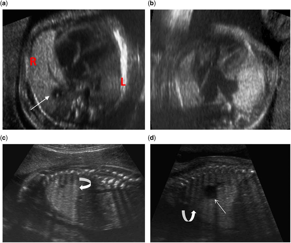
All but one fetus (Case 1) (88.9%) presented with a heterogeneous ultrasound appearance of the lung parenchyma, not related to ultrasound artefacts or fetal presentation. In these eight patients, the dilated lobar pulmonary arteries were also visible on the four-chamber view (Fig 1). The only fetus (Case 1) with normal homogeneous lung ultrasound appearance had no visible dilated segmental pulmonary artery on the four-chamber view.
( a – d ) Case no. 7, a 22-week of gestational age fetus. ( a ) Four-chamber view demonstrating a leftward deviated cardiac axis, dilatation of the right inferior pulmonary artery (white arrow), and hyperechoic lung (R) compared with the left lung (L). ( b ) Three-vessel view showing important dilatation of pulmonary artery branches and hyperechogenic left lung. ( c and d ) Abnormal heterogeneity of the fetal lung (curved arrows) and dilatation of the inferior pulmonary artery (white arrow) are demonstrated in parasagittal views.

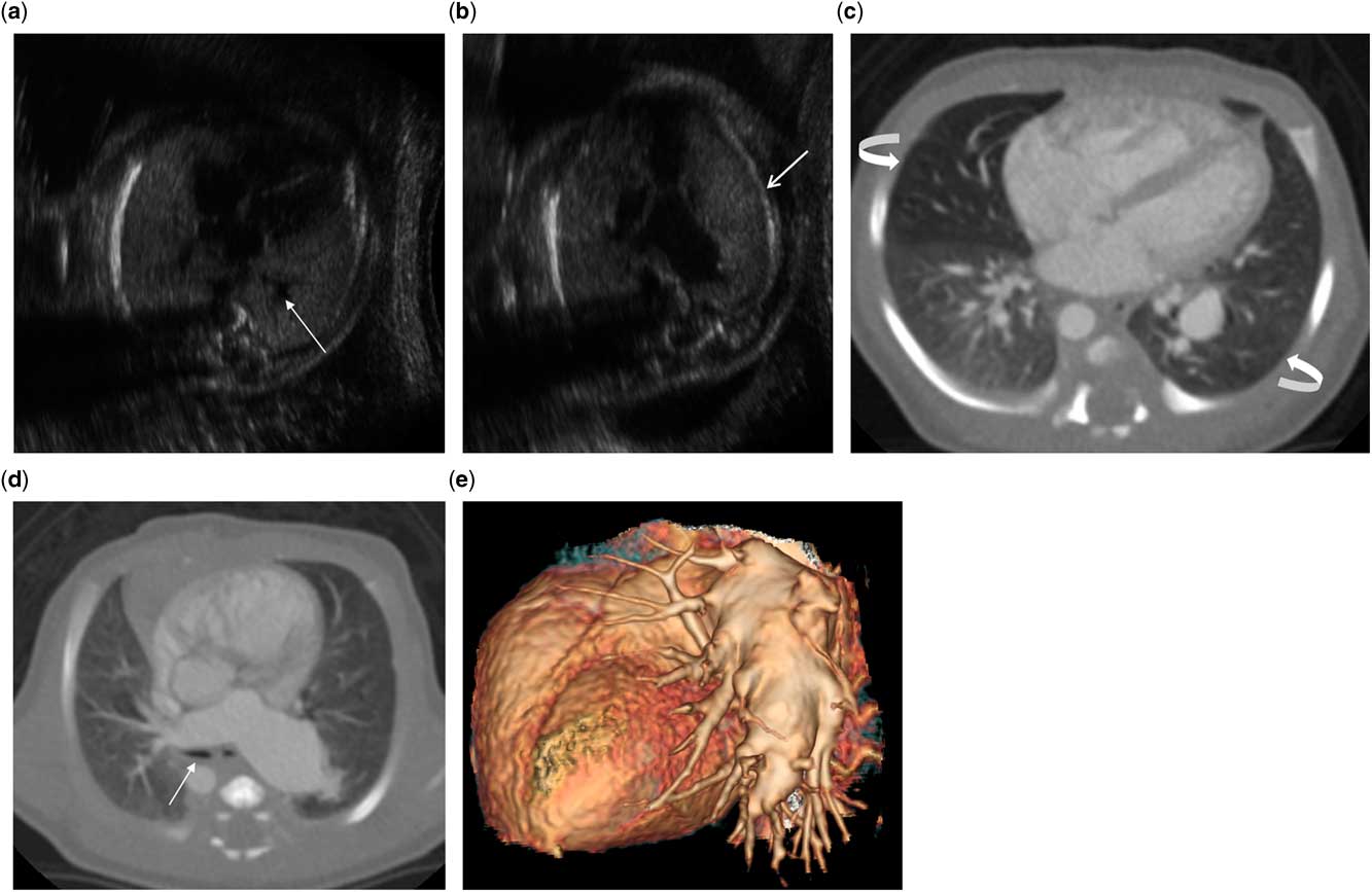
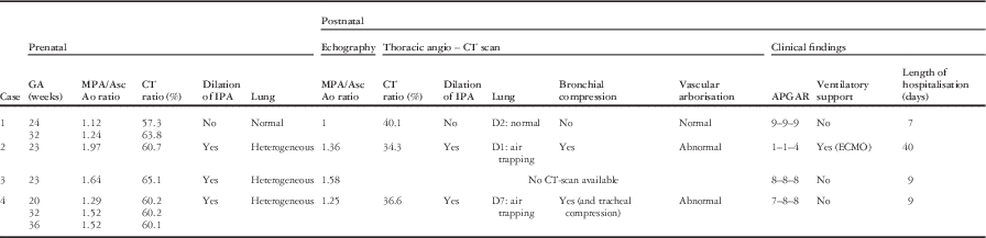
Correlation between prenatal and postnatal data of the four neonates is shown in Table 3. For three of them, a postnatal CT was performed for preoperative assessment of the cardiac malformation and/or respiratory symptoms (Patients 1, 2, and 4). In patient 1, there was no detectable anomaly of the lung parenchyma on prenatal ultrasound images, and the chest CT performed at day 2 did not show any parenchymal anomaly. For the two other cases, the postnatal appearance of the lung parenchyma correlated well with its prenatal aspect. In Case 2, at 23 weeks of gestation, the fetus presented with an abnormal echogenicity of both lungs that corresponded to air trapping on the postnatal CT performed at day 1. In Case 4, there were visible anomalies of the left lung at 20 weeks of gestation. The postnatal CT performed at day 7 demonstrated air trapping in the left inferior lobe (Fig 2). In Cases 2 and 4, an abnormal branching pattern of the pulmonary vessels was visible on postnatal CT.
( a – e ) Case no. 4 ( a ), a 20-week of gestational age fetus. ( a ) Four-chamber view demonstrating a leftward deviated cardiac axis and dilatation of the left inferior pulmonary artery (white arrow). ( b ) Three-vessel view showing important dilatation of pulmonary artery branches mainly the left one and left lung parenchymal heterogeneity (white arrow). Postnatal thoracic CT angiography (CTA) at 7 days. ( c ) These transverse views show air trapping of the left lung and middle pulmonary lobe, as demonstrated by parenchymal hypodensity associated with reduced vessel size (curved arrows), and ( d ) demonstrates important dilatation of the pulmonary arterial branches with compression of the bronchi (white arrow). ( e ) Sagittal view of a volume-rendering CTA image showing the dilated pulmonary arteries and the abnormal pulmonary branching at the segmental level.

Pre and postnatal correlation.

CT ratio=cardiothoracic ratio; ECMO=extracorporeal membrane oxygenation; GA=gestational age; IPA=inferior pulmonary artery; MPA/Asc Ao ratio=main pulmonary artery/ascending aorta ratio
Discussion
Unlike classical forms of tetralogy of Fallot, with a tendency to have hypoplastic pulmonary arteries, tetralogy of Fallot with absent pulmonary valve is characterised by major expansion of pulmonary arteries. The outcome depends not only on the cardiac malformation but also importantly on the presence of respiratory distress secondary to bronchial compression by the dilated lung arteries. Outcome is still poor in cases with respiratory distress with a survival rate of 17%.Reference Galindo, Gutiérrez-Larraya and Martinez 14 For this reason, it is mandatory to find prenatal criteria for the severity of bronchial compression. This could help in providing wiser prenatal counselling and to anticipate the need for ventilatory support immediately after birth.
Postnatally, airway obstruction in children with CHD has already been described, but the most common abnormality is extrinsic compression of the lower airway by dilated pulmonary arteries or repaired aortic arch.Reference Lee, Cheung, Leung, Ng and Tsoi 15 , Reference An, Choi and Kwon 16 The typical clinical presentation of patients with tetralogy of Fallot with absent pulmonary valve syndrome is severe respiratory distress related to bronchial compression by dilated pulmonary arteries.Reference Driscoll, Shaddy and Feltes 17
In tetralogy of Fallot with absent pulmonary valve, the dilatation of the pulmonary arteries is not limited to the pulmonary trunk or the lobar level. Indeed, several detailed histological studies have described an abnormal branching pattern of the distal pulmonary arteries.Reference Rabinovitch, Grady and David 12 – Reference Yeager, Van Der Velde, Waters and Sanders 18 Rabinovitch et al have noted that the number of arteries is increased at the segmental level.Reference Rabinovitch, Grady and David 12 , Reference Milanesi, Talenti, Pellegrino and Thiene 13 More distally, arteries are dilated, entwining and compressing the structurally normal adjacent bronchi. Instead of a single, segmental artery, tufts of vessels are present, resembling the branching pattern of a weeping willow instead of the usual dichotomous birch-tree pattern. The relationship between the dilatation of the distal vascular tree and bronchomalacia can be explained by this particular branching pattern. Volpe et al. showed in his prenatal series of 21 cases of tetralogy of Fallot with absent pulmonary valve that bronchomalacia was highly probable when cardiomegaly and marked branch pulmonary dilatation were present.Reference Volpe, Paladini and Marasini 19 In fact, this pattern of abnormal branching was visible postnatally in the CT scans performed in our series (Cases 2 and 4).
It is interesting to note that the major dilatations involve the left and right pulmonary branches even more than the pulmonary trunk. Indeed, in our series, the z-score of the main pulmonary artery varied between +1.45 and +4.86, wherease it varied between +2.6 and +10.12 for the left and right branches (Fig 2). In Case 9, the left and right branches reached z-scores of +7.21 and +2.44, respectively. It seems logical to assume that the more peripheral the vascular tree dilatation, the greater the effect on the pulmonary parenchyma. This was observed in our series, with eight fetuses (88.9%) already having significant dilatation of the pulmonary arteries, as detected by the visualisation of a dilated pulmonary artery on the four-chamber view. These eight fetuses also had abnormal lung echogenicity that could be secondary to bronchial compression by dilated pulmonary arteries.
After birth, the appearance of the lung parenchyma in tetralogy of Fallot with absent pulmonary valve includes air trapping due to a “ball-valve” effect in the segments corresponding to the compressed bronchi. This hyperinflation can even result in a “congenital lobar hyperinflation” with amniotic fluid retention in the first few hours of life.Reference Fink, Edis and Massie 20 On the other hand, it is also recognised that lung segments or lobes with air-trapping appear overinflated and hyperechogenic on prenatal ultrasound, as documented in congenital lobar emphysema, and congenital high airway obstruction syndrome – for instance, the radiological presentation of congenital lobar emphysema has been well depicted by several authors both prenatally and postnatally.Reference Beydon, Larroquet and Coulomb 21 – Reference Pariente, Aviram, Landau and Hershkovitz 23 The affected zones are hyperechogenic on the second-trimester fetal ultrasound and then hyperlucent on postnatal CT scan. By analogy, it seems reasonable to think that lung regions of air trapping visible after birth could already be identified during fetal life. Indeed, in two of our three cases for which a postnatal CT scan was available, this hypothesis was confirmed.
In our series, all fetuses with significantly dilated left and/or right pulmonary arteries also had abnormal heterogeneous echogenicity of the lung parenchyma. The only fetus with normal homogeneous lung echogenicity had had a lower degree of arterial dilatation and a relatively good outcome. Thus, the observation of abnormal heterogeneous lung echogenicity in fetuses with tetralogy of Fallot with absent pulmonary valve confirms the impact of vascular dilatation on the pulmonary parenchyma even before birth; and this may have prognostic implications for the prenatal counselling.
In our series, the full-term fetuses appeared to have a lower pulmonary trunk/aorta ratio compared with interrupted cases. This can be explained by the fact that an interruption of pregnancy was “proposed” when severe dilatation and/or when abnormal lung echogenicity were detected at the time of the ultrasound. It is, however, interesting to note that the only case (Case 2) that was carried to full term and did not survive the perinatal period had a pulmonary trunk/aorta ratio similar to the fetuses that were interrupted. It is thus probable that a higher ratio increases the risk of pulmonary complications.
In our study, the ratio between the pulmonary trunk and the aorta did not seem to vary significantly throughout pregnancy, even though an early diagnosis was made. This observation is in agreement with the absence of significant change in age-adjusted pulmonary arterial z-scores observed in a previous study.Reference Wertaschnigg, Jaeggi and Chitayat 10 This measure could thus help in determining the risk of airway compression of the affected fetus early in pregnancy, which is meaningful for early pregnancy counselling.
Finally, for Volpe et al., cardiomegaly is one of the rare proved prognostic factors available until now in tetralogy of Fallot with absent pulmonary valve syndrome detected in utero. In our series, cardiomegaly seemed not to play an important prognostic role.Reference Volpe, Paladini and Marasini 19
This study has some limitations. Owing to its retrospective nature, evaluation of the lung parenchyma on prenatal ultrasound was possible only on the available images. It was not possible to determine precisely the affected lobe. It was difficult to make a precise correlation of the affected lobes prenatally and postnatally. The number of cases was limited, owing to the rarity of tetralogy of Fallot with absent pulmonary valve syndrome. Finally, postnatal correlations were limited to the four fetuses that were not interrupted.
In conclusion, our series suggests that detection of abnormal lung echogenicity in a fetus with tetralogy of Fallot with absent pulmonary valve already implies significant bronchial compression secondary to pulmonary arterial dilatation. This pattern can be observed early during pregnancy. Its detection may have poor prognostic implications.
Acknowledgements
None.
Financial Support
This research received no specific grant from any funding agency or from commercial or not-for-profit sectors.
Conflicts of Interest
None.
Ethical Standards
The authors assert that all procedures contributing to this study comply with the ethical standards of the relevant national guidelines on human experimentation (Ethical Conduct for Research Involving Humans – Health Canada) and with the Helsinki Declaration of 1975, as revised in 2008, and has been approved by the institutional committee (Comité d’éthique de la recherche – CHU Sainte-Justine approved on 22 May, 2014).