Introduction
For anatomical reasons, the anterior laryngeal midline is the commonest route of carcinomatous invasion of the laryngeal framework.Reference Micheau, Luboinsky, Lanchi and Cachin1, Reference Rifai and Khattab2 The oncological demarcation between the anterior glottis and the supraglottis is called the ‘X-space’.Reference Bagatella and Bignardi3 The X-space is situated in the anterior commissure, covering the gap between the thyroid cartilage and the laryngeal mucosa. It is composed of dense fibrous tissue, which (controversially) offers resistance to the spread of cancer but, at the same time, represents a route along which cancer can spread.Reference Bagatella and Bignardi3
When the tumour extends beyond the X-space, there are no barriers to submucosal cancer spread.Reference Rifai and Khattab2, Reference Bagatella and Bignardi3 Furthermore, transthyroid carcinomatous invasion is also facilitated by: the close proximity between the vocal fold mucosa and the laryngeal framework (only 2–3 mm); the absence of inner perichondrium on the thyroid cartilage in this area; and the capability of subglottic tumours to penetrate through the cricothyroid membrane (the weak point in the laryngeal framework).Reference Rifai and Khattab2
Carcinomatous transgression of the thyroid cartilage can lead to an initial presentation of laryngeal carcinoma as a pre-thyroid neck mass, in the form of: an abscess,Reference Op de Beeck, Hermans, Delaere, Van den Bogaert and Marchal4 a thyroid goitre,Reference Donangelo, Coelho, Buescu and Vaisman5 and, as in our case, a prelaryngeal pseudolaryngocoele.
We present an unreported case of a man with laryngeal carcinoma who initially presented with a large, prelaryngeal, air-filled cavity. The formation of this pseudolaryngocoele was due to carcinomatous erosion of the thyroid cartilage, allowing air to escape from the larynx and be retained in the prelaryngeal soft tissues by a single-ball valve effect. As the air-filled sac was not a dilatation of the laryngeal saccule, we describe this abnormality as a pseudolaryngocoele.
Case report
A 47-year-old male smoker was referred to our ENT department with a two-month history of progressive, midline neck swelling with an associated mild dyspnoea and husky voice.
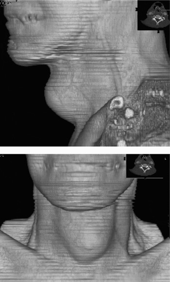
On examination, the patient had a 6 × 4 cm, soft, painless, irreducible, midline neck mass attached to the thyroid cartilage, which moved on deglutition (Figure 1). He also had a 2 cm, fixed, right-sided, level II lymph node. Flexible endoscopic examination of the larynx showed an exophytic glottic lesion involving the anterior commissure, extending to the anterior third of the right false vocal fold. Attempted fine needle aspiration of the anterior neck lesion demonstrated an air-containing structure, which temporarily collapsed but regained its original size within five minutes.
Volume-rendering computed tomography scan demonstrating the initial clinical presentation with a large, anterior neck mass.

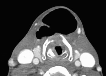
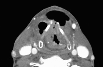
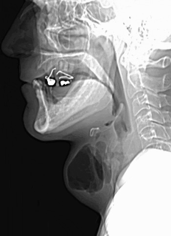
An urgent computed tomography (CT) scan of the neck was reported as showing a 1.5 cm, anterior glottic/supraglottic mass which eroded through the thyroid cartilage, with signs suggesting a fistula connecting the larynx with a 7 cm, air–fluid collection ventral to the cartilages (Figures 2 and 3). Multiple, air-containing dilatations were also found on the larynx, compatible with a diagnosis of other fistulous tracts internal to the thyroid cartilage (Figure 4). Associated lymphadenopathy was also found on the right at levels II and III.
Axial computed tomography scan showing a mass eroding the thyroid cartilage and an anterior, air-filled lesion (pseudolaryngocoele).

Axial computed tomography scan showing a laryngeal mass and gas collections anterior to the thyroid cartilage, with a fistulous tract in the anterior commissure.

Plain, lateral, soft tissue X-ray of the neck, showing an air–fluid level inside a large, cavitated structure anterior to the laryngeal cartilages, with additional gas collections.

Rigid endoscopic examination confirmed the presence of an anterior transglottic lesion involving the anterior commissure and extending to the laryngeal ventricles. The lesion was debulked with CO2 laser, and biopsy specimens were sent for analysis. Histological analysis reported an invasive, moderately differentiated squamous cell carcinoma, which was graded at T4a N2a M0 (stage IVa; tumour–node–metastasis grading).
The multidisciplinary team reached a consensus agreement to pursue a curative approach, which consisted of radical surgery followed by post-operative chemo-radiotherapy. The patient underwent total laryngectomy with modified right neck dissection and elliptical skin excision around the needle puncture site. The surgical specimen had a transthyroid cartilage sinus originating at the level of the laryngeal tumour in the anterior part of the glottis. The anterior cervical pseudolaryngocoele was dissected subcutaneously and excised in conjunction with the specimen.
Histological examination of the larynx confirmed the diagnosis of squamous cell carcinoma. However, due to previous laser surgery, it was not possible to identify tumour at the level of the entry of the laryngeal sinus.
Four weeks after surgery, the patient underwent post-operative radiotherapy. Twelve months after treatment, the patient remained free of tumour recurrence.
Discussion
Virchow first described a laryngocoele in 1867.Reference Close, Merkel, Burns, Deaton and Schaefer6 It is defined as an abnormal dilatation of the saccule of the laryngeal ventricle, either through the thyro-hyoid membrane (external), into the vellecula (internal), or as a mixed type.Reference Close, Merkel, Burns, Deaton and Schaefer6–Reference Akbas, Unal and Pata8 Laryngocoeles can be further divided into congenital (enlargement of the saccule) or acquired (by increased, sustained intralaryngeal pressure).Reference Akbas, Unal and Pata8
There is a well documented association between laryngocoeles and carcinoma of the larynx.Reference Close, Merkel, Burns, Deaton and Schaefer6, Reference Celin, Johnson, Curtin and Barnes7 The reported incidence of laryngocoele with squamous cell carcinoma varies between 4.9 and 28.8 per cent.Reference Celin, Johnson, Curtin and Barnes7 The incidence of occult laryngeal carcinoma in excised laryngocoeles is reported as 4.6 per cent.Reference Celin, Johnson, Curtin and Barnes7 On the other hand, undiagnosed laryngocoeles have been found in 18 per cent of laryngectomies.Reference Micheau, Luboinsky, Lanchi and Cachin1
Retrospective studies of the laryngeal CT scans of patients without symptoms of laryngocoele have demonstrated an incidence of occult laryngocoele of 12.5 per cent (62.7 per cent were internal, 5.9 per cent external, 11.8 per cent mixed and 19.6 per cent diagnosed as saccular dilatations). Post-scan biopsies performed on those patients have shown that 28.8 per cent were associated with laryngeal cancer and only 8.6 per cent were unrelated.Reference Close, Merkel, Burns, Deaton and Schaefer6
In the detection of laryngocoele, CT scanning is more sensitive than clinical examination and direct laryngoscopy.Reference Close, Merkel, Burns, Deaton and Schaefer6 Despite this, cancer-related laryngocoeles are infrequently detected during clinical examination and radiological investigation; they are usually only identified post-operatively upon histological examination of the larynx. These discrepancies between high incidence and low rates of pre-operative detection are explained by the small size of these lesions and their common intra-thyroid location.Reference Micheau, Luboinsky, Lanchi and Cachin1, Reference Close, Merkel, Burns, Deaton and Schaefer6, Reference Celin, Johnson, Curtin and Barnes7
There is still controversy regarding the pathophysiological relationship between laryngocoele and laryngeal cancer.Reference Celin, Johnson, Curtin and Barnes7, Reference Akbas, Unal and Pata8 Some authors suggest that laryngocoeles occur secondary to tumours arising from the lining of the saccule.Reference Celin, Johnson, Curtin and Barnes7–Reference Cassano, Lombardo, Marchese-Ragona and Pastore9 Other theories describe intra-laryngocoele dysplasia as the origin of laryngeal cancer.Reference Cassano, Lombardo, Marchese-Ragona and Pastore9 A more accepted theory suggests that the partial airway obstruction produced by a laryngeal tumour increases the intraluminal pressure above the lesion, and the combined carcinoma and inflammatory tissue mass acts as a single, valve-like mechanism, leading to the formation of an air-sac containing structure.Reference Celin, Johnson, Curtin and Barnes7–Reference Cassano, Lombardo, Marchese-Ragona and Pastore9 As the air-filled sac is not a dilatation of the laryngeal saccule, it cannot be considered as a true laryngocoele; hence, it is referred to as a pseudolaryngocoele.
In the present case, the erosion of the tumour through the thyroid cartilage created a potential weak area, facilitating the insufflation of air into laryngeal and pre-thyroid soft tissue and consequent formation of a pseudolaryngocoele. However, the creation of a persistent air-filled structure can only be explained in the presence of a ball-valve system; in our case, this is likely to have been produced by the carcinoma or the related inflammatory changes. The unlikeliness of the above sequence may be the reason why this unique presentation of laryngeal carcinoma has not previously been described in the literature.
• This paper describes a case of squamous cell carcinoma of the larynx presenting as a large pseudolaryngocoele, arising through a thyroid cartilage defect
• The anatomy of the anterior commissure region and its effect on the spread of laryngeal carcinoma is reviewed in order to explain the pathophysiology of this unusual presentation
• The authors highlight the need for a high index of suspicion of malignancy if a laryngocoele or pseudolaryngocoele is detected clinically
The elevated incidence of laryngocoele in cases of laryngeal cancer highlights the importance of having a high index of suspicion of malignancy if a laryngocoele or pseudolaryngocoele is detected clinically or radiologically. It also underlines the importance of actively looking for laryngocoele or pseudolaryngocoele in patients with metastatic neck nodes from an unknown primary carcinoma, as a potential indication of a laryngeal location of the primary lesion.