Introduction
Listeria monocytogenes, a Gram-positive, facultative intracellular rod is one of the six species in the genus Listeria, which also includes Listeria ivanovii, Listeria innocua, Listeria seeligeri, Listeria welshimeri, and Listeria grayi (Lorber, Reference Lorber1997). L. monocytogenes and L. ivanovii are the only pathogenic species in the genus (Czuprynski et al., Reference Czuprynski, Kathariou, Poulsen, Gyles, Prescott, Songer and Thoen2010). L. ivanovii is responsible for few cases of listeriosis and is limited to reproductive disorders in ruminant species. L. monocytogenes is ubiquitous in the environment and causes disease in animals and people due to contamination of foods. Disease typically manifests as sepsis, abortion, and central nervous system dysfunction but can also occur as enteritis in humans.
The L. monocytogenes species can be classified into three lineages (reviewed in Kathariou, Reference Kathariou2002; Cheng et al., Reference Cheng, Siletzky, Kathariou and Liu2008). Lineage 1 contains serotypes 1/2b, 3b, and 4b. Lineage 2 contains serotypes 1/2a, 3a, 1/c, and 3c. Lineage 3 contains serotypes 4a, 4c, and a few serotype 4b strains. The vast majority of human listeriosis outbreaks are attributed to serotypes 1/2a, 1/2b, and 4b (Schuchat et al., Reference Schuchat, Swaminathan and Broome1991; Tappero et al., Reference Tappero, Schuchat, Deaver, Mascola and Wenger1995; Rocourt et al., Reference Rocourt, Jacquet and Reilly2000; Vazquez-Boland et al., Reference Vazquez-Boland, Kuhn, Berche, Chakraborty, Dominguez-Bernal, Goebel, González-Zorn, Wehland and Kreft2001). In the United States, L. monocytogenes strains associated with human outbreaks are identified by molecular subtyping using pulsed-field gel electrophoresis. The resulting band patterns are published on PulseNet, a web-based network managed by the Centers for Disease Control and Prevention (Swaminathan et al., Reference Swaminathan, Barrett, Hunter and Tauxe2001). This network shares information with PulseNet International, which encompasses Africa, Asia (Pacific), Canada, Europe, Latin America and the Caribbean, and the Middle East.
Molecular typing is used to place L. monocytogenes strains isolated from human outbreaks into epidemic clone (EC) groups EC1, EC1a, and EC2 (Swaminathan and Gerner-Smidt, Reference Swaminathan and Gerner-Smidt2007; Cheng et al., Reference Cheng, Siletzky, Kathariou and Liu2008). Strains belonging to EC1 and EC1a are from earlier documented human listeriosis outbreaks in the late 1970s and early 1980s associated with coleslaw and Mexican-style cheese, respectively (reviewed in Cheng et al., Reference Cheng, Kim, Lee, Siletzky and Kathariou2010). EC2 strains of L. monocytogenes strains were first recognized in a multistate hot-dog associated outbreak in 1998–1999 and are implicated in the majority of recent outbreaks of listeriosis in the USA (MMWR, 1998, 1999; Cheng et al., Reference Cheng, Siletzky, Kathariou and Liu2008; Graves et al., Reference Graves, Hunter, Ong, Schoonmaker-Bopp, Hise, Kornstein, DeWitt, Hayes, Dunne, Mead and Swaminathan2005).
L. monocytogenes infection of animals results in clinical disease almost exclusively in ruminant and pseudo-ruminant species (Gray and Killinger, Reference Gray and Killinger1966; Whitehead, Reference Whitehead2009). Other monogastric and hind-gut fermenting species can become infected with and shed L. monocytogenes, but rarely show signs of clinical disease (reviewed in Czuprynski et al., Reference Czuprynski, Kathariou, Poulsen, Gyles, Prescott, Songer and Thoen2010). One exception is the rabbit, which is used as an infection model and displays similar clinical syndromes to humans and ruminants during experimental and natural L. monocytogenes infection (Lecuit, Reference Lecuit2007). Commercial fresh water fisheries occasionally recover L. monocytogenes in product (Ben Embarek, Reference Ben Embarek1994; Wang et al., Reference Wang, Zhang, Austin and Boyle1998). Poultry operations have also reported encephalitis in broiler chickens and turkey poults (Cooper, Reference Cooper1989; Huff et al., Reference Huff, Huff, Dutta, Johnson and Nannapaneni2008). Turkey processing plants commonly have problems with contamination by L. monocytogenes, which suggests that the turkeys shed the bacteria. However, the source of contamination has yet to be determined and is thought to be multifactorial involving human, environmental, and animal sources (reviewed in Czuprynski et al., Reference Czuprynski, Kathariou, Poulsen, Gyles, Prescott, Songer and Thoen2010). The most recognizable clinical syndromes in cattle and sheep are abortion storms and ‘circling disease,’ due to meningioencephalitis. L. monocytogenes also is a common cause of neonatal septicemia. L. monocytogenes is a rare cause of mastitis in commercial dairies; many lactating animals shed the pathogen without overt clinical signs of mastitis (reviewed in George, Reference George and Smith2009; Czuprynski et al., Reference Czuprynski, Kathariou, Poulsen, Gyles, Prescott, Songer and Thoen2010).
Human listeriosis shares two of the common syndromes with the animal disease: abortion storms and neonatal septicemia. The latter often leads to neonatal meningitis (reviewed in Lecuit, Reference Lecuit2007). Severe clinical diseases in humans are almost exclusively found in at risk populations that include pregnant women, infants, and immunocompromised adults (Goulet and Marchetti, Reference Goulet and Marchetti1996). Healthy adults who consume contaminated foods typically are asymptomatic or have mild to moderate self-limiting gastroenteritis (Vazquez-Boland et al., Reference Vazquez-Boland, Kuhn, Berche, Chakraborty, Dominguez-Bernal, Goebel, González-Zorn, Wehland and Kreft2001; Wing and Gregory, Reference Wing and Gregory2002).
L. monocytogenes virulence factors
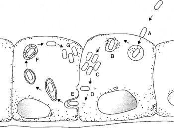
L. monocytogenes has been used as a model for studying the pathophysiology of intracellular bacterial pathogens. L. monocytogenes can invade and multiply within a variety of cell types including macrophages, dendritic cells, and various types of parenchymal cells (Fig. 1) (Gray et al., Reference Gray, Freitag and Boor2006). The most important virulence factors for L. monocytogenes include internalin A (InlA) and internalin (InlB), a hemolysin (listeriolysin O), two phospholipases, and a protein (ActA) responsible for intracellular motility. As L. monocytogenes bacterial cells come into contact with host cells they use internalin proteins, InlA and InlB, respectively to mediate cell entry via E-cadherin and c-Met tyrosine kinase on the host cells (Fig. 1(a)) (Cossart et al., Reference Cossart, Pizarro-Cerda and Lecuit2003; Khelef et al., Reference Khelef, Lecuit, Bierne and Cossart2006). Recognition of InlA by E-cadherin is specific to primates, guinea pigs, ruminants, and gerbils (Lecuit et al., Reference Lecuit, Dramsi, Gottardi, Fedor-Chaiken, Gumbiner and Cossart1999; Bakardjiev et al., Reference Bakardjiev, Stacy, Fisher and Portnoy2004), and facilitates invasion by L. monocytogenes. There are many more internalin genes in the L. monocytogenes genome (currently there are 24 described), but many of these have undetermined roles (Swaminathan and Gerner-Smidt, Reference Swaminathan and Gerner-Smidt2007).
Illustration of the events of listerial cell invasion, intracellular growth, intracellular movement, and cell-to-cell spread. (a) L. monocytogenes uses proteins (nlA and InlB, respectively, to mediate cell entry via E-cadherin and c-Met tyrosine kinase on the host cells. (b) After internalization, the listerial cells escape the phagosome by expressing LLO and phosphatidylinositol-specific phospholipase C. (c) In the cytoplasm, L. monocytogenes cells multiply with a generation time of approximately 1 h at 37°C. (d) Listerial cells move to the periphery of the infected cell via action of the bacterial surface protein, ActA, which orchestrate addition of actin monomers to the caudal end of the bacteria forming actin filaments that propel it rostrally. (e) Spread from cell to cell occurs by the bacteria being propelled into the cell membrane, forming an invagination of the adjacent cell membrane resulting in the listerial cell being contained within a double-membrane vacuole. (f) To escape the double-membrane vacuole, L. monocytogenes uses a phosphatidylcholine-dependent phospholipase C (lecithinase) and LLO. (G) The process is repeated. This figure was reproduced from Albritton et al. (Reference Albritton, Cochi and Feeley1984).

Once the listerial cells are internalized, they escape the phagosome by expressing listerolysin O (LLO) and phosphatidylinositol-specific phospholipase C (Fig. 1(b)) (Schluter et al., Reference Schluter, Domann, Buck, Hain, Hof, Chakraborty and Deckert-Schlüter1998; Decatur and Portnoy, Reference Decatur and Portnoy2000; Glomski et al., Reference Glomski, Gedde, Tsang, Swanson and Portnoy2002; Kohda et al., Reference Kohda, Kawamura, Baba, Nomura, Ito, Kimoto, Watanabe and Mitsuyama2002). L. monocytogenes cells multiply in the cytoplasm with a generation time of approximately 1 h at 37°C (Fig. 1(c)) (Fernandez et al., Reference Fernandez, George, Sills and Peck1997). As the bacteria multiply, they move to the periphery of the infected cell via action of the bacterial surface protein, ActA, which orchestrate addition of actin monomers to the caudal end of the bacteria. This results in formation of actin filaments that propel the bacterial cells rostrally (Fig. 1(d)) (Beckerle, Reference Beckerle1998; Darji et al., Reference Darji, Bruder, zur Lage, Gerstel, Chakraborty, Wehland and Weiss1998; Moors et al., Reference Moors, Levitt, Youngman and Portnoy1999). As the bacterial cell is propelled into the cell membrane, it forms an invagination of the adjacent cell membrane resulting in the listerial cell being contained within a double membrane vacuole (Fig. 1(e)). To escape from this double-membrane vacuole, L. monocytogenes uses a phosphatidylcholine-dependent phospholipase C (lecithinase) and LLO (Fig. 1(f)). The process of intracellular mobility and multiplication is then repeated (Fig. 1(g)) (Portnoy et al., Reference Portnoy, Schreiber, Connelly and Tilney1989; Schluter et al., Reference Schluter, Domann, Buck, Hain, Hof, Chakraborty and Deckert-Schlüter1998).
Nearly all of the L. monocytogenes virulence factors are encoded by genes that cluster on a well-defined area on the bacterial chromosome (Kathariou, Reference Kathariou2002). Their expression is regulated by a transcriptional activator, positive regulatory factor A (PrfA) (Renzoni et al., Reference Renzoni, Cossart and Dramsi1999; Ferreira et al., Reference Ferreira, Sue, O'Byrne and Boor2003; Chaturongakul et al., Reference Chaturongakul, Raengpradub, Wiedmann and Boor2008). After transcriptional activation of the virulence genes, PrfA also acts to enhance transcription of InlA, InlB, and LLO via a variety of promoters in the PrfA box (Gray et al., Reference Gray, Freitag and Boor2006). The action of PrfA is temperature-dependent and repressed at temperatures <25°C (Leimeister-Wachter et al., Reference Leimeister-Wachter, Domann and Chakraborty1992). However, bacterial growth continues at cold temperatures without expression of virulence genes. PrfA therefore is part of L. monocytogenes ability to sense various environments, such as soil, vegetation, or refrigerated dairy products, and differentiate these from the intracellular environment of mammals. Clinical isolates of L. monocytogenes are more able than non-pathogenic strains to detect environmental changes and resume log phase growth after moving from a cold to a warm environment (Avery and Buncic, Reference Avery and Buncic1997). PrfA activity is up-regulated in low pH environments (pH of 4.5–5.5), to which L. monocytogenes cells are exposed several times during the infection cycle. Examples include many types of fermented foods (which are acidic), passage through the stomach, and within the acidified phagosome of the infected cell (Cotter and Hill, Reference Cotter and Hill2003).
Immune response to L. monocytogenes
The host immune response to L. monocytogenes infection is thought to be effective in healthy adults, as listeriosis is a relatively rare cause of food-borne disease (estimated 1600 cases per year in the USA) (Lund and O'Brien, Reference Lund and O'Brien2011). Because of this, L. monocytogenes has been widely used as a model to investigate interactions of intracellular pathogens with the host innate and adaptive immune responses.
The first line of defense against L. monocytogenes infection is through innate mechanisms that include the low pH environment of the stomach, the intestinal microflora, antimicrobial peptides, and several types of leukocytes (Lecuit et al., Reference Lecuit, Vandormael-Pournin, Lefort, Huerre, Gounon, Dupuy, Babinet and Cossart2001; Czuprynski et al., Reference Czuprynski, Kathariou, Poulsen, Gyles, Prescott, Songer and Thoen2010). Neutrophils, or polymorphonuclear leukocytes (PMNs), respond to bacterial infection. Together with monocytes and macrophages, they ingest and kill a portion of the bacterial population (Czuprynski et al., Reference Czuprynski, Henson and Campbell1984, Reference Czuprynski, Noel, Doyle and Schultz1989; Portnoy et al., Reference Portnoy, Schreiber, Connelly and Tilney1989). Cells of the innate immune response recognize L. monocytogenes in part via receptors for pathogen-associated molecular patterns (PAMPs) on the bacterial cell surface. These PAMPs include lipoteichoic acid, peptidoglycan, and flagellin (Hayashi et al., Reference Hayashi, Smith, Ozinsky, Hawn, Yi, Goodlett, Eng, Akira, Underhill and Aderem2001; Machata et al., Reference Machata, Tchatalbachev, Mohamed, Jansch, Hain and Chakraborty2008). Toll-like receptor 2 (TLR2) and TLR5 have been reported to bind listerial PAMPs lipoteichoic acid and flagellin, respectively (Torres et al., Reference Torres, Barrier, Bihl, Quesniaux, Maillet, Akira, Ryffel and Erard2004). After recognition by TLR2 and TLR5, signaling mediated by myeloid differentiation factor 88 (MyD88) prompts macrophages to produce pro-inflammatory cytokines such as interleukin-1 (IL-1), IL-6, and tumor necrosis factor-α (TNFα; Way et al., Reference Way, Kollmann, Hajjar and Wilson2003; Posfay-Barbe and Wald, Reference Posfay-Barbe and Wald2009). These cytokines, in turn, activate the arachidonic acid inflammatory cascade to produce leukotrienes (specifically LTB4), which attract additional neutrophils to the site of infection (Grone et al., Reference Grone, Scheffer and Konig1992).
The innate immune response sets the stage for an adaptive immune response to L. monocytogenes. Protective immunity is dependent on CD8+ T cells and peaks 7–10 days after infection (Berg et al., Reference Berg, Crossley, Murray and Forman2003). CD4+ T cells also are activated and mediate delayed type hypersensitivity but are less important for clearance of listerial cells (Pamer, Reference Pamer2004). Antigen specific CD8+ and CD4+ T cells, as well as natural killer cells, produce interferon-γ (IFNγ), which is important for activation of macrophages and differentiation of T helper 1 (Th1) cells. Products of Th1 cells are responsible for increasing major histocompatibility (MHC) I and MHC II antigen presentation, continued expansion of CD8+ T cells, and inhibition of CD4+ T cell expansion (Pamer, Reference Pamer2004). Recently, attention has been paid to a potential role of Th17 cells (CD4+) and gamma-delta T cells, which both produce IL-17 early in the course of L. monocytogenes infection (Hamada et al., Reference Hamada, Umemura, Shiono, Tanaka, Yahagi, Begum, Oshiro, Okamoto, Watanabe, Kawakami, Roark, Born, O'Brien, Ikuta, Ishikawa, Nakae, Iwakura, Ohta and Matsuzaki2008; Orgun et al., Reference Orgun, Mathis, Wilson and Way2008). The roles of Th17 cells and IL-17a are considered in detail in the following section.
Immune response to L. monocytogenes infection during pregnancy
The protective immune response to L. monocytogenes infection typically seen in healthy adults may be less effective in pregnant women, who have an approximately 20-fold greater risk of acquiring listeriosis (Southwick and Purich, Reference Southwick and Purich1996). The reasons for the failure of a pregnant mother to protect her fetus are unclear, but may be partially due to changes in the maternal immune response during pregnancy. Previously it was thought these changes were chiefly due to a shift in the Th1:Th2 cell ratio toward a humoral (Th2) and away from a cell-mediated (Th1) response. The latter is potentially harmful to the fetal allograft (Wegmann et al., Reference Wegmann, Lin, Guilbert and Mosmann1993). The switch to a Th2 humoral response is thought to be due to in part to local expression of progesterone and progesterone-induced blocking factor by placental trophoblast cells (Kelemen et al., Reference Kelemen, Paldi, Tinneberg, Torok and Szekeres-Bartho1998). The change in Th cell populations is important, because cytokines associated with the Th1 cell response (e.g. IFNγ and TNFα) are critical for protective cellular immunity to L. monocytogenes (Pamer, Reference Pamer2004). However, more recent studies suggest that attributing increased susceptibility to listeriosis simply on Th1:Th2 cell ratios may be an over-simplification of the complex systemic and local immune response in the gravid uterus (Chaouat et al., Reference Chaouat, Zourbas, Ostojic, Lappree-Delage, Dubanchet, Ledee and Martal2002).
The recent description of pro-inflammatory Th17 cells may provide additional insight into fetal inflammation due to L. monocytogenes infection. Rapid Th17 cell proliferation is more frequently associated with extracellular bacterial infections than with infections by intracellular bacteria and viruses (Lin et al., Reference Lin, Slight and Khader2010). However, intracellular bacteria including Mycobacterium tuberculosis and Staphylococcus aureus have also been reported to induce Th17 cells in humans and mice, respectively (Hawkridge et al., Reference Hawkridge, Scriba, Gelderbloem, Smit, Tameris, Moyo, Lang, Veldsman, Hatherill, Merwe, Fletcher, Mahomed, Hill, Hanekom, Hussey and McShane2008; Scriba et al., Reference Scriba, Kalsdorf, Abrahams, Isaacs, Hofmeister, Black, Hassan, Wilkinson, Walzl, Gelderbloem, Mahomed, Hussey and Hanekom2008; Lin et al., Reference Lin, Ibrahim, Xu, Farber, Avanesian, Baquir, Fu, French, Edwards and Spellberg2009). Th17 cells have also been implicated in fetal inflammation and placental necrosis in pregnant women (Gargano et al., Reference Gargano, Holzman, Senagore, Thorsen, Skogstrand, Hougaard, Rahbar and Chung2008). The inflammatory response to L. monocytogenes infection during pregnancy (Kaufmann, Reference Kaufmann1993; Unanue, Reference Unanue1996; Milon, Reference Milon1997; Mucida et al., Reference Mucida, Park, Kim, Turovskaya, Scott, Kronenberg and Cheroutre2007) has been hypothesized to be influenced by Th17 cell proliferation. Orgun et al. showed that L. monocytogenes-infected mice produce a significant Th17 response when IFNγ and the type 1 IFN receptor were experimentally neutralized in non-pregnant mice (Orgun et al., Reference Orgun, Mathis, Wilson and Way2008). These results suggest that when Th1 cell proliferation is repressed during pregnancy, Th17 cell production during L. monocytogenes infection could be significant.
The main effector cytokine of Th17 cells is IL-17a, which in turn influences the production of chemokines (CXCL1, CXCL2, CXCL5 and CXCL8) and cytokines (GM-CSF granulocyte macrophage colony-stimulating factor and G-CSF granulocyte colony stimulating factor) that are responsible for recruitment, differentiation, and activation of neutrophils (Kawaguchi et al., Reference Kawaguchi, Adachi, Oda, Kokubu and Huang2004; Kolls and Linden, Reference Kolls and Linden2004). IL-17a has been linked to several immune-mediated and autoimmune diseases including rheumatoid arthritis, asthma, systemic lupus erythematosus, allograft rejection, and anti-tumor immune responses (Aggarwal and Gurney, Reference Aggarwal and Gurney2002). There is a potential role for IL-17a in listeriosis during pregnancy because the hallmark histopathological lesion is neutrophilic inflammation (Redline and Lu, Reference Redline and Lu1988; Takahashi et al., Reference Takahashi, Naito, Katabuchi and Higashi1991). The fetus is essentially an allograft that can be recognized as such by the maternal immune system. IL-17a has been detected in the human placenta during inflammation, and is associated with miscarriage and other pregnancy abnormalities (Gargano et al., Reference Gargano, Holzman, Senagore, Thorsen, Skogstrand, Hougaard, Rahbar and Chung2008). IL-17a has been found in the mouse placenta during normal pregnancy, but its presence in the placenta during L. monocytogenes infection has not been investigated (Ostojic et al., Reference Ostojic, Dubanchet, Chaouat, Abdelkarim, Truyens and Capron2003).
Another important product of Th17 cells is IL-22, which has been implicated in host defense, inflammation, and tissue repair (Wolk et al., Reference Wolk, Kunz, Witte, Friedrich, Asadullah and Sabat2004; Boniface et al., Reference Boniface, Bernard, Garcia, Gurney, Lecron and Morel2005; Sa et al., Reference Sa, Valdez, Wu, Jung, Zhong, Hall, Kasman, Winer, Modrusan, Danilenko and Ouyang2007). IL-22 is part of the IL-10 family and regulates function of parenchymal cells, such as epithelial cells and hepatocytes (Andoh et al., Reference Andoh, Zhang, Inatomi, Fujino, Deguchi, Araki, Tsujikawa, Kitoh, Kim-Mitsuyama, Takayanagi, Shimizu and Fujiyama2005). In certain circumstances IL-22 works synergistically with IL-17a and is reported to have functions that are both pro- and anti-inflammatory (Sonnenberg et al., Reference Sonnenberg, Nair, Kirn, Zaph, Fouser and Artis2010). IL-22 has been found in human placenta, and was shown to be significantly elevated in experimental L. monocytogenes infection (Xu et al., Reference Xu, Presnell, Parrish-Novak, Kindsvogel, Jaspers, Chen, Dillon, Gao, Gilbert, Madden, Schlutsmeyer, Yao, Whitmore, Chandrasekher, Grant, Maurer, Jelinek, Storey, Brender, Hammond, Topouzis, Clegg and Foster2001; Yang et al., Reference Yang, Yang, Htut, Ouyang, Hanidu, Li, Sellati, Jiang, Zhang, Li, Zhao, Ting, Mayer, Unkeless, Labadia, Hodge, Li and Xiong2008). Intravenous L. monocytogenes infection of IL-22(−/−) mice showed that bacterial clearance is not dependent on IL-22 (Zenewicz et al., Reference Zenewicz, Yancopoulos, Valenzuela, Murphy, Karow and Flavell2007; Graham et al., Reference Graham, Carr, Sieve, Indramohan, Break and Berg2011). A recent report demonstrated that IL-22 is not required for clearance of listerial cells from pregnant mice infected orally with L. monocytogenes (Poulsen et al., Reference Poulsen, Faith, Steinberg and Czuprynski2013).
Clinical listeriosis during pregnancy
Despite major differences in diet and placental anatomy, pregnancy is a common risk factor for listeriosis in both humans and ruminant species leading to maternal illness, fetal infection, and abortion (George, Reference George and Smith2009; Lamont et al., Reference Lamont, Sobel, Mazaki-Tovi, Kusanovic, Vaisbuch, Kim, Uldbjerg and Romero2011). ‘Abortion storms’ occur in humans and ruminants. In both cases, outbreaks occur following ingestion of a contaminated foodstuff. An important difference between human and ruminant listeriosis outbreaks is that human outbreaks are amplified by intra- and inter-state distribution of contaminated foods, while ruminant outbreaks tend to remain within a herd (Linnan et al., Reference Linnan, Mascola, Lou, Goulet, May, Salminen, Hird, Yonekura, Hayes, Weaver, Audurier, Plikaytis, Shirley, Kleks and Broome1988). Abortion does not occur in all infected women during pregnancy and is observed infrequently in pregnant cattle in which meningioencephalitis is more common (Hume, Reference Hume1976; George, Reference George and Smith2009). In a review of 191 cases of human listeriosis during pregnancy, 32% had mild flu-like symptoms, 65% reported fever, and 29% were asymptomatic (Mylonakis et al., Reference Mylonakis, Paliou, Hohmann, Calderwood and Wing2002).
Listeriosis in early pregnancy results in abortion or fetal resorption (Mylonakis et al., Reference Mylonakis, Paliou, Hohmann, Calderwood and Wing2002). L. monocytogenes infection later in gestation results in abortion, stillbirth, or premature birth (Reiss and Krebs, Reference Reiss and Krebs1951; Johnston et al., Reference Johnston, Morton, Wong and Roy1955; Mylonakis et al., Reference Mylonakis, Paliou, Hohmann, Calderwood and Wing2002). Listeriosis in ruminants commonly occurs in the third trimester. It is not known whether L. monocytogenes is a risk factor for early embryonic death, because definitive diagnostics are costly and fetuses typically are not submitted to a veterinary diagnostic laboratory (George, Reference George and Smith2009). Transmission from mother to fetus is most likely due to fetal inhalation of infected amniotic fluid, transplacental infection (hematogenous), or ascending colonization of the fetoplacental unit from the birth canal (Becroft et al., Reference Becroft, Farmer, Seddon, Sowden, Stewart, Vines and Wattie1971; Lennon et al., Reference Lennon, Lewis, Mantell, Becroft, Dove, Farmer, Tonkin, Yeates, Stamp and Mickleson1984).
Premature labor is one outcome of L. monocytogenes infection during pregnancy. Prematurity for any reason is a high-risk factor for short- and long-term neonatal complications, including death (Goldenberg et al., Reference Goldenberg, Culhane, Iams and Romero2008). Infants born prematurely due to maternal listeriosis are at a high risk of neonatal sepsis (reviewed in (Lamont et al., Reference Lamont, Sobel, Mazaki-Tovi, Kusanovic, Vaisbuch, Kim, Uldbjerg and Romero2011)). One study reported 68% of mothers diagnosed with listeriosis had neonates who also developed listeriosis (Mylonakis et al., Reference Mylonakis, Paliou, Hohmann, Calderwood and Wing2002). From a review of 222 cases of listeriosis during pregnancy that resulted in neonatal listeriosis, 68.2% of neonates recovered, 12.7% developed long-term neurologic deficits, and 24.5% died (Mylonakis et al., Reference Mylonakis, Paliou, Hohmann, Calderwood and Wing2002). Neonatal listeriosis manifests as either early or late-onset disease. Early onset listeriosis is apparent within 1–3 days of birth, with an average of 36 h (Relier, Reference Relier1979). Neonates with early onset listeriosis tend to be born prematurely; 44–89% of those mothers had been diagnosed with L. monocytogenes and often had suffered recently from a flu-like illness (Linnan et al., Reference Linnan, Mascola, Lou, Goulet, May, Salminen, Hird, Yonekura, Hayes, Weaver, Audurier, Plikaytis, Shirley, Kleks and Broome1988; McLauchlin, Reference McLauchlin1990). The most common clinical presentation of early onset neonatal listeriosis is septicemia, with pneumonia being less common (Linnan et al., Reference Linnan, Mascola, Lou, Goulet, May, Salminen, Hird, Yonekura, Hayes, Weaver, Audurier, Plikaytis, Shirley, Kleks and Broome1988; McLauchlin, Reference McLauchlin1990). Late onset neonatal listeriosis affects full-term infants and develops at 5–14 days of life (Albritton et al., Reference Albritton, Cochi and Feeley1984; McLauchlin, Reference McLauchlin1990). Unlike early onset neonatal listeriosis, infants with late onset neonatal listeriosis usually are born to asymptomatic mothers with negative L. monocytogenes diagnostic tests (Linnan et al., Reference Linnan, Mascola, Lou, Goulet, May, Salminen, Hird, Yonekura, Hayes, Weaver, Audurier, Plikaytis, Shirley, Kleks and Broome1988; McLauchlin, Reference McLauchlin1990). Meningitis is the most common clinical syndrome associated with late onset neonatal listeriosis (Smith et al., Reference Smith, Lieberman, Allen and Barson1983; Posfay-Barbe et al., Reference Posfay-Barbe and Wald2009). Transmission of L. monocytogenes to the neonate can occur in utero, as described above, during passage through the birth canal, or by ingestion of contaminated colostrum and breast milk (Svabic-Vlahovic et al., Reference Svabic-Vlahovic, Pantic, Pavicic and Bryner1988). Human breast milk currently is not recognized as a risk factor for L. monocytogenes transmission from a mother to her nursing infant. However, this is not the case for ruminant species, which are well known to shed L. monocytogenes in their milk (George, Reference George and Smith2009).
Animal models for L. monocytogenes infection during pregnancy
L. monocytogenes has been associated with human disease since the early 1920s and is recognized as a cause of neonatal disease since the mid 1940s (Lecuit, Reference Lecuit2007). Laboratory animal models of listeriosis were studied for years prior to recognition of L. monocytogenes as a human food-borne pathogen and were responsible for a significant amount of information regarding the pathogenesis of intracellular pathogens and cell-mediated immunity against them (Miki and Mackaness, Reference Miki and Mackaness1964). Each of the animal infection models currently used has advantages and disadvantages due to microbial variables and the complex series of events that lead to clinical listeriosis.
Models of L. monocytogenes infection during pregnancy need to consider the variability of placental structure. Mammals tend to fit into three broad categories of placentation: (1) epitheliochorial (horse, pig, and cow); (2) endothelialchorial (carnivores); and (3) hemochorial (rodents and primates) (reviewed in Carter, Reference Carter2007). Epitheliochorial placentation has the most complete separation of fetal and maternal blood, endothelialchorial has moderate separation, and hemochorial has the least. Epitheliochorial placentation and long gestation time limits the use of ruminant species as models for human listeriosis.
Pregnant non-human primates have placental tissue with the greatest similarity to humans (Caldecott and Miles, Reference Caldecott and Miles2005; Lecuit, Reference Lecuit2007). Rhesus macaque monkeys have been used as a model for L. monocytogenes fetal infections, as they exhibit similar responses as humans including abortion, stillbirth, and neonatal death (reviewed in Williams et al., Reference Williams, Castleman, Lee, Mote and Smith2009). The pathological lesions seen with L. monocytogenes infection are essentially the same in humans and non-human primates (Chalifoux and Hajema, Reference Chalifoux and Hajema1981). However, there are ethical considerations regarding the use of non-human primates, and their use to study infections during pregnancy is often cost-prohibitive (Williams et al., Reference Williams, Castleman, Lee, Mote and Smith2009).
Guinea pigs have been used to study L. monocytogenes since the 1970s and provide one of the first models to characterize virulence of different L. monocytogenes strains (Williams et al., Reference Williams, Castleman, Lee, Mote and Smith2009). More recently, guinea pigs have gained renewed attention after it was discovered that their E-cadherin amino acid sequence is the same as that of humans (Lecuit et al., Reference Lecuit, Dramsi, Gottardi, Fedor-Chaiken, Gumbiner and Cossart1999; Schubert et al., Reference Schubert, Urbanke, Ziehm, Beier, Machner, Domann, Wehland, Chakraborty and Heinz2002). Although guinea pigs are considered an acceptable model for human infection via the gastrointestinal tract, they require a high infective dose and it is difficult to quantify microbial load in their tissues (Bakardjiev et al., Reference Bakardjiev, Stacy, Fisher and Portnoy2004; Williams et al., Reference Williams, Irvin, Chmielewski, Frank and Smith2007). Furthermore the utility of the guinea pig as a L. monocytogenes infection model is limited because the guinea pig Met receptor, responsible for binding InlB, is dissimilar to the human Met receptor. Some have suggested infection of pregnant gerbils as a potential listeriosis model as they have the same E-cadherin site and similar Met receptors as humans (Disson et al., Reference Disson, Grayo, Huillet, Nikitas, Langa-Vives, Dussurget, Ragon, Le Monnier, Babinet, Cossart and Lecuit2008). Intravenous infection of pregnant gerbils with L. monocytogenes has been shown to cause fetal death, but oral infection has yet to be reported (Disson et al., Reference Disson, Grayo, Huillet, Nikitas, Langa-Vives, Dussurget, Ragon, Le Monnier, Babinet, Cossart and Lecuit2008).
The mouse has been used for L. monocytogenes infection studies since the 1950s (Osebold and Sawyer, Reference Osebold and Sawyer1957). Denk et al. (Reference Denk, Flamm and Kovac1966) first described the pregnant mouse as an animal model to study the effects of listeriosis on the fetus. The advantages to using mice include their short generation time, their relatively small size, and the availability of genetic, immunological, and molecular biological tools (e.g. antibodies, genetic mutant strains, and cytokines). The genetic background of inbred mouse strains is an important factor for L. monocytogenes infection studies. Innate resistance to L. monocytogenes is regulated principally by the Hc locus on chromosome 2 (Cheers and McKenzie, Reference Cheers and McKenzie1978; Gervais et al., Reference Gervais, Stevenson and Skamene1984; Czuprynski et al., Reference Czuprynski, Canono, Henson and Campbell1985). Mouse strains with the resistant allele (e.g. C57BL/6) at the Hc locus are significantly more resistant to intravenous, intraperitoneal, or intragastric L. monocytogenes infection than are strains with the susceptible allele (e.g. A/J) (Gervais et al., Reference Gervais, Stevenson and Skamene1984; Czuprynski et al., Reference Czuprynski, Canono, Henson and Campbell1985, Reference Czuprynski, Faith and Steinberg2003). Interestingly, this genetically mediated difference in resistance to L. monocytogenes infection in the mouse is not so apparent in pregnancy. Recently, our laboratory has shown that there is no significant difference in susceptibility of C57BL/and A/J mice to infection with L. monocytogenes at 10–14 days of gestation (Poulsen et al., Reference Poulsen, Faith, Steinberg and Czuprynski2011). The mouse infection model is criticized by some because murine E-cadherin differs from humans at a critical amino acid residue and hence does not interact with L. monocytogenes InlA. Specifically, the difference is at the 16th amino acid in the chain of the peptide that is a proline in human and glutamic acid in murine E-cadherin (Lecuit et al., Reference Lecuit, Dramsi, Gottardi, Fedor-Chaiken, Gumbiner and Cossart1999). A transgenic mouse has been developed that expresses human E-cadherin in intestinal tissue, but not other tissues including placenta. Infection of the pregnant mouse results in more severe clinical symptoms than those seen in human infection (e.g. meningitis and stillbirth) including high mortality in adult mice and high fetal mortality with severe inflammation and necrosis of the fetus (Williams et al., Reference Williams, Castleman, Lee, Mote and Smith2009). However, several studies have shown that intragastric infection of pregnant mice leads to fetal infection and fetal death in the mouse, with inflammatory lesions that resemble those of L. monocytogenes infection in women carrying more than one fetus (Lammerding et al., Reference Lammerding, Glass, Gendron-Fitzpatrick and Doyle1992; Poulsen et al., Reference Poulsen, Faith, Steinberg and Czuprynski2011).
In vitro models for L. monocytogenes infection of placental trophoblasts
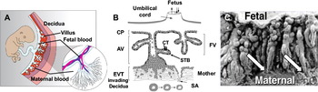
During pregnancy, listerial cells have trophism for placental cells at the maternal–fetal interface (Czuprynski et al., Reference Czuprynski, Kathariou, Poulsen, Gyles, Prescott, Songer and Thoen2010). Despite extensive work with L. monocytogenes to elucidate its pathogenesis, little is known about the underlying mechanisms of fetal and placental infection. As stated earlier, humans have a hemochorial villous placenta with minimal barriers between maternal and fetal structures (Fig. 2) (Carter, Reference Carter2007). Syncytiotrophoblast and extravillous trophoblast cells are located on the villous parts of the fetal placenta. Syncytiotrophoblasts line the villi, which are immersed in maternal blood. At the tip of the villi, extravillous trophoblast cells anchor the fetal placenta to the decidual tissue, and are essential in altering the uterine vasculature to increase maternal blood flow to the fetus (reviewed in Whitley and Cartwright, Reference Whitley and Cartwright2009; Harris, Reference Harris2011).
Human placenta structure. (A) Developing fetus with placental villi bathed in maternal blood in the intervillous space. These tissues are attached to the decidual tissue in the uterus. The inset illustrates an enlargement of a fetal villus, which facilitates oxygen exchange and nutrient transport to the fetal circulation via the umbilical arteries (blue) and vein (red). (B) Enlargement of panel A shows different trophoblast cell types. Extravillous trophoblast cells anchor the fetal placenta to the decidual tissue, influence the maternal immune system to allow fetal tolerance, and alter maternal blood flow in the spiral arteries (SA) to increase nutrient availability to the fetus. Extravillous trophoblasts (EVT) arise from the chorionic plate (CP) in trophoblast cell columns to form anchoring villi (AV) and floating villi (FV). Villous cytotrophoblast cells (CTB) differentiate from the syncytiotrophoblast (STB) layer overlying the villus. The syncytiotrophoblast cell layer is responsible for gas and nutrient exchange from the maternal blood in the intervillous space to the fetal blood in capillaries located in the villous stroma. (C) Scanning electron micrograph of the maternal-fetal interface of a rhesus monkey showing anchoring villi (arrows) of fetal placenta attached to maternal decidual tissue (reproduced from [King and Mais, 1982]).

Previously, it was shown that L. monocytogenes can infect primary cultures of extravillous trophoblast cells and immortalized lines of these cells (JEG-3, JAR, and BeWo cells) (Robbins et al., Reference Robbins, Skrzypczynska, Zeldovich, Kapidzic and Bakardjiev2010; Zeldovich et al., Reference Zeldovich, Robbins, Kapidzic, Lauer and Bakardjiev2011). Entry of listerial cells into the trophoblast cells on placental villi is mediated by E-cadherin–InlA interaction (Lecuit et al., Reference Lecuit, Dramsi, Gottardi, Fedor-Chaiken, Gumbiner and Cossart1999). However, different laboratories have suggested alternative trophoblast target cells for L. monocytogenes entry. Lecuit et al. showed syncytiotrophoblast cells to be the point of entry into the placenta to infect the fetus (Lecuit et al., Reference Lecuit, Nelson, Smith, Khun, Huerre, Vacher-Lavenu, Gordon and Cossart2004). On the other hand, Robbins et al. (Reference Robbins, Skrzypczynska, Zeldovich, Kapidzic and Bakardjiev2010) have shown that syncytiotrophoblast cells constitute a barrier to transmission of L. monocytogenes from the maternal bloodstream to the placenta. These authors also found that primary cultures of extravillous trophoblast cells can become infected but restrict growth and intracellular spread of L. monocytogenes (Robbins et al., Reference Robbins, Skrzypczynska, Zeldovich, Kapidzic and Bakardjiev2010). All of the published work on L. monocytogenes infection of placental cells focused on the ability of the bacterial cells to infect trophoblasts and the mechanism by which listerial cells spread to the fetus. The effect of L. monocytogenes infection on trophoblast function, and how this might affect the ability of the placenta to support the continued growth of the fetus, is yet to be examined.
Conclusion
Despite strict guidelines set by government agencies to decrease L. monocytogenes contamination of food, this pathogen will likely continue to cause significant clinical disease in the future. With improved surveillance in developing countries and a higher proportion of the population being at greater risk for infection (>60 years of age and immunocompromised individuals), the incidence of listeriosis may increase, similar to what has been reported in the United Kingdom during the past decade (Mook et al., Reference Mook, O'Brien and Gillespie2011). Future research should focus on knowledge gaps regarding L. monocytogenes pathogenesis in high-risk populations. Animal models, despite their limitations, will undoubtedly be used to address these issues. Translational research, including pathogenesis of listeriosis in veterinary species may provide novel insights into risk factors for transmission of listeriosis in late onset neonatal listeriosis and the perinatal period.
Acknowledgments
The authors would like to thank Ms. Julie A Monahan and Dr. Thaddeus G Golos for their help illustrating Figure 2.