Introduction
Traumatic tympanic membrane perforations are commonly encountered in otology clinics, and may be caused by barotrauma, thermal or caustic burns, or blunt or penetrating injuries by instruments to the external auditory canal.Reference Afolabi, Aremu, Alabi and Segun-Busari1,Reference Sagiv, Migirov, Glikson, Mansour, Yousovich and Wolf2 Barotraumas are often the result of blast and non-blast injuries sustained in fights, assaults by security agents, road traffic injuries, or slaps to the head. Previous studies showed that most traumatic tympanic membrane perforations have everted or inverted edges,Reference Lou, Lou and Zhang3,Reference Lou, Tang and Yang4 and it has been suggested that such edges may prolong the closure time and result in middle-ear cholesteatoma.Reference Armstrong5–Reference Saito, Kazama and Yazawa7
Most cases of traumatic tympanic membrane perforation resulting from barotrauma tend to heal spontaneously, while some traumatic tympanic membrane perforations show prolonged closure time and may fail to close. Some research groups have suggested that early edge approximation facilitates eardrum healing and shortens closure time.Reference Armstrong5–Reference Jung, Yun, Kim, Joo, Song and Rah9 However, there have been no previous detailed studies on the correlation between healing outcome and the size of the everted or inverted edge. Typically, barotraumas in peacetime are caused by a slap or the impact of a fist on the ear. This study was performed to evaluate the healing and hearing outcomes related to the everted or inverted edge area on slap- and fist-induced large tympanic membrane perforations.
Materials and methods
Ethical considerations
The study was reviewed and approved by the Institutional Ethics Review Board of Wenzhou Medical University Affiliated to the Yiwu Central Hospital. Informed consent was obtained from all participants prior to enrolment in the study.
Patients
We recruited 120 consecutive patients who visited the ENT department following a diagnosis of traumatic tympanic membrane perforation between June 2007 and June 2012. These 120 patients fulfilled the following criteria: (1) slap- or fist-induced tympanic membrane perforation with an everted or inverted edge; (2) no history of middle-ear disease or pre-existing myringosclerosis; (3) duration of trauma of less than 3 days, without middle-ear infection; and (4) tympanic membrane perforation affecting 50–75 per cent of the pars tensa of the ear. The exclusion criteria were: middle-ear infection or severe vertigo at the time of the hospital visit, suspicion of ossicular chain damage or granulation tissue hyperplasia, any additional (particularly craniocerebral) injury associated with an inability to walk independently, and patient relocation from the region served by our hospital associated with loss to follow up.
Each patient was endoscopically examined after the removal of cerumen and/or blood clots in the external auditory canal using a cotton bud soaked in povidone-iodine solution, and the site and size of the perforation were documented. The size of tympanic membrane perforations was analysed using ImageJ image processing software (National Institutes of Health, Bethesda, Maryland, USA). The perforation position was categorised as: anterior half (including antero-inferior and antero-superior regions), posterior half (including postero-inferior and postero-superior regions) or kidney-shaped (including antero-inferior and postero-inferior regions).
Standard pure tone audiometric testing was performed at the initial and final visits, and at six months after treatment. Pure tone averages were determined for air and bone conduction at 0.5, 1, 2 and 4 kHz.
The principal investigator, aided by a registered nurse, allocated patients to treatment groups using simple random sampling. Consecutive subjects who fulfilled the inclusion criteria, and who had signed the consent form, were assigned random numbers generated by SPSS for Windows statistical software (version 19.0; SPSS, Chicago, Illinois, USA). On the basis of the numbers generated, patients were allocated to an edge approximation group (n = 60) or a spontaneous healing (n = 60) group. Everted and inverted edges were defined as reversed edges in this study for descriptive convenience. The edge approximation group was then divided into subgroup A (reversed edge of more than 50 per cent of the total perforation) and subgroup B (reversed edge of less than 50 per cent of the total perforation).
Edge approximation group
Using an endoscope, lidocaine (2 per cent) and adrenaline (1:100 000 diluted) were injected into the bony cartilaginous junction of the external auditory canal. The everted and inverted edges, if not crusted and still visible, were moistened with a cotton ball soaked in Ringer's solution, and then aligned with the original position as much as possible using a Rosen needle or a small vacuum suction tip. A modified absorbable sponge sheet (Gelfoam; Pfizer, New York, USA) was placed under the tympanic membrane remnant, as an underlay, to provide a scaffold for healing. The healing process could be easily observed endoscopically when the sponge was removed and replaced with a fresh piece of sponge at every visit.
Spontaneous healing group
No intervention was provided, but all patients underwent regular follow up.
Follow up
Follow-up appointments were scheduled at the following time points: every other day in the 1st week after the initiation of treatment; twice a week until 1 month after commencing treatment; once a week until perforation closure or 6 months, whichever came first; and at 24 months. The absorbable sponge was removed and replaced with a fresh piece under endoscopic guidance, at each visit. The tympanic membrane was repeatedly examined and photographed endoscopically. Colour photographs were taken at each visit by an independent clinician who was blinded to the group allocation.
The primary outcomes, assessed at six months, were closure rate, infection rate and average closure time. The secondary outcome was the cholesteatoma status of the middle ear. Computed tomography (CT) of the temporal bone was scheduled to determine the cholesteatoma status of the middle ear at 24 months after perforation closure.
Statistical analysis
Data are given as means ± standard deviation or percentages. The three groups were compared by one-way analysis of variance for parameters including average age and average closure time. The Kruskal–Wallis test was used to compare the groups in terms of perforation duration. The chi-square test was used for analysing categorical data (including injury side, perforation position, everted or inverted edge, and closure rate). The two-sample t-test or Mann–Whitney U test was used to compare the results of two groups. P-values between 0.05 and 0.005 were considered suggestive of significance; only p-values of less than 0.005 were considered significant.Reference Laccourreye and Maisonneuve10 All statistical analyses were performed using SPSS software (version 19.0).
Results
Demographic data
Of the 120 patients, 2 in the spontaneous healing group were lost to follow up during the initial six-month period. Therefore, only 118 patients were included in the final healing and hearing outcome analysis.
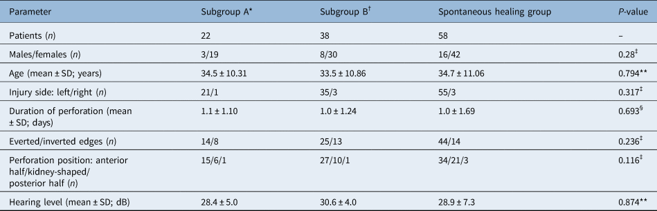
These 118 patients consisted of 27 males and 91 females, with an average age of 34.7 ± 5.2 years (range, 21–58 years). The injury was in the left ear in 111 patients and in the right ear in 7 patients. Of the 118 patients, 83 had everted edges, while 35 had inverted edges. The demographic data for the study population are shown in Table 1.
Demographic data

P-values between 0.05 and 0.005 considered ‘suggestive’; only p-values less than 0.005 considered significant. *Reversed edge of more than 50 per cent of the total perforation; †reversed edge of less than 50 per cent of the total perforation. ‡Chi-square test; **one-way analysis of variance; §Kruskal–Wallis test. SD = standard deviation
There was no correlation between perforation size and reversed edge area. All three groups were comparable in terms of sex, age, injury side, perforation position, perforation duration, hearing level, and everted and inverted edge status (p > 0.05).
Healing and hearing outcomes
The outcomes of patients who completed the six-month follow-up period are summarised in Table 2. Only one perforation in the edge approximation subgroup B (reversed edge of less than 50 per cent of the total perforation) was infected. The perforation closure rates in the edge approximation subgroup A (reversed edge of more than 50 per cent of the total perforation), edge approximation subgroup B and spontaneous healing group were 90.9 per cent (20 out of 22), 92.1 per cent (35 out of 38) and 84.5 per cent (49 out of 58), respectively. The difference in closure rate among the three groups was not statistically significant (p = 0.393).
Healing and hearing outcomes

P-values between 0.05 and 0.005 considered ‘suggestive’; only p-values less than 0.005 considered significant. Regarding closure time statistical comparisons: subgroup A versus subgroup B, p = 0.847; subgroup A versus spontaneous healing group, p < 0.005; and subgroup B versus spontaneous healing group, p < 0.005. *Reversed edge of more than 50 per cent of the total perforation; †reversed edge of less than 50 per cent of the total perforation. ‡Chi-square test; **one-way analysis of variance. SD = standard deviation
The average perforation closure time was 33.15 ± 4.90 days, 32.25 ± 6.58 days and 43.91 ± 14.58 days for edge approximation subgroup A, edge approximation subgroup B and the spontaneous healing group, respectively. The average closure time was significantly longer in the spontaneous healing group than in the edge approximation subgroups A and B (p < 0.005). However, the average closure time was not significantly different between edge approximation subgroups A and B (p = 0.847).
The mean hearing improvement was 11.7 ± 2.6 dB for edge approximation subgroup A, 12.4 ± 3.1 dB for edge approximation subgroup B, and 11.9 ± 2.0 dB for the spontaneous healing group in the perforated ear at six months after closure, indicating that the treatment method did not affect hearing gain (p = 0.691).
At 24–29 months after perforation closure, four patients in the edge approximation subgroup A, two patients in the edge approximation subgroup B, and three patients in the spontaneous healing group were lost to follow up. Computed tomography of the temporal bone was performed in 47 patients (out of 53, 88.7 per cent) in the edge approximation group and in 46 patients (out of 49, 93.4 per cent) in the spontaneous healing group. The CT findings showed normal pneumatisation of the mastoid system and middle ear, and no middle-ear cholesteatoma in any patient evaluated.
Endoscopic characteristics
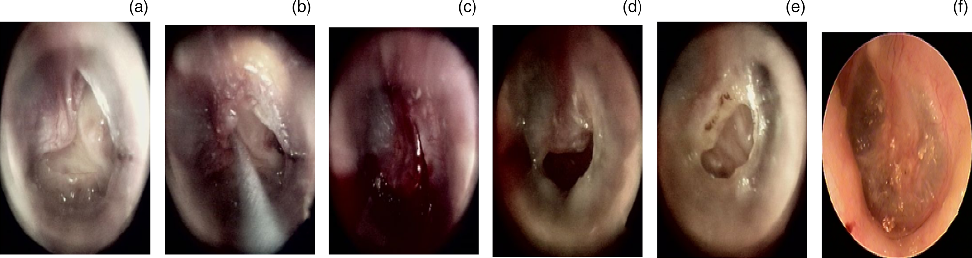
In the 60 patients in the edge approximation group, the perforation size was significantly reduced after edge approximation. However, the reversed edges had started to retract toward the annulus or malleus within 2–3 days, which became obvious at 4–5 days after edge approximation, and the perforations again increased in size. The reversed edges retracted completely toward the annulus or malleus (regardless of the size of the everted and inverted edges) within one week, and the epithelial layer then started to proliferate (Figure 1). In addition, hyperplasia of the epithelial layer gradually increased at everted and inverted edges.
Healing of a large perforation after edge approximation: (a) 3 days after perforation, (b) edge approximation, and (c) 1 day, (d) 3 days, (e) 6 days and (f) 27 days after treatment.

In the 58 patients in the spontaneous healing group, the reversed edges gradually became necrotic and formed a black crust within one week. The crust eventually migrated toward the external auditory canal. Hyperplasia of the epithelial layer gradually increased within one week at the everted and inverted edges.
Discussion
The tympanic membrane and middle ear are sensitive to variations in external pressure. Most cases of traumatic tympanic membrane perforation in peacetime are caused by a slap or fist to the ear. The severity of tympanic membrane perforation is affected by the direction, intensity and duration of the shockwave, and pneumatisation of the temporal bone and Eustachian tube.Reference Aktaş and Kutlu11 Some perforated tympanic membranes yield large amounts of debris, while others have an everted or inverted edge. However, the reversed edge area is not proportional to the perforation size.
• This study evaluated healing and hearing outcomes related to everted or inverted edge area on slap- and fist-induced tympanic membrane perforations
• The reversed edge area did not affect outcomes, regardless of edge approximation of everted or inverted edges
• The approximated edges had hardly any impact on eardrum healing
• Short-term follow up showed that cases with reversed edges did not develop middle-ear cholesteatoma
• The treatment method did not affect hearing gain in patients with slap- or fist-induced tympanic membrane perforations
Most traumatic tympanic membrane perforations tend to heal spontaneously. However, an everted or inverted edge may result in the failure of tympanic membrane perforation healing and the development of middle-ear cholesteatoma.Reference Armstrong5–Reference Saito, Kazama and Yazawa7 Early edge approximation has been reported to restore the original anatomy and reduce the perforation size, thereby facilitating the healing process and shortening the closure time of traumatic tympanic membrane perforations.Reference Meyerhoff, Marple, Roland, Roland, Marple and Meyerhoff6–Reference Jung, Yun, Kim, Joo, Song and Rah9 However, in the present study, the closure rate was not significantly different between edge approximation subgroup A (reversed edge of more than 50 per cent of the total perforation), edge approximation subgroup B (reversed edge of less than 50 per cent of the total perforation) and the spontaneous healing group. In addition, the closure time was not significantly different between edge approximation subgroups A and B, and there was no significant correlation between the healing outcome and the reversed edge area.
Although edge approximation may provisionally reduce the tympanic membrane perforation size, Gelfoam patches were also used, to provide a scaffold for the healing process, as the repaired edge continued to retract along the annulus or malleus regardless of the size of the everted or inverted edge. Tympanic membrane perforations are not similar to skin and cornea trauma, and reversed edges cannot be sutured. Therefore, repaired edges show gradual retraction under the influence of tension, ossicular chain movement and pulling of the middle-ear muscle.
In this study, although the edge approximation group showed a shorter closure time compared to the spontaneous healing group, this short healing time was not a function of edge approximation and was instead a result of the Gelfoam patch scaffold. Previous studies have shown that a perforated edge did not affect the healing outcome of biomaterial patching.Reference Park, Kim, Lee and Lee8,Reference Jung, Yun, Kim, Joo, Song and Rah9,Reference Camnitz and Bost12,Reference Jun, Oh, Yoo, Han, Chang and Jung13 It is well known that the use of Gelfoam patches aids in eardrum healing. Gelfoam patches stimulate the exposed epithelial layer and fibrous layer, and facilitate proliferation of granulation tissue, thereby accelerating the healing of traumatic tympanic membrane perforations and reducing the healing time.Reference Niklasson and Tano14,Reference Lou, Tang, Chen and Xiao15 In addition, this study showed that the area of reversed edges did not affect the hearing outcome.
Previous studies have shown that cases of traumatic tympanic membrane perforation with everted and inverted edges may progress to middle-ear cholesteatoma.Reference Armstrong5–Reference Saito, Kazama and Yazawa7 In this study, we performed CT of the temporal bone to evaluate the cholesteatoma status of the middle ear at 24 months after perforation closure. The CT findings showed normal pneumatisation of the mastoid system and middle ear, and no middle-ear cholesteatoma, in 88.7 per cent of patients evaluated in the edge approximation group and in 93.4 per cent of patients in the spontaneous healing group, at 24–29 months after perforation closure. In the spontaneous healing group, the everted and inverted edges gradually formed a crust, and then migrated toward the external auditory canal. In the edge approximation group, the approximated edge also retracted completely to the perforation edge, subsequently forming a crust and migrating toward the external auditory canal, but not entering into the tympanic cavity.
The formation of middle-ear cholesteatoma has mainly been reported in traumatic tympanic membrane perforations as a result of blast-induced perforations,Reference Kronenberg, Ben-Shoshan, Modan and Leventon16 but not of slap- or fist-induced perforations. We speculated that, in traumatic tympanic membrane perforations, the development of middle-ear cholesteatoma may be due to debris of the ruptured tympanic membrane or the presence of foreign bodies in the tympanic cavity, rather than to everted and inverted edges. However, further follow-up studies are necessary.
Conclusion
This study suggested that for slap- or fist-induced tympanic membrane perforations, the healing and hearing outcomes are not significantly affected, regardless of whether the edges are approximated. The edge approximation has hardly any beneficial impact on the healing of the eardrum. Short-term follow up showed that patients with tympanic membrane perforations with reversed edges did not develop middle-ear cholesteatoma.
Acknowledgements
This study was financially supported by the Science and Technology Agency of Zhejiang Province, the Health and Medicine Agency of Zhejiang Province, and the Science and Technology Agency of Yiwu, China (grant numbers 2013C33176, 2015KYB420, and 2015-3-06).
Competing interests
None declared