Introduction
Detecting early stage head and neck squamous cell carcinomas (SCCs) is difficult, so diagnosis is generally made at a late disease stage in most patients. The resulting large lesion size is associated with a large extent of surgical resection and significantly shortened survival times. The lesion often prevents swallowing and is associated with loss of voice function, which seriously reduces patient quality of life. Therefore, an early diagnosis of head and neck SCCs is clinically important. Early stage cancers on the laryngopharyngeal mucosal surface are characterised by relatively superficial lesions. Radiological methods such as computed tomography and magnetic resonance imaging usually fail to identify these tumours, and endoscopy is the major diagnostic technique. Recent developments in endoscopic illumination have generated novel endoscopic imaging systems, including narrow band and autofluorescence imaging endoscopy, that have improved the detection of early stage malignant lesions.Reference Subramanian and Ragunath 1 – Reference Ni and Wang 4 This study was designed to compare the effectiveness of narrow band and autofluorescence imaging endoscopy for diagnosing malignant laryngopharyngeal tumours.
Materials and methods
Patients
Patients admitted for examination to the Endoscopy Department of the Cancer Hospital, Chinese Academy of Medical Sciences, from May 2010 to October 2010 were enrolled in the study. All patients with laryngopharyngeal space occupying lesions (as suggested by radiological examination) with a high malignant potential, those with cervical lymph node metastatic SCCs with an unknown primary lesion or those with a hoarse voice requiring electronic laryngoscopy for diagnosis were included. Exclusion criteria were lidocaine allergy, haemorrhagic disease and an inability to provide informed consent. The study was approved by the hospital's medical ethics committee, and informed consent was obtained from each patient prior to examination.
Laryngopharyngeal examination
Prior to endoscopic examination, patients were placed in a horizontal, supine position and the surface of the nasal cavity was anaesthetised and lubricated with 2 per cent lidocaine hydrochloride gel. The endoscope was then introduced through the nasal passage for sequential observation of the nasopharynx, oropharynx, hypopharynx and larynx. The entire pharynx and larynx were first observed under ordinary white light. The mode was then changed to narrow band imaging, and each anatomical partition was observed in turn; any lesions present were photographed. Owing to incompatibility between narrow band and autofluorescence imaging endoscopy, the original endoscope was withdrawn and replaced by an endoscope with an autofluorescence imaging function. This was introduced into the pharynx and the larynx, and abnormal lesions were photographed under autofluorescence imaging. Abnormal lesions detected under white light image, narrow band imaging or autofluorescence imaging endoscopy were biopsied, fixed in 10 per cent formalin and submitted for pathological examination. The EVIS LUCERA 260 system was used for white light image endoscopy, with a BF-260 electronic bronchoscope for narrow band imaging and a BF-F260 electronic bronchoscope for autofluorescence imaging (Olympus Medical Systems, Tokyo, Japan).
Lesion classification under different endoscopy modes
Under white light image endoscopy, laryngopharyngeal lesions were classified as (1) malignant if they showed significant uplift, ulcers and a rough lesion surface or as (2) benign if they exhibited congestion, oedema, leukoplakia, or small nodules or polyps. Under narrow band imaging, lesions were classified as (1) malignant if they had brown spots or twisted cord-like structures (such as serpentine, earthworm or tadpole shaped) and clear boundaries on the mucosal surface or as (2) benign if there was a clearly visible vascular texture on the mucosal surface, leukoplakia coverage without significant microvascular exposure, or small punctate structures on the mucosal surface. Under autofluorescence imaging, lesions were classified as (1) malignant if they were coloured dark purple and had relatively clear boundaries or as (2) benign if the surface was coloured green or light purple. The endoscopic diagnosis of each lesion was compared with the pathological findings for surgical samples (or biopsies, if no surgery was conducted), which is considered the ‘gold standard’ for diagnosis. Severe atypical hyperplasia and/or lesion severity of carcinoma in situ or above were classified as malignant.
Statistical analysis
Statistical analysis was conducted using SPSS for Windows, version 13.0 software (SPSS Inc, Chicago, Illinois, USA). The sensitivity, specificity and accuracy for diagnosing laryngopharyngeal malignant tumours were calculated for all three endoscopy modes. The χ2 test was used for comparing inter-group differences; a p value of less than 0.05 was considered statistically significant.
Results
Patient demographics and histopathological diagnosis
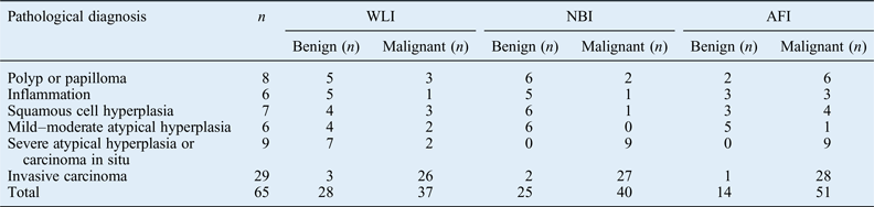
The study group comprised 50 patients (43 men and 7 women) with a median age of 55 years (range 33–86 years). Endoscopy examinations and biopsies were performed in the endoscopy clinic; no complications were observed. Pathological findings confirmed that 29 patients had SCCs (17 laryngeal cancers, 10 hypopharyngeal cancers and 2 oropharyngeal cancers), 4 had severe atypical hyperplasia and carcinoma in situ, 2 had mild or moderate atypical hyperplasia, 2 had squamous cell hyperplasia, 4 had polyps and papillomas, 2 had inflammation and 7 had no abnormality. A total of 22 patients also had an abnormal lesion in another part of the pharynx or larynx: pathology findings for biopsy specimens indicated five were severe atypical hyperplasia or carcinoma in situ, four were mild or moderate atypical hyperplasia, five were squamous cell hyperplasia, four were polyps or papillomas, and four were inflammation. Table I shows the classification of these benign and malignant lesions by white light image, narrow band imaging and autofluorescence imaging endoscopy.
Pathological diagnosis and endoscopy classification of laryngopharyngeal lesions

WLI = white light image endoscopy; NBI = narrow band imaging endoscopy; AFI = autofluorescence imaging endoscopy
Features of lesions under narrow band imaging and autofluorescence imaging endoscopy
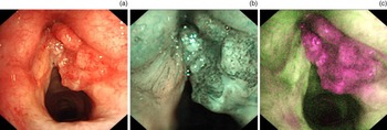
Under narrow band imaging endoscopy, malignant lesions had closely arranged brown spots in the lesion area and clearer lesion boundaries compared with white light image endoscopy: bigger spots were more clearly defined and more likely to be malignant. With cancer progression, punctate capillaries in the lesion area developed into structures resembling twisted cords (serpentine, earthworm or tadpole shaped; Fig. 1). Severe atypical hyperplasia and carcinoma in situ were characterised by large brown spots, while mild and moderate atypical hyperplasia was associated with significantly smaller visible spots that were further apart and in a less regular pattern. As leukoplakia was present on the lesion surface, simple hyperplasia was characterised by a white pseudomembranous covering and an absence of spots and inflammation by mild telangiectasia on the mucosal surface.
(a) Photomicrograph showing a right vocal fold squamous cell carcinoma under white light image endoscopy. (b) Under narrow band imaging, spots and twisted cord-like microvessels can be observed on the lesion surface. (c) Under autofluorescence imaging endoscopy, the lesion is shown as bright purple.

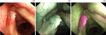
Under autofluorescence imaging endoscopy, normal laryngopharyngeal mucosa was typically indicated by bright green fluorescence; in contrast, mucosal inflammation or mild–moderate atypical hyperplasia was indicated by a pale purple fluorescence. In severe atypical hyperplasia or carcinoma in situ, dark purple fluorescence was clearly seen on the mucosal surface. Invasive carcinoma had a similar presentation to carcinoma in situ, namely, deep purple fluorescence on the lesion surface with clearer lesion contours and boundaries. Benign laryngopharyngeal lesions, such as polyps, papillomas and epithelial keratosis induced leukoplakia, were often characterised by purple fluorescence and thus easily misdiagnosed as malignant lesions (Fig. 2).
(a) Photomicrograph showing left vocal fold leukoplakia pathologically diagnosed as squamous cell hyperplasia with hyperkeratosis and parakeratosis in partial epithelium under white light image. (b) Under narrow band imaging, the surface is shown as white with no signs of malignancy. (c) Under autofluorescence imaging endoscopy, the surface is shown as bright purple and malignant lesion features were identified as false positives.

Diagnostic efficiency of all three endoscopy modes
For the statistical analysis, pathologically diagnosed severe atypical hyperplasia, carcinoma in situ and invasive carcinoma were classified as malignant lesions, while polyps, papillomas, inflammation, squamous cell hyperplasia and squamous epithelium with mild–moderate atypical hyperplasia were classified as benign lesions. The sensitivity and specificity for diagnosing malignant lesions were 73.7 per cent and 66.7 per cent, respectively, for ordinary white light image endoscopy; 94.7 per cent and 85.2 per cent, respectively, for narrow band imaging endoscopy; and 97.4 per cent and 48.1 per cent, respectively, for autofluorescence imaging endoscopy. There was no significant difference in sensitivity for diagnosing malignant lesions between narrow band imaging and autofluorescence imaging endoscopy, but both modes were more sensitive than white light image endoscopy (χ2 = 12.676, p = 0.002). Narrow band imaging endoscopy had a significantly higher specificity than both white light image and autofluorescence imaging endoscopy (χ2 = 8.333, p = 0.016). Thus, narrow band endoscopy was significantly more accurate for diagnosing benign and malignant lesions compared with white light image and autofluorescence imaging endoscopy (χ2 = 8.366, p = 0.01; see Table II for details).
Diagnostic capability of endoscopy modes for laryngopharyngeal malignant lesions*

* Defined as severe atypical hyperplasia, carcinoma in situ and invasive carcinoma. WLI = white light image endoscopy; NBI = narrow band imaging endoscopy; AFI = autofluorescence imaging endoscopy
Discussion
The main pathological subtype of malignant laryngopharyngeal tumour is SCC, which usually grade-increasing progresses gradually from a low-grade intraepithelial neoplasia through a high-grade intraepithelial neoplasia to carcinoma in situ. The slow progression provides a time window for the early detection, diagnosis and treatment of SCC. However, early detection of laryngopharyngeal SCC in the clinic has proven to be difficult because the lesions are superficial or small. Laryngoscopy is a useful method for diagnosing laryngopharyngeal diseases. In clinical practice, diagnosis by conventional electronic or fibre laryngoscopy is based on the overall morphological characteristics of lesions within or underlying the mucosa. This method mainly relies on visual observation under white light to identify lesions with significant morphological or colour changes (such as raised lumps or depressed ulcers); however, such small, flat lesions are difficult to diagnose and may often be missed. Cancer development on the mucosal surface may only be indicated by a reddened mucosa, which is difficult to distinguish visually from the surrounding normal pink mucosa. However, changing the endoscopic light source can overcome this problem. For example, narrow band imaging and autofluorescence imaging endoscopy are both designed to detect colour changes associated with abnormal lesions. Narrow band imaging endoscopy and autofluorescence imaging endoscopy are reported to have improved lesion detection rates and can identify early malignant lesions.Reference Ni, He, Xu, Gao, Lu and Yuan 5 – Reference Shi, Jin, Li, Wei and Xu 7 However, no studies have yet compared these two endoscopy modes for otorhinolaryngology tumours. The present study compared the clinical effectiveness of narrow band imaging endoscopy and autofluorescence imaging endoscopy to provide clinicians with reference data for endoscopy mode selection.
Narrow band imaging was first used for the endoscopic diagnosis of precancerous and early oesophageal cancerous lesions. In this technique, removing red light from the visible spectrum enables the transmission of light with central wavelengths of 415 nm (blue) and 540 nm (green). These narrow wavelengths have limited ability to penetrate into the tissue, thus highlighting the microstructure of the mucosal surface and microvessels.Reference Gono 8 – Reference Emura, Saito and Ikematsu 11 During squamous epithelium cancerisation, changes to the intraepithelial papillary capillary loop (capillaries at the terminal of lesion mucosal surface) may include significant expansion, extension, twisting and angiogenesis. These alterations are displayed on the endoscopy screen as a brown colour, while the surrounding normal mucosa is shown as light green. The visual contrast enables the detection of superficial lesions that are difficult to identify by normal white light image endoscopy. In addition, visualisation of morphological changes in the microvascular intraepithelial papillary capillary loop on the mucosal surface enables neoplastic and non-neoplastic lesions to be more accurately distinguished.Reference Inoue, Kaga, Ikeda, Sato, Sato and Minami 12
Autofluorescence imaging endoscopy was first used to detect early precancerous lesions of the tracheal mucosa.Reference He, Wang, Wu, Feng, Cao and Chen 13 – Reference Zaric, Becker, Perin, Stojanovic, Jovelic and Eri 15 This technique detects endogenous fluorophores (collagen, elastin, reduced nicotinamide adenine dinucleotide and flavin adenine dinucleotide) that are mainly aggregated in the submucosa of tissues; they represent the biochemical and structural characteristics of specific tissues. Upon irradiation of the mucosal surface with both blue (wavelength, 390–470 nm) and green (wavelength, 540–560 nm) light, the blue light may generate autofluorescence within the tissue and the green light is reflected. Autofluorescence at a wavelength of 470–690 nm is detected by a charge-coupled device, converted into electrical signals by the video processor and displayed on the endoscopy monitor. For normal submucosa, blue light excitation results in strong green autofluorescence; however, a thickened mucous layer in the lesion, along with increased blood flow in the tumour and reduced collagen fluorophores in submucosal connective tissues, leads to a weaker autofluorescence signal and a colour change to purple or bright purple. Therefore, the difference in colour can be used for direct diagnosis of lesions in a clinical setting.
-
• Early diagnosis of head and neck squamous cell carcinomas is difficult
-
• Image-enhanced endoscopy can improve the early detection of malignant laryngopharyngeal tumours
-
• Narrow band imaging had a high sensitivity and specificity for diagnosing malignant lesions
-
• Autofluorescence imaging endoscopy had a high sensitivity but lower specificity for diagnosing malignant lesions
-
• Overall, narrow band imaging was the best endoscopy mode for diagnosing laryngopharyngeal tumours
The present study identified the typical characteristics of laryngopharyngeal SCC under narrow band imaging endoscopy as clearly visible brown spots and twisted, expanded abnormal microvessels on the lesion surface. Under autofluorescence imaging endoscopy, the surface of malignant lesions was indicated by bright purple fluorescence. Both of these endoscopy modes were significantly better than white light image endoscopy at detecting superficial early laryngopharyngeal lesions and their boundaries, including severe atypical hyperplasia and carcinoma in situ. Thus, they may be useful in the early detection and treatment of malignant laryngopharyngeal tumours. Narrow band imaging had improved detection of vessels on the mucosal surface. Terminal microvessels on the mucosal surface have different morphological features in benign polyps, atypical hyperplasia and various cancer stages; these can be used to distinguish the different types of lesions and provide a clear differential diagnosis for benign and malignant lesions.Reference Folkman, Watson, Ingber and Hanahan 16 However, the diagnostic accuracy of narrow band imaging is reduced if microvessels on the mucosal surface are covered by leukoplakia or pseudomembrane. Overcoming this problem requires careful observation of the lesion surface by narrow band imaging. Malignant tumours are characterised by invasiveness and destruction of the original, adjacent and distant organs. Thus, visual examination of the lesion surface can identify capillaries exposed by vascular proliferation at the edge of malignant lesions, which are not seen in benign lesions, thus improving the differential diagnosis capacity of narrow band imaging endoscopy. The colour displayed under autofluorescence imaging endoscopy closely correlates with mucosal thickness. However, both benign and malignant lesions may have increased mucosal thickness and thus exhibit the same colour change. Therefore, specificity is lower for autofluorescence imaging endoscopy than for narrow band imaging endoscopy in the differential diagnosis of benign and malignant lesions.
Conclusion
This study found that narrow band imaging was better than autofluorescence imaging endoscopy and white light image endoscopy in both the diagnosis and differential diagnosis of malignant laryngopharyngeal lesions.
Acknowledgements
This study was supported by the National Natural Science Foundation of China (grant number 81470122) and Beijing Natural Science Foundation (grant number 7162162).