Introduction
The family Haploporidae Nicoll, 1914 comprises a cosmopolitan and taxonomically diverse group of intestinal digenean trematodes that parasitize teleost fishes inhabiting estuarine, marine, and freshwater environments in tropical and subtropical regions (Atopkin et al Reference Atopkin, Semenchenko, Solodovnik, Ivashko and Vinnikov2021; Overstreet and Curran Reference Overstreet, Curran, Jones, Bray and Gibson2005). Members of this family are morphologically characterized by the presence of a hermaphroditic sac and an external seminal vesicle (Overstreet and Curran Reference Overstreet, Curran, Jones, Bray and Gibson2005). The Haploporidae includes eight subfamilies: Haploporinae Nicoll, 1914; Megasoleninae Manter, 1935; Waretrematinae Srivastava, 1937; Chalcinotrematinae Overstreet & Curran, Reference Overstreet, Curran, Jones, Bray and Gibson2005; Forticulcitinae Blasco-Costa, Balbuena, Kostadinova & Olson, Reference Blasco-Costa, Balbuena, Kostadinova and Olson2009; Cadenatellinae Gibson & Bray, 1982; Hapladeninae Andres, Pulis, Curran & Overstreet, 2018; Pseudohaploporinae Atopkin, Besprozvannykh, Ha, Nguyen, Nguyen & Chalenko, 2018 (Atopkin et al Reference Atopkin, Besprozvannykh, Beloded, Ha, Nguyen and Nguyen2022).
The Chalcinotrematinae subfamily comprises a group of digenean endoparasites that infect cyprinodontiform, cichliform, characiform, gobiiformes, mugiliform, and siluriform fishes with a geographical distribution encompassing South America, North America, South and East Asia, and Africa (Andrade-Gómez et al Reference Andrade-Gómez, Sereno-Uribe and García-Varela2019; Overstreet and Curran Reference Overstreet, Curran, Jones, Bray and Gibson2005). The chalcinotrematin currently includes 43 species, classified into six genera: Chalcinotrema Teixeira de Freitas, 1947 (4 spp.), Megacoelium Szidat, Reference Szidat1954 (2 spp.), Paralecithobotrys Teixeira de Freitas, 1948 (3 spp.), Intromugil Overstreet & Curran, Reference Overstreet, Curran, Jones, Bray and Gibson2005 (4 spp.), Unicoelium Thatcher & Dossman, 1975 (1 sp.), and Saccocoelioides Szidat, Reference Szidat1954 (29 spp.) (Andrade-Gómez et al Reference Andrade-Gómez, Sereno-Uribe and García-Varela2019; Overstreet and Curran Reference Overstreet, Curran, Jones, Bray and Gibson2005). Members of these genera are morphologically characterized by having vitelline follicles surrounding the testis, a uterus that extends from the hindbody into the forebody, short prepharynx, and eggs which may or may not contain miracidia with eye-spots (Curran et al Reference Curran, Pulis, Andres and Overstreet2018; Overstreet and Curran Reference Overstreet, Curran, Jones, Bray and Gibson2005). Among these, the genus Megacoelium comprises two species currently known parasitising armour catfishes (Loricariidae) in South America, M. plecostomi Szidat, Reference Szidat1954 (type species) from Hypostomus plecostomus in Argentina and M. spinicavum Thatcher & Varella, Reference Thatcher and Varella1981 from Pterygoplichthys pardalis (type host) and P. multiradiatus in Brazil and Venezuela, respectively (Moravec and Prouza Reference Moravec and Prouza2024). To date, no molecular data are available for any species of Megacoelium, limiting inferences about their phylogenetic relationships within the Haploporidae.
The genus Saccocoelioides comprises haploporid digeneans that parasitize freshwater and brackish/marine fishes in the Americas and South and East Asia and is considered the most diverse genus within the Chalcinotrematinae (Andrade-Gómez et al Reference Andrade-Gómez, Sereno-Uribe and García-Varela2019), currently including 29 valid species, with 15 distributed in the Neotropical region (Martorelli et al Reference Martorelli, Montes, Barneche, Cardarella and Curran2022; Montes et al Reference Montes, Croci, Barneche, Ferrari, Reig Cardarella and Martorelli2024a,Reference Montes, Arredondo, Croci, Barneche, Balcazar and Reig Cardarellab). Species of this genus are characterized by having a small to moderately sized fusiform body, a weakly developed ventral sucker, extensive uterine coils occupying most of the hindbody, and vitelline follicles located laterally and posterior to the testis (Overstreet and Curran Reference Overstreet, Curran, Jones, Bray and Gibson2005). Recent molecular analyses have revealed that species currently assigned to Saccocoelioides do not form a single monophyletic lineage, with species distributed in at least two well-supported clades (Curran et al Reference Curran, Pulis, Andres and Overstreet2018; Martorelli et al Reference Martorelli, Montes, Barneche, Cardarella and Curran2022; Montes et al Reference Montes, Croci, Barneche, Ferrari, Reig Cardarella and Martorelli2024a,Reference Montes, Arredondo, Croci, Barneche, Balcazar and Reig Cardarellab). The first, referred to as the ‘minute clade’, includes morphologically slender, small-bodied species with a narrow vitelline distribution. In contrast, the ‘robust clade’, comprises species with a more robust body, denser tegumental spination, smaller eggs, and a massive uterus that extends over a large portion of the posterior body region (Curran et al Reference Curran, Pulis, Andres and Overstreet2018; Montes et al Reference Montes, Croci, Barneche, Ferrari, Reig Cardarella and Martorelli2024a,Reference Montes, Arredondo, Croci, Barneche, Balcazar and Reig Cardarellab). However, these molecular groups do not fully correspond to the morphological classification proposed in earlier studies, indicating that morphology alone may be insufficient to resolve their relationships (Montes et al Reference Montes, Arredondo, Croci, Barneche, Balcazar and Reig Cardarella2024b).
In this study, we provide the first molecular data for the genus Megacoelium, based on specimens of M. spinicavum recovered from P. pardalis in the Peruvian Amazon. Using an integrative approach that combines detailed morphological assessment and molecular phylogenetic analyses, we aim to elucidate the phylogenetic position of Megacoelium within Haploporidae. Additionally, we offer supplemental data on M. spinicavum based on newly collected specimens. Our findings contribute to a better understanding of the evolutionary history and host-parasite associations of chalcinotrematine digeneans in Neotropical freshwater ecosystems, and highlight the relevance of incorporating molecular tools in the systematics of poorly known digenean lineages.
Materials and Methods
Collection of Samples and Morphological Study
Four specimens of P. pardalis were obtained in November 2024 from artisanal fishermen in the Shapajal seasonal lagoon (08° 46′ 59″ S, 074° 08′ 20″ W), near the Tamaya River, Ucayali Region, Peru (Figure 1). Host identification followed Dávila et al. (Reference Dávila, Riveiro, Silva, de Loayza, Chávez, Ruiz and Renno2018). Fish hosts were examined for helminth infections, and five digeneans were recovered from the intestine of a single specimen. Digeneans were rinsed in 0.9% saline solution and fixed in hot 70% ethanol. Three individuals were stained with iron acetocarmine, dehydrated in a graded ethanol series, cleared in methyl salicylate, and mounted in Canada balsam for light microscopy. Observations and photographs were made using a Nikon™ Eclipse SI compound microscope, and drawings were prepared with the aid of a drawing tube. Measurements are given in micrometres (μm), unless otherwise indicated, as straight-line distances between the extreme points of each structure, and are presented as ranges followed by the mean and the number (n) of structures measured in parentheses. One specimen was post-fixed in 10% buffered formalin, dehydrated through an ethanol series, subjected to critical point drying with carbon dioxide, sputter-coated with gold, and examined under a scanning electron microscope (Inspect S50-FEI) at an accelerating voltage of 5–10 kV.
Sampling location of fish specimens in Ucayali Region, Peru. (Star): 08° 46′ 59″ S–074° 08′ 20″ W. Data from the World Cities Database, provided by Simplemaps (Pareto Software, LLC).
Source: Map created using Open Source QGIS. Bar = 2 km.

Specimens were deposited in the Helminthological Collection of the Museum of Natural History at the San Marcos University (MUSM-HEL), Lima, Peru.
Molecular Analysis
Genomic DNA was extracted from a portion of a single digenean specimen using the TIANamp Genomic DNA Kit (TianGen, Beijing, China) following the manufacturer’s protocol. A partial fragment of the 28S rDNA was amplified by polymerase chain reaction (PCR) using the forward primer digl2 (5′−AAG CAT ATC ACT AAG CGG−3′) and the reverse primer 1500R1 (5′–GCT ACT AGA TGG TTC GAT TAG–3′) (Tkach et al., Reference Tkach, Lotz, Swiderski and Esteban2002). A partial fragment of the mitochondrial cytochrome c oxidase subunit I gene (cox1) was amplified using the forward primer Cox5_Schisto (5′–TCT TTR GAT CAT AAG CG–3′) and the reverse primer JB5 (5′–AGC ACC TAA ACT TAA AAC ATA ATG AAA ATG–3′) (Lockyer et al., Reference Lockyer, Olson, Ostergaard, Rollinson, Johnston, Attwood, Southgate, Horak, Snyder, Le, Agatsuma, McManus, Carmichael, Naem and Littlewood2003; Derycke et al., Reference Derycke, Remerie, Vierstraete, Backeljau, Vanfleteren, Vincx and Moens2005; Fernandes et al., Reference Fernandes, Dos Santos, de Vasconcelos Melo, Achatz, Greiman, Bonilla and Tkach2021). PCR amplifications were carried out in a final volume of 25 μL, containing 12.5 μL of PCRBIO Taq Mix Red (PCR Biosystems), 0.3 μL of each primer (10 μM), 1.2 μL of template DNA, and nuclease-free water to volume. Amplification conditions included an annealing temperature of 55 °C for 28S and 45 °C for cox1. PCR products were purified using sparQ PureMag Beads (Quantabio, USA) according to the manufacturer’s instructions, and sequenced in PSOMAGEM Inc. (Brooklyn, New York, USA) with the Sanger sequencing method.
The 28S rDNA and cox1 sequences were assembled using ProSeq 2.9 beta (Filatov, Reference Filatov2002). The 28S rDNA dataset included sequences of species belonging to the clade comprising the subfamilies Chalcinotrematinae, Haploporinae, and Pseudohaploporinae, as proposed by Atopkin et al. (Reference Atopkin, Besprozvannykh, Beloded, Ha, Nguyen and Nguyen2022), whereas the cox1 dataset included sequences selected according to Montes et al. (Reference Montes, Croci, Barneche, Ferrari, Reig Cardarella and Martorelli2024a,Reference Montes, Arredondo, Croci, Barneche, Balcazar and Reig Cardarellab). Both datasets were aligned using Clustal W (Larkin et al Reference Larkin, Blackshields, Brown, Chenna, McGettigan, McWilliam, Valentin, Wallace, Wilm, Lopez, Thompson, Gibson and Higgins2007). Subsequently, each aligned dataset was analyzed with the software JModelTest2 (Santorum et al., Reference Santorum, Darriba, Taboada and Posada2014), which compares different models of DNA substitution in a hierarchical hypothesis-testing framework to select a base substitution model that best fits the data for each gene. The optimal nucleotide substitution models were TVM+I+G for the 28S rDNA sequences and TIM3+I+G for cox1 sequences, selected with the corrected Akaike information criterion (AICc; Posada and Buckley Reference Posada and Buckley2004). Next, the best model was implemented in MrBayes 3.2.7a (Huelsenbeck and Ronquist Reference Huelsenbeck and Ronquist2001) for Bayesian Inference (BI) analysis. For the BI analysis, unique random starting trees were used in the Metropolis-coupled MCMC (Ronquist et al Reference Ronquist, Teslenko, van der Mark, Ayres, Darling, Höhna, Larget, Liu, Suchard and Huelsenbeck2012). The analysis was performed for a total of 50,000,000 generations. Visual inspection of log-likelihood scores against generation time indicated that the log-likelihood values reached a stable equilibrium before the 100,000th generation. Thus, a burn-in of 1000 samples was conducted; every 100th tree was sampled from the MCMC analysis, obtaining a total of 100,000 trees, and tree topology represented the 50% majority rule consensus trees. Support for nodes in the BI tree topology was obtained by posterior probability. The phylogenetic analyses were conducted in the CIPRES Science Gateway V. 3.3 platform (http://www.phylo.org/ (accessed 2025)) (Miller et al Reference Miller, Pfeiffer and Schwartz2011). Phylogenetic trees were visualized and edited in Figtree 1.4.4 (Rambaut Reference Rambaut2009). Following Muff et al. (Reference Muff, Nilsen, O’Hara and Nater2022), we defined the following five categories for BI nodal support as: PP = 1: Fully supported; PP = 0.99–0.90: Strongly supported; PP = 0.89–0.80: Moderated support; PP = 0.79–0.70: Weakly supported; PP < 0.69: Not supported.
The proportion (p) of absolute nucleotide sites (p-distance) (Nei and Kumar Reference Nei and Kumar2000) was calculated to compare the genetic distance between sequences used. The p-value matrix was obtained using MEGA 12 (Kumar et al Reference Kumar, Stecher, Li, Knyaz and Tamura2018), employing the bootstrap method (5000 replicates) and uniform rate nucleotide substitution (transition + transversions). Newly generated sequences and other sequences used in this study are given in Table 1.
Collection data and GenBank accession numbers for sequences analysed in this study; new sequences in bold

*Outgroups.
Results
Haploporidae Nicoll, 1914
Chalcinotrematinae Overstreet & Curran, Reference Overstreet, Curran, Jones, Bray and Gibson2005
Megacoelium Szidat, Reference Szidat1954
Megacoelium spinicavum Thatcher & Varella, Reference Thatcher and Varella1981
Line drawing of Megacoelium spinicavum Thatcher & Varella, Reference Thatcher and Varella1981 (ventral view) from Pterygoplichthys pardalis (Siluriformes: Loricariidae) from the Ucayali Region.

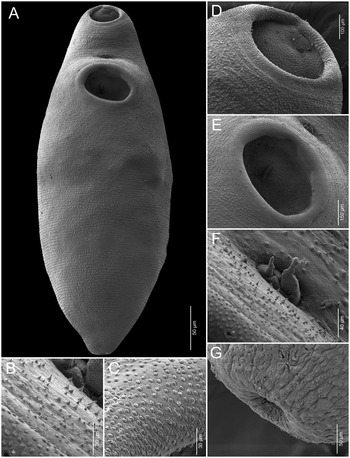
Scanning electron micrographs of Megacoelium spinicavum. A. Whole body, ventral view. B. Sharply pointed spines. C. Button-like spines. D. Oral sucker. E. Ventral sucker. F. Genital pore. G. Excretory pore.

Supplemental data (based on three specimens). Body subfusiform, 3.74–4.38 (4.12; n = 3) mm long, with rounded anterior end, narrowly tapering posterior end; greatest width 1.77–1.80 (1.78; n = 3) mm at level anterior of testis. Body length/body width ratio 2.08–2.48 (2.31; n = 3), body width/body length ratio 0.40–0.48 (0.43; n = 3). Tegument entirely covered with two spine types: small, button-like spines densely over the body surface, including the lumen of both suckers; sharply pointed spines localized in discrete rows anterior to the genital pore. Forebody 1.61–1.80 (1.74; n = 3) mm long, representing 41–43% of body length. Hindbody 2.13–2.58 (2.42; n = 3) mm long, representing 57–60% of body length. Oral sucker subglobular, subterminal, 529–691 × 684–704 (615 × 694; n = 3). Prepharynx absent. Pharynx muscular, 346–368 × 313–331 (356 × 320; n = 3). Intestinal caeca lateral to the ventral sucker and gonads, extending near the posterior margin of the testis. Ventral sucker oval, slightly larger than oral sucker, 748–885 × 756–979 (834 × 898; n = 3). Ventral/oral sucker ratio 1:0.71–0.78 long, 1:0.72–0.90 wide. Gonads tandem. Testis single, median, subtriangular, longer than wide, in posterior half of body, 1569–1660 × 538–595 (1618 × 559; n = 3). Hermaphroditic sac oval, 401–445 × 262–265 (427 × 264; n = 3), including internal seminal vesicle, pars prostatica, metraterm, and hermaphroditic duct; hermaphroditic duct distally bifurcated (Figure 3F). Hermaphroditic sac length/body length ratio 0.10–0.11 (0.10; n = 3). External seminal vesicle fusiform, 309–312 × 118–120 (310 × 119; n = 3). Genital pore opening medially between oral and ventral suckers. Ovary oval, pretesticular, dorsal and slightly lateral to testis, 251–266 × 280–295 (258 × 287; n = 4). Laurer’s canal and Mehlis’ gland not observed. Uterus extending between hermaphroditic sac and posterior margin of testis. Vitellarium composed of two lateral rows of follicles, extending from midlevel of ventral sucker to near posterior end of testis. Eggs 106–117 × 49–57 (111 × 53; n = 3); fully developed eggs containing miracidia with eye spots. Pharynx/egg ratio length 1:3.15–3.26. Excretory vesicle Y-shaped.
Taxonomy Summary
Host: Pterygoplichthys pardalis Castelnau (Siluriformes: Loricariidae), Amazon sailfin catfish.
Locality: Shapajal seasonal lagoon (08° 47′ 00″ S, 74° 08′ 34″ W), near the Tamaya River, Ucayali region, Peru.
Site in host: Intestine.
Infection levels: 5 worms observed, all from a single individual among the 4 hosts examined.
Deposited specimens: 1 voucher specimen (MUSM-HEL 5576), 1 hologenophore (MUSM-HEL 5577).
Representative DNA sequences: PV936516(cox1 mDNA) and PV936470 (28S rDNA).
Molecular Analyses
A partial sequence of the 28S rDNA gene (1223 bp) was obtained from a single specimen of M. spinicavum collected from P. pardalis. Combined with sequences retrieved from GenBank (Table 1), the final dataset comprised 46 terminals and 1116 aligned bp. In the resulting Bayesian phylogenetic tree (Figure 4), M. spinicavum formed a sister branch to Saccocoelioides bacilliformis Szidat, 1973 with weak support (PP = 0.78). This pair, in turn, was the sister group to a fully supported clade (PP = 1) containing S. magnus Szidat, Reference Szidat1954, S. elongatus Szidat, Reference Szidat1954, and S. miguelmontesi Montes, Croci, Barneche, Ferrari, Cardarella & Martorelli, 2024, all within the so-called ‘robust species’ group of Saccocoelioides (sensu Curran et al., Reference Curran, Pulis, Andres and Overstreet2018). The genetic distances between M. spinicavum and these species ranged from 2.5% to 3.7%, corresponding to 28–41 bp (Table 2).
Phylogenetic tree based on 28S region for Megacoelium spinicavum inferred by Bayesian inference (BI) to show their relationships with other species of Haploporidae. GenBank accession numbers are shown, and the scale bar indicates the nucleotide mutations per site.

Pairwise sequence divergences for 28S rDNA gene among Megacoelium spinicavum and Saccocoelioides spp. The proportion (p) of absolute nucleotide sites (p-distance) is shown as percentage (below the diagonal) and the raw number of bp-pairwise differences above the diagonal

*Outgroups.
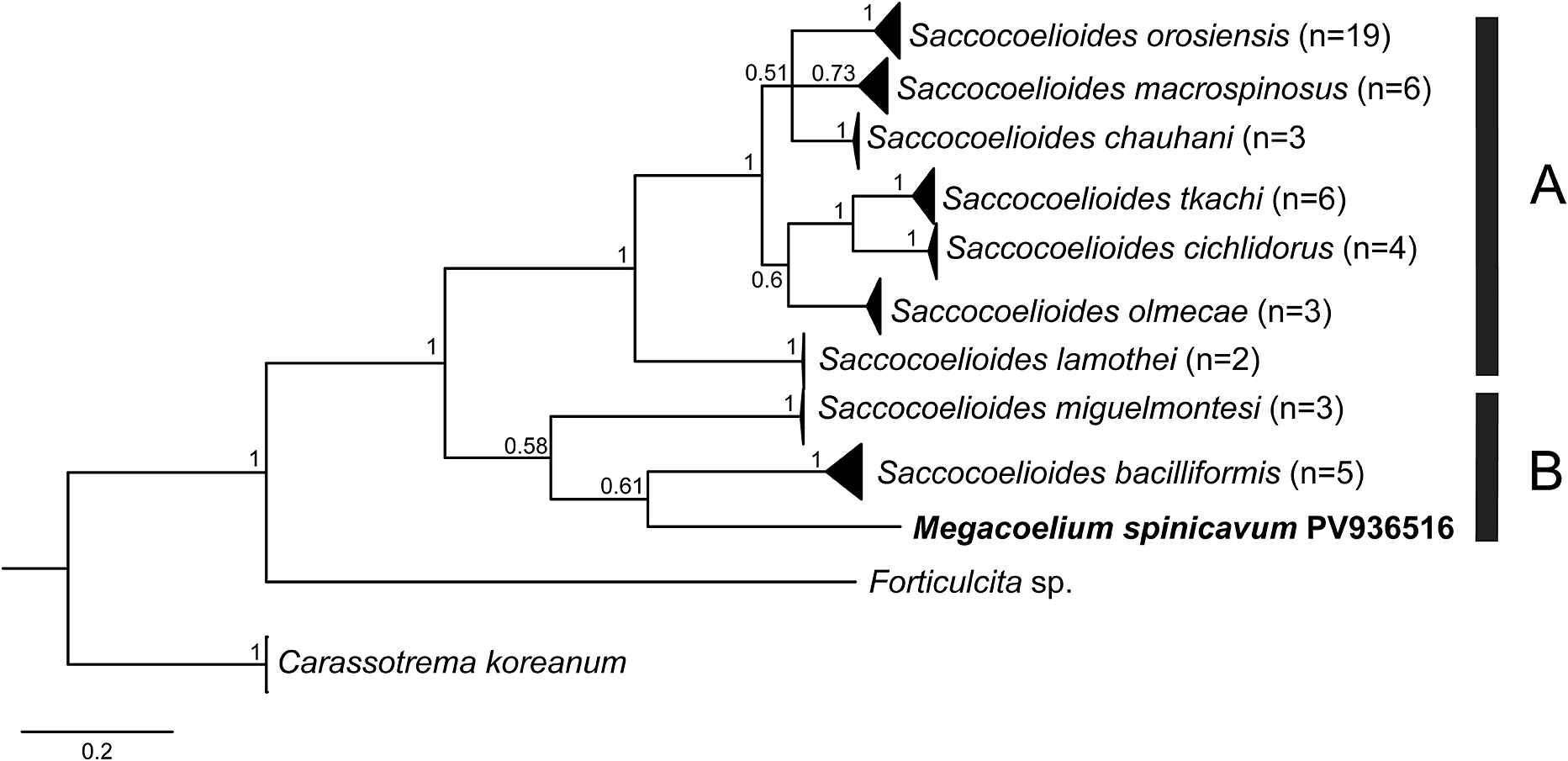
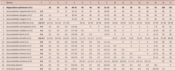
A partial sequence of the cox1 gene (582 bp) was obtained from M. spinicavum. Including GenBank sequences (Table 1), the final alignment contained 55 terminals and 562 bp. In the cox1 phylogenetic tree (Figure 5), M. spinicavum clustered within the ‘robust species’ group, alongside S. bacilliformis and S. miguelmontesi, although with low support (PP = 0.58). Pairwise p-distances and nucleotide differences between M. spinicavum and S. bacilliformis and S. miguelmontesi ranged from 17.5% to 18.4% (98–103 bp) and 19.9% (111 bp), respectively (Table 3).
Phylogenetic tree based on cox1 for Megacoelium spinicavum inferred by Bayesian inference (BI) to show their relationships with other species of Haploporidae. GenBank accession numbers are shown, and the scale bar indicates the nucleotide mutations per site.

Pairwise sequence divergences for Cox1 mtDNA gene among Megacoelium spinicavum and Saccocoelioides spp. The proportion (p) of absolute nucleotide sites (p-distance) is shown as percentage (below the diagonal) and the raw number of bp-pairwise differences above the diagonal

*Outgroups.
Discussion
The present study provides a detailed morphological supplemental data of M. spinicavum, including new line drawings and the first SEM images illustrating tegumental features. In addition, we present the first molecular data for this species, based on partial sequences of the 28S rDNA and the mitochondrial cox1 gene. Megacoelium spinicavum was originally described from P. pardalis (type host) and Pterygoplichthys sp. in Brazil (Thatcher and Varella, Reference Thatcher and Varella1981), and subsequently reported infecting the intestines of the Orinoco sailfin catfish P. multiradiatus (Hancock) in Venezuela (Moravec and Prouza, Reference Moravec and Prouza2024). The present work constitutes the first report of M. spinicavum from the Peruvian Amazon.
Our molecular analyses based on 28S rDNA and cox1 indicate that the sequences of M. spinicavum cluster within the ‘robust species’ clade of Saccocoelioides sensu Curran et al. (Reference Curran, Pulis, Andres and Overstreet2018) (Group B sensu Montes et al Reference Montes, Arredondo, Croci, Barneche, Balcazar and Reig Cardarella2024b), forming a weakly supported clade with Saccocoelioides bacilliformis Szidat, 1973, a species originally described from Psalidodon rutilus (Jenyns) (syn = Astyanax fasciatus) (Characiformes) in Argentina. In the 28S rDNA analysis, this clade (M. spinicavum + S. bacilliformis) was recovered as sister to a second clade that includes S. magnus Szidat, Reference Szidat1954; S. elongatus Szidat, Reference Szidat1954; and S. miguelmontesi Montes, Croci, Barneche, Ferrari, Cardarella & Martorelli, 2024, all of which have been consistently assigned to the ‘robust species’ clade of Saccocoelioides. Our specimens exhibited body lengths greater than 1.7 mm (3.74–4.38 mm) and eggs smaller than the pharynx (pharynx 346–368; eggs 106–117), a feature that resembles morphological conditions previously reported for taxa historically referred to as the ‘robust species’. However, we do not assign our specimens of M. spinicavum to any group composed of species of Saccocoelioides, as these represent a different genus. In contrast, these two lineages were clearly separated from the so-called ‘minute species’ clade of Saccocoelioides (Group A sensu Montes et al. Reference Montes, Arredondo, Croci, Barneche, Balcazar and Reig Cardarella2024b), which included S. nanii Szidat, Reference Szidat1954 (type species of the genus) and nine other Saccocoelioides species characterized by a slender body, a restricted vitelline field, relatively few large eggs, and a uterus confined to the hindbody (Curran et al Reference Curran, Pulis, Andres and Overstreet2018; Martorelli et al Reference Martorelli, Montes, Barneche, Cardarella and Curran2022; Montes et al Reference Montes, Croci, Barneche, Ferrari, Reig Cardarella and Martorelli2024a,Reference Montes, Arredondo, Croci, Barneche, Balcazar and Reig Cardarellab). Rather than supporting a ‘robust group of Saccocoelioides’, our results indicate that this assemblage represents a distinct lineage within Chalcinotrematinae that likely includes multiple genera not referable to Saccocoelioides. This interpretation is consistent with previous studies showing that Saccocoelioides, as currently defined, is not monophyletic (Curran et al Reference Curran, Pulis, Andres and Overstreet2018; Montes et al Reference Montes, Croci, Barneche, Ferrari, Reig Cardarella and Martorelli2024a,Reference Montes, Arredondo, Croci, Barneche, Balcazar and Reig Cardarellab), and requires abandoning the concept of a ‘robust species group’ in favour of a broader, lineage-based framework within Chalcinotrematinae.
Although our phylogenetic analyses place M. spinicavum together with S. bacilliformis within the ‘robust species’ clade of Saccocoelioides, these taxa exhibit marked morphological differences. These differences between both species include: (1) the presence of pigments in the anterior half of the body (absent in M. spinicavum), (2) the extension of intestinal caeca (reaching near the posterior margin of the testis in M. spinicavum vs terminating in the pre-testicular zone in S. bacilliformis), and the position of the testis (in the posterior half of the body in M. spinicavum vs in the middle of the body in S. bacilliformis). Megacoelium spinicavum was originally described as having a densely spinous tegument, with spines not only covering the body surface but also occurring within the lumen of both suckers (Thatcher and Varella Reference Thatcher and Varella1981). This characteristic has been corroborated herein via SEM observation, which revealed two distinct types of tegumental spines: (1) small, button-like spines distributed throughout the body surfaces and within the suckers; and (2) sharply pointed spines localized in discrete rows anteriorly, particularly surrounding the genital pore. In contrast, S. bacilliformis has been described as having a body entirely covered with sharply pointed spines, but this species remains unexamined under SEM.
The results presented here reinforce the hypothesis that Saccocoelioides is not monophyletic and support restricting the genus to the ‘minute species’ clade containing the type species. The ‘robust species’ clade appears to include at least three divergent lineages that may warrant recognition as separate genera: the first comprising S. miguelmontesi, S. elongatus, and S. magnus; the second represented by S. bacilliformis; and the third by M. spinicavum. Megacoelium plecostomi, the type species of the genus, was described as having a sigmoid-shaped testis and a spinous body, although spines within the suckers were not reported (Szidat Reference Szidat1954; Thatcher and Varella Reference Thatcher and Varella1981). In contrast, M. spinicavum possesses a subtriangular testis and conspicuous spines covering the body, including the lumen of both suckers. However, the absence of molecular data for M. plecostomi hinders clarification of its phylogenetic placement. Given the low support recovered for the nodes leading to the clades containing M. spinicavum, it is not feasible at this stage to propose new genera or to synonymize Megacoelium. For this reason, and following a conservative taxonomic approach, we retain M. spinicavum within Megacoelium until additional molecular data, particularly from type species or closely related congeners, are available to clarify the relationships among the major lineages. A comprehensive phylogenetic and morphological analysis is needed to resolve the taxonomic status of Megacoelium and the position of M. plecostomi, including the use of SEM to identify potential synapomorphies supporting formal generic designations.
Supplementary material
The supplementary material for this article can be found at http://doi.org/10.1017/S0022149X25101119.
Data availability
The obtained sequences are deposited in GenBank under the accession numbers PV936516.
Acknowledgements
This work was funded by the Consejo Nacional de Ciencia, Tecnología e Innovación Tecnológica (CONCYTEC) and the Programa Nacional de Investigación Científica y Estudios Avanzados (PROCIENCIA) under the call ‘E041-2024-03 Proyectos de Investigación Básica’ [PE501087644-2024].
Competing interests
The authors declare that they have no conflict of interest.
Ethics approval
Not applicable.
Consent to participate
Not applicable.
Consent for publication
All the authors provided their consent for the publication of this manuscript.