Introduction
For the elderly patients with an affective disorder, electroconvulsive therapy (ECT) is an effective and relatively safe treatment (Stek et al., Reference Stek, Wurff van der, Hoogendijk and Beekman2009). It is generally applied in persons with severe depression accompanied by psychosis, psychomotor retardation, refusal to eat and drink, suicidal tendencies, or in case of pharmacotherapy resistance (Salzman et al., Reference Salzman, Wong and Wright2002). The effects of ECT on neurocognitive functioning in elderly depressed patients are still unclear. It is generally assumed that, compared with younger adults, the elderly patients are more vulnerable to cognitive side effects after ECT and that the side effects are more severe (Zervas et al., Reference Zervas1993). However, most studies of ECT and neurocognitive functioning have focused on younger and mixed-age populations.
In the last decade six studies and two reviews about the cognitive side effects of ECT in older persons have been published. In both reviews (Gardner and O’Connor, Reference Gardner and O’Connor2008; Tielkes et al., Reference Tielkes, Comijs, Verwijk and Stek2008) mixed results were reported with respect to the effects of ECT on verbal memory. Improvement or no significant changes were found for global cognitive functioning, information processing speed, attention, and working memory shortly after ECT compared with pre-treatment functioning. At follow-up, most studies reported improvement of cognitive function (Gardner and O’Connor, Reference Gardner and O’Connor2008; Tielkes et al., Reference Tielkes, Comijs, Verwijk and Stek2008), but follow-up intervals are usually limited to two months or less. Only two studies (Stoudemire et al., Reference Stoudemire, Hill, Morris, Martino-Saltzman and Lewison1993; Brodaty et al., Reference Brodaty, Hickie, Mason and Prenter2000) examined long-term cognitive functioning in elderly depressed patients after ECT with global screening instruments. To date no study has examined the impact of ECT on autobiographical memory in older adults, whereas in younger adults (aged <65 years), an association between ECT and retrograde memory loss was found, especially for public, personal, and autobiographical events (Fraser et al., Reference Fraser, O’Carroll and Ebmeier2008). Also, in previous research, most of the attention was given to the effects of ECT on global cognitive and memory functioning, whereas research on other cognitive domains, such as executive functioning, is probably just as important as for daily functioning (Ingram et al., Reference Ingram, Saling and Schweitzer2008). In conclusion, from a clinical perspective it is highly relevant to investigate the nature and extent of changes across multiple domains of neurocognitive functioning in a sample of elderly depressed patients.
When studying cognitive side effects of ECT, it is important to take into account the most relevant treatment and clinical characteristics that may influence these cognitive side effects, such as electrode placement, age, and the severity of depressive symptoms. Although electrode positioning influences cognitive side effects following ECT, e.g. unilateral ECT being associated with significant smaller decreases in cognitive functioning than bilateral ECT (Semkovska et al., Reference Semkovska, Keane, Babalola and McLoughlin2011), most studies on the effects of ECT on neurocognitive functioning in the elderly patients do not discriminate between unilateral, bilateral, or mixed electrode placement (Gardner and O’Connor, Reference Gardner and O’Connor2008; Tielkes et al., Reference Tielkes, Comijs, Verwijk and Stek2008).
The present study examined cognitive changes in depressed elderly patients (aged ≥55 years) before (pre-ECT), within one week (short-term post-ECT [Stpost ECT]), and six months after completion (long-term post-ECT [Ltpost ECT]) of ECT. Acute post-ECT confusion (e.g. post-ictal or inter-ictal confusion) was not a focus of our study. All patients were prospectively evaluated for mood and neurocognitive outcomes over a course of treatment with brief pulse right unilateral (RUL) ECT or RUL followed by bilateral (BL) ECT. Next to the general cognitive functioning and anterograde memory, we assessed retrograde memory, attention, processing speed, and executive functions. The association between cognitive functioning and the most important treatment and clinical characteristics were studied. With respect to the issue of representativity of our results, we examined the characteristics of patients who were too ill or refused to participate in the study.
Methods
Participants
This naturalistic study of depressed elderly patients was conducted at the academic department of old age psychiatry of GGZinGeest/VU Medical Center in Amsterdam. Patients aged 55 years and older, referred for an acute course of ECT treatment, suffering from a primary major depressive disorder or bipolar depression (with or without psychosis) diagnosed with the Diagnostic and Statistical Manual of Mental Disorders (DSM)-IV criteria by two experienced old age psychiatrists, were included in this study. Patients came for ECT from referrals in the catchment area and from tertiary referrals throughout the Netherlands. Over a five-year period (2002–2007), 72 patients were treated with ECT. At baseline, socio-demographic and clinical data were noted: age, gender, marital status, in/out-patient status, age of onset, present major depressive episode duration, psychosis, number of previous episodes, number of previous admissions, and previous ECT treatment. The scoring system of Verhage (Reference Verhage1964; range 1–7) was used to define patient's level of education. All patients provided written informed consent to participate. The institutional review board of GGZinGeest approved the study.
ECT procedures
Patients received a course of twice weekly ECT. A course started preferably with right unilateral stimuli in order to diminish the effect of ECT on cognition (NICE, 2003). All treatments were administered using the Thymatron system IV (Somatics, LLC, Lake Bluff, Illinois, USA; maximum energy 200%, 1008 mCoulombs) with a brief pulse (1.0 milliseconds) setting. Age determined the energy supplied, e.g. a 75-year-old patient received right unilateral ECT at a dosage of 75%, corresponding to 378 mCoulombs. When treated bilaterally (only bitemporal), half of this dosage was considered adequate (Petrides and Fink, Reference Petrides and Fink1996). Etomidate (0.2–0.3 mg/kg) and succinylcholine (1–2 mg/kg) were used for anaesthetic induction and muscle relaxation respectively. A motor seizure of less than 20 seconds was considered as inadequate. Due to acute life-threatening conditions, four patients were treated bilaterally from the start of the ECT course. Clinical evaluation was carried out weekly. ECT treatment was stopped if patients remitted or showed no further improvement in clinical conditions during the last two weeks of ECT sessions. Switching to bilateral ECT was done when the clinical condition worsened or after six unilateral treatments without effect despite adequate seizures. Worsening was determined by debilitating psychotic features, increased suicidal thoughts, dehydration, or weight loss. The number of ECT sessions and switching to bilateral ECT were determined on clinical grounds by the treating psychiatrist. Psychotropics were tapered off prior to the start of ECT. Antipsychotics were allowed when clinically indicated.
Assessments
Clinical assessment
Two experienced geriatric psychiatrists, blind to the neurocognitive data, confirmed the diagnosis of depression according to the DSM-IV criteria and the presence or absence of psychotic and melancholic features. The Montgomery–Åsberg Depression Rating Scales (MADRS; Montgomery and Åsberg, Reference Montgomery and Åsberg1979; range 0–60 and remission <10) were completed by the same trained research nurse for all the patients before the first ECT, every one to two weeks during ECT, and one week after the final ECT.
The Center for Epidemiological Studies-Depression scale (CES-D; range 0–60; Radloff, Reference Radloff1977) is a self-reported depression scale and was completed together with the neuropsychological assessment before the first ECT (pre-ECT) and then within one week after the final ECT (Stpost ECT) and six months (Ltpost ECT) after the final ECT.
Neuropsychological assessment
Neurocognitive assessment was performed by a neuropsychologist or a supervised neuropsychology trainee. The cognitive domains studied included global cognitive status, episodic memory (verbal, anterograde, and retrograde), verbal fluency, speed of information processing, and executive function/working memory. The specific instruments in the test battery were as follows: the Mini-Mental State Examination (MMSE) (global cognitive function; Folstein et al., Reference Folstein, Folstein and McHugh1975), the Visual Association Test (VAT) (automatic learning; Lindeboom et al., Reference Lindeboom, Schmand, Tulner, Walstra and Jonker2002), a modified version of the auditory verbal learning test (Rey, Reference Rey1964), 10 Words Verbal Learning Test (verbal learning, retention), a self-designed autobiographical memory questionnaire (episodic autobiographical memory), including 18 questions about personal semantic memories and seven questions about public events (range 0–25), Category Fluency Test – Animals (semantic memory; Luteijn and van der Ploeg, Reference Luteijn and van der Ploeg1983), Letter Fluency Test-“D” (executive function; Luteijn and van der Ploeg, Reference Luteijn and van der Ploeg1983), the Digit Span Forwards and Backwards – Wechsler Adult Intelligence Scale III (WAIS III) (attention and working memory; Wechsler, Reference Wechsler2000), the Expanded Mental Control Test (EMCT) (mental control; Lindeboom et al., Reference Lindeboom, Koene and Matto1993), the Stroop Color Word Test (attention and executive functioning; Strauss et al., Reference Strauss, Sherman and Spreen2006), the Trail Making Test (TMT) (attention, visual motor speed, and executive function; Reitan, Reference Reitan1979), and two subtests (Key Search and Rule Shift Cards) of the Behavioral Assessment of the Dysexecutive Syndrome (BADS) (executive function; Wilson et al., Reference Wilson, Evans, Alderman, Burgess, Emslie and Rabbitt1997).
Statistical analysis
All patients who completed a pre- and post-ECT cognitive assessment were included in the analysis. To describe the cognitive changes of ECT, the mean raw scores of each test were calculated together with the standard deviation and range of performance. Paired t-tests were used to analyze differences in pre-ECT, Stpost ECT, and Ltpost ECT neurocognitive outcomes.
In order to quantify clinical relevance, we calculated the effect sizes (Cohen's d) of the Stpost ECT and Ltpost ECT neurocognitive outcomes. A negative effect size indicates that the cognitive performance after ECT was worse than before ECT. Effect sizes are interpreted as suggested by Cohen (Reference Cohen1988): effect size of 0.2–0.5 is considered a small effect, 0.5–0.8 a medium effect, and ≥0.8 a large effect.
Multivariate linear regression analyses were performed to investigate the relation between electrode placement, total number of treatment sessions, severity of depression, age, and neurocognitive outcome. Results were considered statistically significant at p < 0.01. All statistical tests were carried out using IBM SPSS version 20.0 (IBM, 2012).
Results
Clinical and demographic characteristics
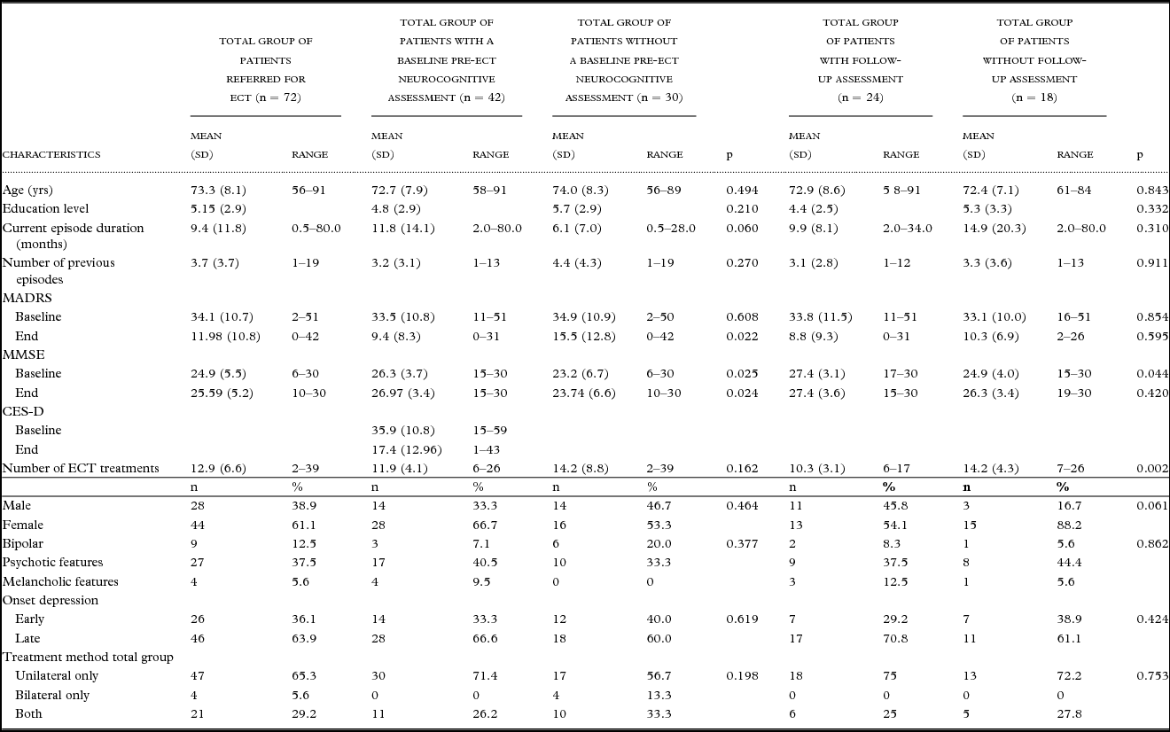
From the 72 ECT patients, 30 (41.6%) were not able to perform an extensive baseline neurocognitive assessment and were excluded from the analyses. This group had a significant lower global cognitive functioning pre-ECT and Stpost ECT (Table 1). The Stpost ECT MADRS score of this group was significantly higher; no difference was found between the groups for the MADRS scores measured before the start of ECT.
Demographic and clinical characteristics

Notes: MADRS: Montgomery–Åsberg Depression Rating Scales; MMSE: Mini-Mental State Examination; CES-D: Center for Epidemiological Studies-Depression scale.
Forty-two patients (65.9% females) received both baseline and post-ECT testing of clinical and neurocognitive parameters. The mean age of this group was 72.7 years (Table 1) and the education level was 4.8. A bipolar disorder was diagnosed in 7.1% of the patients. Psychotic and melancholic features were seen respectively in 40.5% and 9.5% of the patients. In 66.6% of the patients, the onset of depression was late (≥55 years).
In 71.4% of the patients, only unilateral ECT was used, and in 26.2% both unilateral and bilateral ECT treatments were used. The mean total number of ECT treatments was 11.9. The mean MADRS scores decreased from 33.5 to 9.4 and the CES-D scores decreased from 35.9 to 17.4. The mean pre-treatment MMSE score was 26.3 (Table 2). Twenty-four of the 42 patients (57.1%) also performed the Ltpost ECT neurocognitive assessment. Due to refusal or inability to comply with the testing procedures, 18 patients were lost in follow-up. This group had a significantly lower global cognitive functioning pre-ECT and a significantly higher total number of ECT treatments (Table 1).
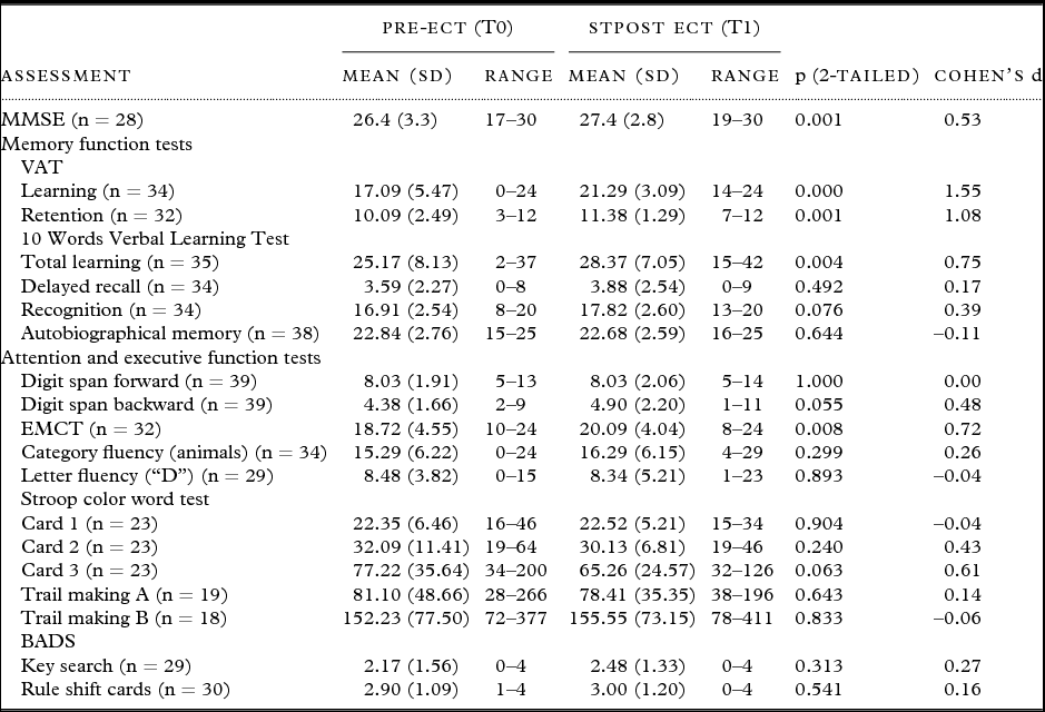
Neuropsychological test scores pre- and Stpost ECT (n = 42)
Notes: STpost ECT: short-term post-ECT; MMSE: Mini-Mental State Examination; VAT:Visual Association Test; EMCT: Expanded Mental Control Test; BADS: Behavioral Assessment of the Dysexecutive Syndrome.
Neurocognitive assessments
Neurocognitive effects STpost ECT
Improvement of neurocognitive functioning was statistically significant for the MMSE, VAT (automatically learning and retention), 10 Words Verbal Learning Test (total learning), and EMCT (Table 2). Effect sizes were medium to large. We found no statistically significant decreases in cognitive functioning. With multivariate linear regression we found neither effect for age nor effect of electrode placement, total number of treatment sessions, or severity of depression on the Stpost ECT neurocognitive measurements.
Neurocognitive effects LTpost ECT
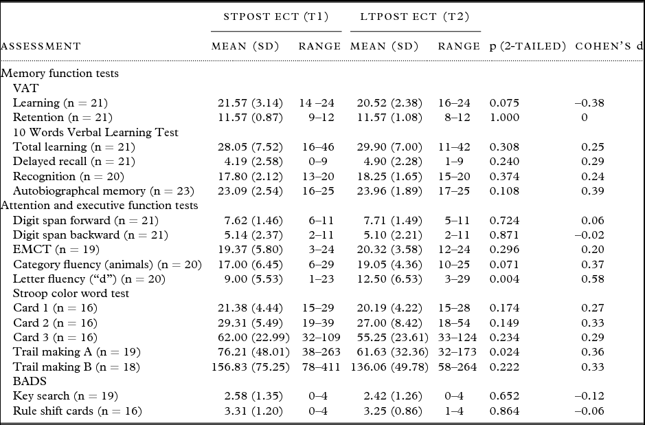
In the group of 24 patients with measurements Ltpost ECT, compared with the Stpost ECT performance, statistically significant improvements were only found for the TMT-A (attention) and the Letter Fluency Test (executive functioning) (Table 3). The effect sizes were small to medium. Cognitive functioning did not decrease in any domain. We found no association between electrode placement, total number of treatment sessions, severity of depression, electrode placement, and Ltpost ECT neurocognitive measurements. No effect was found between age and change in neurocognitive functioning Stpost ECT and Ltpost ECT.
Neuropsychological test scores Stpost ECT and Ltpost ECT (n = 24)
Notes: STpost ECT: short-term post-ECT; LTpost ECT: long-term post-ECT; MMSE: Mini-Mental State Examination; VAT: Visual Association Test; EMCT: Expanded Mental Control Test; BADS: Behavioral Assessment of the Dysexecutive Syndrome.
Discussion
The present study examined whether and to what extent neurocognitive changes occurred within one week (Stpost ECT) and six months (Ltpost ECT) after finishing ECT in severely depressed elderly patients. Global cognitive function and memory, such as anterograde memory and retrograde memory, as well as non-memory cognitive functions, including attention and executive functions, were assessed. Our results show that significant positive neurocognitive changes occurred in some but not all measured neurocognitive domains and our data offer no evidence of cognitive deterioration, given its power to detect a difference.
Global cognitive functioning (MMSE) shows a significant improvement one week after ECT compared with pre-ECT performances. Similar positive findings have been reported shortly after ECT in four recent studies (Tew et al., Reference Tew, Mulsant, Haskett, Dolata, Hixson and Mann2002; Stoppe et al., Reference Stoppe, Louza, Rosa, Gil and Rigonatti2006; O’Connor et al., Reference O’Connor, Gardner, Eppingstall and Tofler2010; Hausner et al., Reference Hausner, Damian, Sartorius and Frolich2011). In their studies, Tew et al. (Reference Tew, Mulsant, Haskett, Dolata, Hixson and Mann2002) and Hausner et al. (Reference Hausner, Damian, Sartorius and Frolich2011) measured global cognitive functioning solely by applying MMSE. Tew et al. (Reference Tew, Mulsant, Haskett, Dolata, Hixson and Mann2002) tested before and one to three days after completing ECT in the RUL ECT group (n = 13). The mean improvement in the MMSE score in their study was higher than in our study, but was not statistically significant, probably due to the small sample size. Hausner et al. (Reference Hausner, Damian, Sartorius and Frolich2011) did their cognitive testing six weeks after completing RUL ECT (n = 13) and found a statistically significantly mean improvement on MMSE, although 44.8% of the patients still had a lower MMSE score than before treatment. This could imply that there is a specific subgroup of patients vulnerable to neurocognitive changes in ECT.
Concerning anterograde memory, total verbal learning (10 Words Verbal Learning Test) also showed significant improvement Stpost ECT compared with pre-ECT performances. After the ECT session, patients were able to recollect more new learned information immediately after they have heard this information. In contrast to our study, O’Connor et al. (Reference O’Connor, Gardner, Eppingstall and Tofler2010) and Stoppe et al. (Reference Stoppe, Louza, Rosa, Gil and Rigonatti2006) found no significant improvement in recollecting new learned verbal information one month after the last ECT session (n = 17). In addition, delayed verbal memory post-ECT, compared with our results, did not significantly improve. After ECT session, patients are not able to consolidate more verbal information. Further comparison with the study of Stoppe et al. (Reference Stoppe, Louza, Rosa, Gil and Rigonatti2006) showed that, in line with our results, no change in attention (digit span forwards), working memory (digit span backwards), and autobiographical memory was found. Our study is the first study that measured automatic learning (VAT) and mental control (EMCT). We found significant improvement in these cognitive functions one week after ECT compared with pre-ECT performances.
Our study is one of three studies in older adults to date that examined long-term effects of ECT on cognitive functioning. We demonstrated that cognitive performances remained stable six months after completing ECT, except for a small to medium improvement on both speed and attention (TMT-A) and on a test appealing to executive functioning (Letter Fluency Test).
In contrast to our results in the depressed elderly patients, many previous studies in younger depressed patients treated with ECT concluded that neurocognitive functioning, such as memory, deteriorates immediately after ECT (Schat et al., Reference Schat, van den Broek, Mulder, Birkenhager, van Tuijl and Murre2007; Sackeim et al., Reference Sackeim2008; Kellner et al., Reference Kellner2010), but this decreased functioning at least improved to the level of pre-ECT performances within one to six months post-ECT. This suggests that cognitive changes after ECT treatment might be age-related, with the elderly patients being less vulnerable to cognitive deterioration than younger adults. In our 55+ years age group, we found no association between age and change in neurocognitive functioning before and post-ECT. This could imply that cognitive outcome after ECT is not at all associated with age in the elderly depressed group (Kohler et al., Reference Kohler, Thomas, Barnett and O’Brien2010).
In this respect and in agreement with our results, there are two recent studies in younger adults (Sackeim et al., Reference Sackeim2008; Sienaert et al., Reference Sienaert, Vansteelandt, Demyttenaere and Peuskens2010) that also found improvement in several cognitive domains. These inconsistent results in younger depressed patients might be due to differences in ECT technique. Sackeim et al. (Reference Sackeim2008) used an ultrabrief pulse width stimulation technique in their study. It has been suggested that ultrabrief pulse ECT causes less cognitive side effects than brief pulse stimulation (Loo et al., Reference Loo, Katalinic, Martin and Schweitzer2012; Verwijk et al., Reference Verwijk, Comijs, Kok, Spaans, Stek and Scherder2012). In the study of Sienaert et al. (Reference Sienaert, Vansteelandt, Demyttenaere and Peuskens2010) ultrabrief pulse as well as lower frequency pulse twice a week was practised. We also assume that the total number of ECT sessions is important when studying cognitive side effects of ECT. Mixed-age population studies have indeed found a relation between the total number of ECT sessions and cognitive decline (Sackeim et al., Reference Sackeim2008). We did not find this effect in our sample.
It is clear that more research is warranted to study the effect of ECT parameters such as pulse width (brief pulse vs. ultrabrief pulse), ECT frequency (three times vs. two times a week), and total number of ECT sessions with respect to changes in cognitive functioning after ECT.
Another possible explanation for conflicting results is the severity of depression and cognitive dysfunction in depression while measuring cognition before and after ECT (Fraser et al., Reference Fraser, O’Carroll and Ebmeier2008). Mixed-age population studies concluded that neurocognitive performance was indeed positively correlated with improvement in depression (Warren and Groome, Reference Warren and Groome1984; Brodaty et al., Reference Brodaty, Berle, Hickie and Mason2001). In our sample, neurocognitive measurements were not influenced by severity of depression or change in depression severity. More recent studies in elderly population also report no influence of depression severity on neurocognitive performance (Tew et al., Reference Tew, Mulsant, Haskett, Dolata, Hixson and Mann2002; Stoppe et al., Reference Stoppe, Louza, Rosa, Gil and Rigonatti2006; O’Connor et al., Reference O’Connor, Gardner, Eppingstall and Tofler2010; Hausner et al., Reference Hausner, Damian, Sartorius and Frolich2011) except that verbal memory recognition was impacted by the severity of depressive symptoms (Stoppe et al., Reference Stoppe, Louza, Rosa, Gil and Rigonatti2006).
Another recent study showed that the severity of a depressive episode is unrelated to the degree of global cognitive or memory difficulties in patients with severe depression (McClintock et al., Reference McClintock2010). Complementary to these results, in another ECT study, Bayless et al. (Reference Bayless, McCormick, Brumm, Espe-Pfeifer, Long and Lewis2010) suggested that it is not the severity but negative symptoms of a depressive episode that are related to cognitive dysfunction. Improvement in negative symptoms after successful treatment may be related to an improvement in cognitive functioning (Kohler et al., Reference Kohler, Thomas, Barnett and O’Brien2010). Possibly, conflicting results in ECT studies may also originate from the heterogeneity of depression in elderly patients, and severity as expressed in total scores of depression rating scales is only part of the story. In our study we had insufficient power to analyze the relation between unipolar and bipolar depression and cognitive function separately. Future studies should take into account not only the type of depression but also the phenomenology of depression (e.g. negative symptoms “loss of interest, lack of initiative”) when investigating neurocognitive functioning in depression.
Limitations
Our results suggest no change in cognitive performance after ECT or even improvement in some cognitive domains. These results seem to be highly encouraging for our patients receiving ECT. But from a more critical perspective, no change or improvement in cognitive performance after ECT means that cognitive performance after ECT is the same as or better than cognitive functioning during a depressive episode. It could be well possible that the cognitive performances of patients who remitted do not reach the same level as in a euthymic state or compared with healthy controls. Without a control group of equally depressed patients who were treated by some other treatment with an equal level of success, it is hard to determine how the positive and negative cognitive effects of ECT work out in practice. For example, studies in drug-free euthymic patients showed residual executive dysfunction in young, middle-aged, and elderly people (Weiland-Fiedler et al., Reference Weiland-Fiedler2004; Gallagher et al., Reference Gallagher, Robinson, Gray, Porter and Young2007; Baba et al., Reference Baba2010). These findings indicate that a pattern of persistent cognitive dysfunction in depression is common, which should be taken into account when interpreting changes in neurocognitive function after ECT, even when cognitive performances improve.
A point of concern is the attrition rate in all studies on this topic. It is hard to examine neurocognitive performance in elderly severely depressed patients. O’Connor et al. (Reference O’Connor, Gardner, Eppingstall and Tofler2010) discussed these low recruitment rates as a major limitation. Patients with the greatest risk of cognitive deficits are not able to participate in these studies, limiting the capacity to generalize findings. In our study we reported a recruitment rate of 58.4%. In comparable studies (Stoppe et al., Reference Stoppe, Louza, Rosa, Gil and Rigonatti2006; O’Connor et al., Reference O’Connor, Gardner, Eppingstall and Tofler2010) a broad range in recruitment rates from 35 to 82% was found. In our study, patients who completed the neurocognitive assessments had a higher MMSE score before the start of ECT and we found no differences with respect to severity of depression. This suggests that patients who are high at risk for developing cognitive decline post-ECT (Hausner et al., Reference Hausner, Damian, Sartorius and Frolich2011) are excluded from our and other studies. The same concern could be the case for the dropout rate for those patients who were not able or refused to comply with the follow-up assessment. Of the recruited patients, 42.9% did not perform an assessment six months post-ECT. This group had a significantly higher number of ECT treatments and is generally associated with more pronounced cognitive side effects (Sackeim et al., Reference Sackeim, Prudic, Fuller, Keilp, Lavori and Olfson2007).
Another limitation is that we had no detailed neurological status of these patients, which could have had a major impact on cognitive status (Flint and Gagnon, Reference Flint and Gagnon2002).
There is a small chance (one out of 19) that we found some spurious findings (type one error) due to multiple comparisons. However, when the chance of incorrectly producing a difference on an individual test (type one error) is reduced, the chance of making a type two error is increased, which implies that no effect or difference is declared, while in fact there is an effect. Thus, by reducing the chance of type one errors for individual tests, the chance of type two errors is increased for all tests. Moreover, if one tests for the significance of an association using variables that are mutually correlated, the Bonferroni correction is even too conservative (Perneger, Reference Perneger1998). As the chance of finding a type one error is very small (one out of 19) and the findings in our study are not unexpected and consistent with other research, we did not use the Bonferroni correction.
The finding that our group of patients seems to have no cognitive changes is probably not a representative of all patients individually. We did not perform analysis on the subgroup of patients who did poorly; and looking into cognitive impairment of individual patients is beyond the scope of this study. If we were able to identify a subgroup of patients that were vulnerable to cognitive side effects, we might have been successful in preventing such a decline. Finally, lack of a standardized diagnostic instrument could be a methodological weakness in our study, although clinical diagnosis of depression was made in all patients by two very experienced geriatric psychiatrists.
Conclusions
In conclusion, our naturalistic clinical follow-up study shows that performance in multiple cognitive domains in elderly depressed patients after ECT remains stable or improves after ECT. Since patients with the poorest cognitive function were not able to participate in an extensive neuropsychological assessment before ECT, these results may not apply to patients with more severe cognitive impairment prior to the start of ECT.
Conflict of interest
None.
Description of authors’ roles
E. Verwijk wrote the paper and carried out the statistical analysis. H.C. Comijs designed the study, supervised the data collection, and assisted with the writing of the paper. R.M. Kok and H.P. Spaans assisted with the writing the paper. C.E.M. Tielkes collected the data and assisted with the writing of this paper. E.J.A. Scherder supervised and assisted with the writing this paper. M.L. Stek supervised and designed the study, supervised the data collection, and assisted with the writing of the paper.