Repair of tetralogy of Fallot often results in pulmonary regurgitation. The resultant chronic volume overload can lead to right ventricular dilatation, biventricular dysfunction, heart failure symptoms, arrhythmias, and sudden death.Reference Murphy, Gersh and Mair1–Reference Bouzas, Kilner and Gatzoulis5 Despite this awareness of the late consequences of chronic pulmonary regurgitation, ventriculotomy with transannular patch remains the most prevalent technique currently used for tetralogy of Fallot repair.Reference Al Habib, Jacobs and Mavroudis6, Reference Sarris, Comas, Tobota and Maruszewski7 Therefore, a substantial portion of the patients who underwent repair of tetralogy of Fallot will require pulmonary valve replacement in the future.
Most studies dealing with pulmonary valve replacement in patients with chronic pulmonary regurgitation have consistently reported subjective improvements in functional class.Reference Geva, Gauvreau and Powell8–Reference Scherptong, Hazekamp and Mulder13 However, there are conflicting results regarding objective improvements in exercise capacity as assessed by cardiopulmonary exercise test. Ghez et alReference Ghez, Tsang and Frigiola14 reported that although most patients showed clinical improvement after pulmonary valve replacement, maximal exercise capacity as assessed by maximal oxygen consumption did not improve after pulmonary valve replacement. Frigiola et alReference Frigiola, Tsang and Bull11 have also found that there was no improvement in maximal oxygen consumption after pulmonary valve replacement.
Magnetic resonance imaging has emerged as a gold standard for evaluating right ventricular volumes and function in patients with pulmonary regurgitation. Many studies using magnetic resonance imaging have confirmed a substantial decrease or normalisation of right ventricular volumes after pulmonary valve replacement.Reference Geva, Gauvreau and Powell8–Reference Frigiola, Tsang and Bull11 Oosterhof et al,Reference Oosterhof, van Straten and Vliegen10 in a study of 71 patients with repaired tetralogy of Fallot, showed decrease in right ventricular volumes of ∼30% after pulmonary valve replacement and reported that they could not find a threshold above which right ventricular volumes did not decrease after surgery. There is a question of whether the reported decrease in right ventricular volumes after pulmonary valve replacement is merely the result of surgical right ventricular reduction – resection or plication of right ventricular outflow tract aneurysm. Although surgical right ventricular reduction at the time of pulmonary valve replacement can possibly result in a greater relative decrease in right ventricular volumes, substantial decrease in right ventricular volumes was also noted in patients who did not undergo surgical right ventricular reduction.Reference Oosterhof, van Straten and Vliegen10
Although pulmonary valve replacement can consistently lead to a substantial decrease of right ventricular volumes, improvement in right ventricular systolic function has not been uniformly demonstrated. Studies reporting no improvement of right ventricular function enrolled patients with already depressed right ventricular function, whereas studies reporting improvement in right ventricular function enrolled patients with preserved right ventricular function.Reference Geva, Gauvreau and Powell8, Reference Frigiola, Tsang and Bull11, Reference Ghez, Tsang and Frigiola14–Reference Harrild, Berul and Cecchin17 This implies that pulmonary valve replacement should be performed before irreversible right ventricular dysfunction occurs.
There have been also conflicting results regarding the impact of pulmonary valve replacement on QRS duration and arrhythmia propensity, and this also might be due to the different characteristics of the patients enrolled in the studies. Studies reporting no change of QRS duration enrolled patients with relatively longer baseline QRS duration or larger right ventricular volumes compared with those reporting improvement in QRS duration.Reference Oosterhof, van Straten and Vliegen10, Reference Harrild, Berul and Cecchin17, Reference Therrien, Siu and Harris18–Reference Gengsakul, Harris and Bradley20 This implies that timely pulmonary valve replacement before severe right ventricular dilatation occurs may have a beneficial effect on QRS duration.
Currently, there are no published data showing a clear long-term survival gain of pulmonary valve replacement. Gengsakul et al,Reference Gengsakul, Harris and Bradley20 in a matched comparison study, reported that there was no difference regarding the composite outcome of death and ventricular tachycardia between patients who had undergone pulmonary valve replacement and patients who had not undergone pulmonary valve replacement. Harrild et alReference Harrild, Berul and Cecchin17 also reported similar results. Long-term follow-up results of carefully designed studies are mandatory to draw a definitive conclusion on this important issue.
Benefits of pulmonary valve replacement have to be weighed against the risks of this procedure. At the present time, pulmonary valve replacement can be performed with low operative mortality. Cheung et al,Reference Cheung, Wong and Cheung21 in a meta-analysis of pulmonary valve replacement after repair of tetralogy of Fallot, reported that the pooled early mortality rate was 2.1% (95% confidence interval 1.1–4.0%). The majority of patients experience an uncomplicated post-operative course, although post-operative morbidities are not negligible.Reference Dos, Dadashev and Tanous22 Most importantly, patients are exposed to the risk of repeat pulmonary valve replacement and this is a weak facet in determining the optimal timing of pulmonary valve replacement.
Options for surgical pulmonary valve replacement
There are several options for pulmonary valve substitute, including bioprostheses, homografts, mechanical valves, and hand-sewn polytetrafluoroethylene valves. Among these, bioprosthetic valves are probably the most widely used, because they are readily available and they do not need permanent anticoagulation therapy. However, most of these bioprostheses will eventually fail and require replacement mainly owing to structural valve deterioration, more specifically leaflet calcification (Fig 1). Although modern design technique and anticalcification treatments applied to currently available bioprosthetic valves have greatly improved the durability of bioprostheses implanted into adult patients,Reference Borger, Ivanov, Armstrong, Christie-Hrybinsky, Feindel and David23, Reference McClure, Narayanasamy and Wiegerinck24 dystrophic calcification leading to early bioprosthetic valve failure is still a great problem in children and young adults. A dominant risk factor predictive of early bioprosthetic pulmonary valve failure is younger age at the time of pulmonary valve replacement, as reported in previous studies.Reference Zubairi, Malik, Jaquiss, Imamura, Gossett and Morrow25, Reference Chen, Sager and Zurakowski26 Currently, the exact cause and mechanism of an accelerated bioprosthetic valve failure in children are not completely understood. Traditionally, active calcium metabolism of rapidly growing children has been regarded as a culprit. Recently, however, some evidences suggest that the greater immune system competence of children and young adults may contribute to an accelerated bioprosthetic valve failure.Reference Konakci, Bohle and Blumer27–Reference Lee, Ahn, Kim, Choi and Kim29 In our study of 181 patients who underwent bioprosthetic pulmonary valve replacement for various congenital heart diseases, we reported that although bioprosthetic pulmonary valve function was maintained stable until 5 years after pulmonary valve replacement, ∼80% of the patients would require re-operation or manifest valve dysfunction by 10 years.Reference Lee, Park and Lee30 We also found that younger age at the time of pulmonary valve replacement was a risk factor for shorter time to repeat pulmonary valve replacement. Jang et al,Reference Jang, Kim, Choi, Lim, Kim and Lee31 in a study of 131 patients who underwent bioprosthetic pulmonary valve replacement after repair of tetralogy of Fallot, reported that freedom from redo pulmonary valve replacement was 66% at 10 years.
Bioprosthetic pulmonary valve made of bovine pericardium showing severe calcification of the leaflets.

Some centres prefer homograft as a valve substitute for pulmonary valve replacement. However, homograft has a drawback of limited availability and also fails in the long term. van de Woestijne et al,Reference van de Woestijne, Mokhles, de Jong, Witsenburg, Takkenberg and Bogers32 in a study of pulmonary valve replacement using homograft after repair of tetralogy of Fallot, reported that freedom from redo pulmonary valve replacement was 70% at 15 years. Oosterhof et alReference Oosterhof, Meijboom and Vliegen33 reported that freedom from homograft dysfunction was 47% at 10 years.
There are limited experiences about implanting mechanical valve in the pulmonary position.Reference Shin, Kim, Ko, Park and Seo34–Reference Stulak, Dearani and Burkhart36 Although mechanical pulmonary valves can be expected to be more durable than bioprosthetic valves and homografts, permanent anticoagulation carries inherent risk of serious bleeding events and can impair the quality of life in children and young adults. Currently, it seems reasonable that mechanical pulmonary valve replacement can be considered in highly selected patients with multiple prior operations or another need for anticoagulation such as presence of a mechanical valve in other positions.
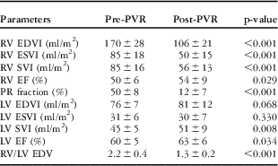
In an effort to overcome the limited durability of bioprosthetic valves in the pulmonary position, Quintessenza et alReference Quintessenza, Jacobs, Morell, Giroud and Boucek37, Reference Quintessenza38 introduced a new method of implanting bicuspid pulmonary valve using polytetrafluoroethylene material. Their rationale for the development of this technique was based on favourable experimental and clinical results of polytetrafluoroethylene monocusp valve.Reference Brown, Ruzmetov, Vijay, Rodefeld and Turrentine39 In the earlier part of their experience, they used polytetrafluoroethylene material with 0.6-mm thickness and experienced a few cases of redo pulmonary valve replacement because of immobile and calcified leaflets.Reference Quintessenza, Jacobs, Chai, Morell and Lindberg40 Freedom from redo pulmonary valve replacement of this valve was ∼70% at 8 years.Reference Quintessenza, Jacobs, Chai, Morell and Lindberg40 Since 2006, they have been using polytetrafluoroethylene membrane with 0.1-mm thickness anticipating improved valve durability owing to non-porous nature of this material, which does not allow cellular in-growth and thickening. Since June, 2009, we have also been implanting this polytetrafluoroethylene bicuspid valve in the pulmonary position (Fig 2). Recently, we reported early results of this technique in 56 patients with various congenital heart diseases.Reference Lee, Lee and Kwak41 In our experience, this valve demonstrated excellent performance for the short term – median follow-up duration of 15 months – as assessed by echocardiography and magnetic resonance imaging (Fig 3). Analysis of the pre-operative and post-operative magnetic resonance imaging data in patients with pulmonary regurgitation showed significant reduction in right ventricular volumes and improvement in biventricular function (Table 1). The median pulmonary regurgitation fraction of this valve was 10%. To date, a total of 92 patients underwent implantation of this valve in our centre with one re-operation due to infective endocarditis. Certainly, long-term follow-up of this valve is mandatory to determine the true value of this technique.
(a) Bicuspid valve made of polytetrafluoroethylene membrane with 0.1-mm thickness. (b) Polytetrafluoroethylene bicuspid valve implanted into the pulmonary position.
Magnetic resonance image showing good coaptation (arrow) of the polytetrafluoroethylene bicuspid pulmonary valve.
Changes in magnetic resonance imaging parameters after pulmonary valve replacement using polytetrafluoroethylene membrane in patients with pulmonary regurgitation (n = 22).Reference Lee, Lee and Kwak41
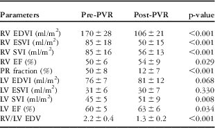
EDV = end-diastolic volume; EDVI = end-diastolic volume index; EF = ejection fraction; ESVI = end-systolic volume index; LV = left ventricular; PR = pulmonary regurgitation; PVR = pulmonary valve replacement; RV = right ventricular; SVI = stroke volume index
Timing of pulmonary valve replacement
Pulmonary valve replacement is clearly indicated when symptoms or decreased exercise tolerance attributable to pulmonary regurgitation are present. However, there is no detailed consensus to guide optimal timing of pulmonary valve replacement in the asymptomatic patients with repaired tetralogy of Fallot and significant pulmonary regurgitation. The guidelines from the American College of Cardiology/American Heart Association state that pulmonary valve replacement is reasonable in patients with severe pulmonary regurgitation in association with moderate to severe right ventricular dysfunction or enlargement.Reference Warnes, Williams and Bashore42 However, the specific thresholds for “moderate to severe right ventricular dysfunction or enlargement” have not been defined.
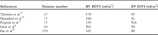
If pulmonary valve replacement can be performed with negligible mortality and morbidity, and durable prosthetic valves are available, pulmonary valve replacement should be recommended as early as possible for all patients with dilated right ventricle. However, because this is not the case, we should decide the “upper threshold” or “point of no return” to which point we can delay pulmonary valve replacement and above which optimal outcome cannot be expected after pulmonary valve replacement (Table 2). Therrien et alReference Therrien, Provost, Merchant, Williams, Colman and Webb15 reported this “point of no return” for the first time. They found that in no patients with a right ventricular end-diastolic volume index exceeding 170 mm/square metre or a right ventricular end-systolic volume index exceeding 85 mm/square metre before pulmonary valve replacement were right ventricular volumes “normalised” after surgery. Oosterhof et alReference Oosterhof, van Straten and Vliegen10 reported that normalisation of right ventricular volumes could be achieved when pre-operative right ventricular end-diastolic volume index was <160 mm/square metre or right ventricular end-systolic volume index was <82 mm/square metre. Geva et al reported that right ventricular end-systolic volume index of <90 mm/square metre was associated with optimal outcome – normal right ventricular size and function.Reference Geva, Gauvreau and Powell8 Frigiola et alReference Frigiola, Tsang and Bull11 suggested the most aggressive policy of performing pulmonary valve replacement when right ventricular end-diastolic volume index was <150 mm/square metre. Recently, in a study of 170 patients who underwent pulmonary valve replacement for chronic pulmonary regurgitation, we found that optimal outcome – normalised right ventricular volumes and function – might not be achieved when pre-operative right ventricular end-diastolic volume index exceeded 163 mm/square metre or right ventricular end-systolic volume index exceeded 80 mm/square metre.Reference Lee, Kim and Lee43 What about the “lower threshold” above which we should consider pulmonary valve replacement? It definitely depends primarily upon the clinical status of an individual patient. In asymptomatic patients, Geva et alReference Geva44 and Dave et alReference Dave, Buechel and Dodge-Khatami9 recommended pulmonary valve replacement when right ventricular end-diastolic volume index exceeded 150 mm/square metre.
Summary of the reported cut-off values of pre-operative right ventricular volume indexes for optimal outcome after pulmonary valve replacement in patients with chronic pulmonary regurgitation.
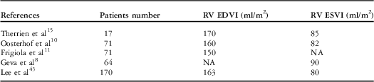
EDVI = end-diastolic volume index; ESVI = end-systolic volume index; NA = not available; RV = right ventricular
Although many studies identified cut-off values of right ventricular end-diastolic volume index as an indication for pulmonary valve replacement, Geva et alReference Geva, Gauvreau and Powell8 and Henkens et alReference Henkens, van Straten and Schalij45 stressed the importance of right ventricular end-systolic volume index in determining the timing of pulmonary valve replacement. We have also found that higher pre-operative right ventricular end-systolic volume index was a sole independent risk factor for suboptimal outcome.Reference Lee, Kim and Lee43 Other factors influencing timing of pulmonary valve replacement include moderate or severe tricuspid regurgitation, sustained tachyarrhythmia, severe branch pulmonary arterial stenosis, and large right ventricular outflow tract aneurysm.Reference Geva44
Summary
Pulmonary valve replacement for patients with chronic pulmonary regurgitation after repair of tetralogy of Fallot can be performed safely with low operative mortality and morbidity. Pulmonary valve replacement in these patients consistently leads to improvement in functional class and a substantial decrease or normalisation of right ventricular volumes. Although currently there are no evidences showing a long-term survival benefit of pulmonary valve replacement, timely pulmonary valve replacement before severe right ventricular dilatation and/or dysfunction occurs may have a beneficial effect on right ventricular function and QRS duration, thus improving long-term survival. Suboptimal durability of currently used bioprosthetic valves is a weak facet in determining the optimal timing of pulmonary valve replacement. Although currently there is no detailed consensus to guide optimal timing of pulmonary valve replacement in asymptomatic patients, evidences that suggest the optimal timing of pulmonary valve replacement based on magnetic resonance imaging parameters are accumulating. Accordingly, the optimal timing of pulmonary valve replacement in asymptomatic patients will be further refined.