Introduction
Most of the functions of the larynx are dependent upon uninterrupted vocal fold motion, which requires an intact recurrent laryngeal nerve. This nerve can be injured during surgery as a result of thermal damage, stretching, cutting, compression or vascular compromise.Reference Tessema, Pitman, Roark, Berzofsky, Sharma and Schaefer 1
In such cases, reinnervation can be attempted in order to restore neural transmission through the recurrent laryngeal nerve. Successful reinnervation may restore normal phonation and airway protection. Several reinnervation techniques have been reported, including direct anastomosis, nerve grafting (e.g. using the phrenic nerve or ansa cervicalis), and the creation of a laryngeal nerve muscle pedicle.Reference Meller 2 – Reference Kaya 4 Nerve anastomosis allows the bulk and tension of the vocal fold to be mostly restored; however, remobilisation of the vocal fold may be poor.Reference Ioachim, Assimakopoulos, Goussia, Peschos, Skevas and Agnantis 5
This study examined the short-term effects of primary nerve anastomosis, performed to reinnervate the recurrent laryngeal nerve, by undertaking laryngeal electromyography and histopathological analysis of the affected vocal fold.
Materials and methods
Experimental animals and surgical procedure
Twenty Zealand rabbits were used in this study. The project was approved by Ege University's Local Ethical Council of Animal Experiments. All animals were maintained in a facility at Ege University's Animal Laboratory. They were humanely treated and all institutional and national guidelines were observed.
The animals were anaesthetised with 35 mg/kg intramuscular ketamine and divided into two groups. In the first group (n = 10), the right recurrent laryngeal nerve was transected and a 1-cm segment was excised. In the second group (n = 10), the right recurrent laryngeal nerve was transected and then sutured through its epineurium with 10/0 Prolene™ to form an end-to-end primary anastomosis. In both groups, the left laryngeal nerves were left untouched.
On the first day of the study, after the above surgical procedure, the larynx was provoked with a cotton sponge and, during spontaneous breathing, the vocal fold movements of the animals in each group were observed with an endoscope and recorded. On the 21st day, under ketamine anaesthesia, the vocal fold movements were again observed with an endoscope during spontaneous breathing, and recorded.
Laryngeal electromyography
This was carried out on the first and 21st day in both groups, during spontaneous breathing under anaesthesia. A Medelec Sapphire 4 Me electromyography device (Medelec, New Delhi, India) was used for electromyographic recordings. To prepare for electromyography, one electrode was placed on the animal's left ear, then a concentric needle electrode was placed in the right posterior cricoarytenoid and thyroarytenoid muscles as previously described.Reference Mu and Yang 6
For thyroarytenoid muscle monitoring, the needle electrode was passed through the middle of the cricothyroid membrane, then moved laterally and upwards until the thyroarytenoid muscle was reached.
For posterior cricoarytenoid muscle monitoring, the larynx was pushed to one side and the thyroid cartilage was palpated at its posterior aspect. The cricoarytenoid muscle was reached by penetrating the posterior aspect of the lower third of the thyroid cartilage.Reference Maronian, Robinson, Waugh and Hillel 7
On the 21st day, following completion of the second laryngeal electromyography session, all animals were sacrificed to enable histopathological analysis.
Histopathological procedure
Following sacrifice, the larynx was completely excised with the tongue pedunculus, under direct inspection. The specimen was infused with 10 per cent formalin and fixed with paraffin. Four-micrometre thick tissue sections were then taken, stained with haematoxylin and eosin, and examined by light microscopy.
Examination took particular note of: (a) vocal fold epithelial changes; (b) histological changes in the posterior cricoarytenoid and thyroarytenoid muscles; and (c) histological changes in the severed areas of the recurrent laryngeal nerve.
Atrophy in the posterior cricoarytenoid and thyroarytenoid muscle cells was graded as: 0, no atrophy; 1, minimal atrophy; 2, light atrophy; 3, medium atrophy; or 4, pronounced atrophy.
Statistical analysis
The Mann–Whitney U test was used for statistical analysis. The chi-square test was used to compare histological changes in the posterior cricoarytenoid and thyroarytenoid muscles. The chi-square test was also used to compare nuclear changes in basal cells. The McNemar–Bowker test was used to compare the right and left vocal folds. A p value of less than 0.05 was considered statistically significant.
Results and analysis
Vocal fold endoscopy
In group one, the right vocal fold was observed to be in the paramedian position, and was immobile during both spontaneous breathing and laryngeal provocation. In group two, the right vocal fold was also observed to be in the paramedian position during both spontaneous breathing and laryngeal provocation; however, perceptible vibratory movements were observed in all animals in this group on the 21st day.
Laryngeal electromyography
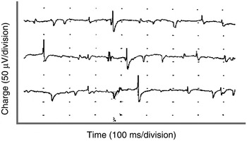
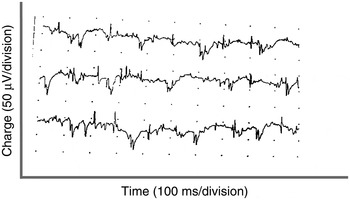
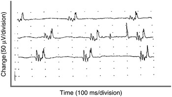
In the first group, total denervation potentials were observed in the right posterior cricoarytenoid and thyroarytenoid muscles, in all animals (Figures 1 and 2). In addition to these findings, two animals in this group showed right posterior cricoarytenoid muscle motor unit potentials of polyphasic character, short length and short duration, with an upward deflection synchronised with spontaneous respiration, indicating muscle regeneration (Figure 3).
Laryngeal electromyographic trace showing denervation findings in the right posterior cricoarytenoid muscle during spontaneous breathing, in a group one animal.

Laryngeal electromyographic trace showing denervation findings in the right thyroarytenoid muscle during spontaneous breathing, in a group one animal.

Laryngeal electromyographic trace showing regeneration potentials in a group one animal, with signs indicating muscle regeneration (see text).

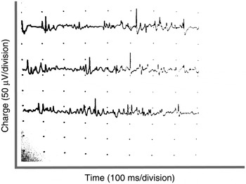
In the second group, fibrillations and positive acute wave formations were identified in the right posterior cricoarytenoid muscle of all animals, indicating denervation potentials. All animals in this group had motor unit potentials of polyphasic character, short length and short duration, as well as normal amplitude motor unit potentials, both signs of regeneration (Figure 4).
Laryngeal electromyographic trace showing regeneration potentials in the right posterior cricoarytenoid muscle of a group two animal.

Histopathological findings
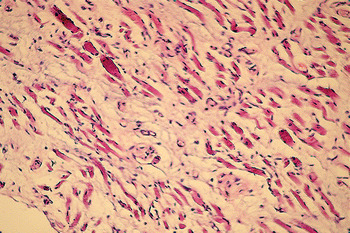
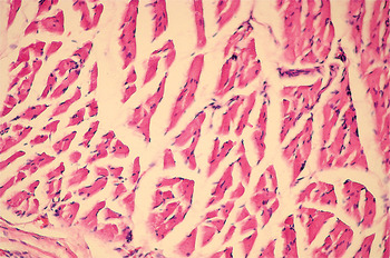
In the group one animals, with two exceptions, signs of atrophy were observed in the right posterior cricoarytenoid and thyroarytenoid muscles: grade 4 atrophy was observed in two animals, grade 3 atrophy in four and grade 2 atrophy in two (Figure 5). No atrophy was observed in two animals. There was no atrophy of the left posterior cricoarytenoid or thyroarytenoid muscles in any animal (Figure 6). In group two, grade 2 atrophy was observed in one animal and grade 1 atrophy in eight. No atrophy was observed in one animal. When the right posterior cricoarytenoid and thyroarytenoid muscle atrophy in the first and second groups was compared, a statistically significant difference was found (p = 0.018).
Photomicrograph showing right thyroarytenoid muscle atrophy in a group one animal. (H&E; ×100)

Photomicrograph showing normal muscle histology in the left thyroarytenoid muscle. (H&E; ×100)

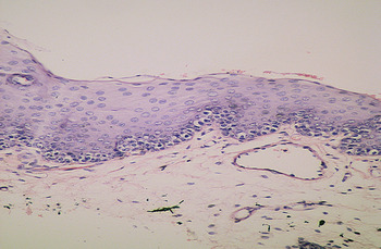
The right and left vocal fold mucosa was also examined histopathologically. In group one on the right side, hyperkeratosis was observed in two animals and parakeratosis in four. Four group one animals showed no signs of hyperkeratosis or parakeratosis, but hyperplasia was encountered in two of these four (Figure 7). Nuclear changes in the basal layer of the vocal fold epithelium were seen in four group one animals. The left vocal fold mucosa showed no signs of hyperkeratosis, parakeratosis or hyperplasia in any group one animal.
Photomicrograph showing basal cellular nuclear changes and hyperplasia in the vocal fold of a group one animal. (H&E; ×100)

In the second group on the right side, only one animal showed signs of vocal fold mucosa parakeratosis. In the same group, hyperplasia was found in three animals, and basal cell nuclear changes were seen in only one.
When the vocal fold mucosal changes of the first and second groups were compared, a statistically significant difference was found for hyperkeratosis and parakeratosis (p = 0.019). There was no statistically significant difference between the two groups regarding hyperplasia and basal cell nuclear changes (p = 0.301).
In the second group, a neuroma was observed in the right recurrent laryngeal nerve of one animal, and hyperplasia of the right recurrent laryngeal nerve in another.
Discussion
Many methods have been reported for the treatment of vocal fold paralysis, including reinnervation techniques. The present study examined the short-term effects of recurrent laryngeal nerve reinnervation, using laryngeal electromyography and histopathological analysis of the vocal folds in a rabbit model.
Signs of neurotransmission were observed in all reinnervated animals, while signs of denervation were observed in the posterior cricoarytenoid muscle in all animals in which anastomosis had not been performed. It is well known that, even under ideal conditions for nerve anastomosis, it is difficult to restore muscle function to normal levels. The degree of nerve recovery is associated with the number, amplitude and myelination level of fibrils returning to the original motor end-plate. In addition to this, the regenerated axons that return to the appropriate muscles are smaller and less myelinated than the original fibrils. Some fibrils are not regenerated and some return to other muscle groups.
A large number of recurrent laryngeal nerve fibres innervate the thyroarytenoid, lateral cricoarytenoid and interarytenoid muscles, all adductor muscles. Approximately 25 per cent of recurrent laryngeal nerve fibres innervate the posterior cricoarytenoid muscle, an abductor muscle.Reference Maronian, Robinson, Waugh and Hillel 7 When recurrent laryngeal nerve damage occurs, nerve regeneration takes place if primary anastomosis is performed.
However, during such regeneration the abductor and adductor fibres within the recurrent laryngeal nerve can become mismatched, resulting in abnormal reinnervation. This is termed synkinesis.Reference Crumley and McCabe 8 Such dysfunctional (‘synkinetic’) recurrent laryngeal nerve reinnervation can result in chronic vocal fold paralysis due to the simultaneous contraction of antagonistic muscles, as first reported by Siribodhi et al. Reference Siribodhi, Sundmaker, Atkins and Bonner 9
There is no consensus over the definition of synkinesis or its electromyographic signs. Similarly, there is no universally accepted method of diagnosing synkinesis: opinions differ and research is ongoing.Reference Maronian, Robinson, Waugh and Hillel 7 In our study, like other researchers, we monitored the activation of the thyroarytenoid muscle during spontaneous respiration, in order to detect synkinetic reinnervation.Reference Crumley 10 Activation of the thyroarytenoid muscle is not expected during spontaneous respiration, and if it occurs this may be a sign of synkinetic reinnervation.Reference Crumley 10 We made the assumption that such thyroarytenoid activation did indeed represent synkinetic reinnervation. We determined motor unit discharges in the course of spontaneous respiration in the thyroarytenoid muscle of six of the group two animals (i.e. with end-to-end recurrent laryngeal nerve anastomosis). Other animal studies have shown that synkinetic reinnervation occurs in 66–88 per cent of vocal fold paralysis cases. An equivalent incidence of synkinetic reinnervation is believed to occur in humans.Reference Flint, Downs and Coltrera 11 – Reference Rosen, Bailey, Johnson and Newlands 14 Based on the above research assumption, we found a synkinesis incidence of 60 per cent. Although comparable to previous reports, this incidence is lower than expected. This may be because the post-reinnervation period in our study was short, and also because multiple muscle sampling was not performed. (Hiroto et al. have reported that the incidence of synkinetic reinnervation is increased when multiple muscle sampling is performed.)Reference Hiroto, Hirano and Tomita 15
It has been reported that, following reinnervation, muscle atrophy decreases, muscle tone improves, vocal fold tension recovers and voice quality improves.Reference Ushio 16 Denervation atrophy is only prevented with reinnervation.Reference Crumley 17
In the reinnervated vocal fold, although regular, physiological vocal fold movements are not observed, the volume, tension and muscle tone of the vocal fold are protected. Our study demonstrates that reinnervation prevents muscle atrophy to a significant degree, even if normal vocal fold function does not fully recover.
In our second group, we identified a neuroma of the recurrent laryngeal nerve in one animal and hyperplasia of the recurrent laryngeal nerve in another. In our first group, in which the recurrent laryngeal nerve was transected and a 1-cm segment removed, regeneration was also observed. Nerve regeneration may explain the presence of neural hyperplasia found in some of our study animals (with neuroma development in one animal).
Animal studies have proven that the recurrent laryngeal nerve has a strong tendency to regenerate after it has been cut. In humans, high incidences of both laryngeal muscle regeneration and nerve reinnervation have been reported in patients with vocal fold paralysis.Reference Hirano, Nozoe, Shin, Maeyama, Hirano, Kirchner and Bless 18
In a clinically paralysed vocal fold, muscle activity may still be present due to incomplete nerve lesions, reinnervation or both. Spontaneous laryngeal reinnervation may occur due to nerve regeneration from healthy remnants of the recurrent laryngeal nerve, from severed elements of the nerve, or from nerves of the surrounding area. Animal studies have examined neural regeneration around denervated laryngeal muscles, but no precise findings have been reported.Reference Lewis, Crumley, Blanks and Pitcock 19 Animal study findings have clearly shown that the recurrent laryngeal nerve has a marked capacity for regeneration following transection and injury.Reference Crumley 10 , Reference Shindo, Herzon, Hanson, Cain and Shagal 20 Studies in humans have found nerve regeneration following surgical transection or strong ligation of the recurrent laryngeal nerve.Reference Netterville, Stone, Rainey, Zealear and Ossof 21 Crumley and McCabe have reported laryngeal muscle reinnervation and regeneration following tight suturing and severing of the recurrent laryngeal nerve.Reference Crumley and McCabe 8 However, in Damrose and colleagues' electromyographic study in humans, no muscle activity was observed in a paralysed vocal fold despite evidence of laryngeal reinnervation.Reference Damrose, Huang, Blumin, Blackwell, Sercarz and Berke 22 These authors stated that spontaneous recurrent laryngeal nerve regeneration is rare in humans. However, others have emphasised that, although no vocal fold mobility exists in cases of spontaneous reinnervation, the vocal fold volume and muscle tone are maintained and a better voice quality is therefore obtained, even after total denervation.Reference Blitzer, Jahn and Keidar 23
In our study, we observed significantly less hyperkeratosis and parakeratosis of the vocal fold epithelium in group two (i.e. the group undergoing primary, end-to-end anastomosis). We believe that the observed hyperkeratosis and parakeratosis were due to compensatory efforts to overcome glottic insufficiency, leading to excessive mechanical irritation of the vocal folds.Reference Rosen, Bailey, Johnson and Newlands 14 In our study, muscle atrophy was prevented to a large extent in group two (the reinnervated group). Since muscle atrophy was prevented in this group, it could be expected that the incidence of glottic failure would decrease. Thus, one would expect fewer compensatory vocal fold movements and therefore a reduction in mechanical vocal fold irritation, in comparison with animals with paralysed vocal folds. In Rodgers and colleagues' study of the histopathological effects of Teflon injection into the paralysed vocal folds of dogs, varying degrees of squamous metaplasia were observed in the vocal fold epithelium.Reference Rodgers, Abdul-Karim and Strauss 24
Previously published animal studies have usually produced hyperkeratosis or parakeratosis of the vocal folds or larynx using irritants or inhaled substances.Reference Rodgers, Abdul-Karim and Strauss 24 , Reference Renne and Gideon 25 We found no previously published information on the formation of hyperkeratosis or parakeratosis in the vocal folds or larynx of animals with paralysed vocal folds. In humans, smoking is known to significantly affect the formation of hyperkeratosis. Gastroesophageal reflux or chronic laryngeal irritation also have proven effects on hyperkeratosis formation.Reference Garcia, Krishna and Rosen 26 Although there are no published studies on hyperkeratosis or parakeratosis in humans with vocal fold paralysis, such hyperkeratosis has been speculated to be due to exposure to excessive mechanical trauma caused by attempts to compensate for glottic failure.Reference Rosen, Bailey, Johnson and Newlands 14 Most histopathological vocal fold changes have been shown to be reversible, resolving when the provocative agent is removed. In human postmortem studies examined retrospectively to determine the histopathological effects of vocal fold paralysis on the vocal fold, muscle atrophy was usually found but pathological changes in the vocal fold epithelium were not mentioned.Reference Bridger 27 , Reference Quiney and Michaels 28 In our study, histopathological changes in the vocal fold epithelium were observed three weeks after the induction of vocal fold paralysis. Since compensatory mechanisms will develop in cases of vocal fold paralysis, findings in the vocal fold epithelium may regress over time. This may be the reason why hyperkeratosis was not found in the human postmortem studies mentioned above.
Formal histopathological examination of the vocal fold epithelium requires an additional surgical intervention. As this second examination may damage the vocal folds, and would be conducted purely for academic rather than therapeutic reasons, it would be hard to justify in humans. This may explain the lack of studies on the histopathological effects of vocal fold paralysis on the vocal fold epithelium. However, we believe that it is essential to perform long-term, consecutive studies of the histopathological changes of paralysed vocal fold epithelium, in order to obtain more detailed information.
-
• In the reported rabbit model of recurrent laryngeal nerve transection, normal vocal fold function did not return
-
• However, primary, end-to-end nerve anastomosis prevented muscle atrophy
-
• After induction of vocal fold paralysis, hyperkeratosis and parakeratosis were observed in the vocal fold epithelium
-
• Recurrent laryngeal nerve anastomosis should be performed as soon as possible after damage to prevent the side effects of denervation
Laryngeal electromyography is a valuable adjunct in the study of vocal fold dysfunction. It yields objective and reproducible data, and may help establish the pathophysiology and prognosis of laryngeal nerve pathology.Reference Hartl and Brasnu 29 – Reference Khan, Pearlman, Bianchi and Hauck 32