Introduction
Chronic fatigue syndrome (CFS) is characterized by unexplained, profound disabling and long-lasting fatigue that is of new or definite onset, that is not the result of ongoing exertion and that is not substantially alleviated by rest. The fatigue must be accompanied by at least 4 or more of the following case-defining symptoms during at least 6 months of consecutive illness: sore throat, tender cervical or axillary lymph nodes, muscle pain, multi-joint pain, post-exertional malaise, unrefreshing sleep, headaches and impaired memory or concentration (Fukuda et al. Reference Fukuda, Straus, Hickie, Sharpe, Dobbins and Komaroff1994). The suggestion that CFS may be related to a dysfunction of the hypothalamic–pituitary–adrenal (HPA) axis derives from the similarity in symptoms between CFS patients and patients with Addison's disease or after bilateral adrenalectomy (Cleare, Reference Cleare2003). Moreover, the HPA axis offers an interesting physiological basis for the link between CFS, immunological disturbances and physical or psychological stress (Gold & Chrousos, Reference Gold and Chrousos2002; Gerrity et al. Reference Gerrity, Papanicolaou, Amsterdam, Bingham, Grossman, Hedrick, Herberman, Krueger, Levine, Mohagheghpour, Moore, Oleske and Snell2004; Van Houdenhove & Egle, Reference Van Houdenhove and Egle2004). In the past, stress has generally been associated with HPA axis hyperactivity, resulting in hypercortisolism. However, chronic stress can also lead to hypocortisolism, as is the case in several stress-related disorders (Heim et al. Reference Heim, Ehlert and Hellhammer2000a; Gold & Chrousos, Reference Gold and Chrousos2002; Fries et al. Reference Fries, Hesse, Hellhammer and Hellhammer2005). In CFS, the overall balance of evidence points to reduced cortisol output in at least a proportion of the patients, despite the negative reports and the poor quality of many studies (Cleare, Reference Cleare2003).
In addition to measurements of basal hormone concentrations, dynamic challenges of the HPA axis have been undertaken for several reasons: to investigate at which level there may be abnormalities in the control of the HPA axis, to detect more subtle disturbances of the axis and to provide a more reliable and standardized indicator of the HPA axis disturbance (Cleare, Reference Cleare2003). Blunted responses of cortisol and/or adrenocorticotropic hormone (ACTH) to awakening and to several challenge tests [corticotropin-releasing factor (CRF), insulin tolerance test, naloxone, social stress and exercise] have been reported in CFS (Demitrack et al. Reference Demitrack, Dale, Straus, Laue, Listwak, Kruesi, Chrousos and Gold1991; Scott et al. Reference Scott, Medbak and Dinan1998a, Reference Scott, Burnett, Medbak and Dinanb; Gaab et al. Reference Gaab, Huster, Peisen, Engert, Heitz, Schad, Schurmeyer and Ehlert2002a; Roberts et al. Reference Roberts, Wessely, Chalder, Papadopoulos and Cleare2004). Suggested mechanisms for the findings in these challenge studies in CFS include a deficiency of hypothalamic CRF or vasopressin and a reduced supra-hypothalamic stimulus to the HPA axis, resulting in hypocortisolism (Demitrack et al. Reference Demitrack, Dale, Straus, Laue, Listwak, Kruesi, Chrousos and Gold1991; Demitrack & Crofford, Reference Demitrack and Crofford1998; Scott et al. Reference Scott, Medbak and Dinan1998a, Reference Scott, Medbak and Dinan1999; Gaab et al. Reference Gaab, Huster, Peisen, Engert, Heitz, Schad, Schurmeyer and Ehlert2002a). Another possible mechanism of hypocortisolism is enhanced sensitivity to the negative feedback of glucocorticoids, also referred to as dexamethasone (Dex) hypersuppression (Heim et al. Reference Heim, Ehlert and Hellhammer2000a; Fries et al. Reference Fries, Hesse, Hellhammer and Hellhammer2005). As to this mechanism, Gaab et al. (Reference Gaab, Hustern, Peisen, Engert, Schad, Schurmeyer and Ehlert2002b) have demonstrated that patients with CFS show an enhanced and prolonged suppression of salivary free cortisol after the administration of a low dose of Dex (0.5 mg). Further support for enhanced sensitivity for glucocortocoids in CFS comes from in vitro studies, showing that lower concentrations of Dex were needed to inhibit interleukin production and proliferation of peripheral blood mononuclear cells from subjects with CFS (Visser et al. Reference Visser, Blauw, Hinloopen, Brommer, de Kloet, Kluft and Nagelkerken1998, Reference Visser, Graffelman, Blauw, Haspels, Lentjes, de Kloet and Nagelkerken2001a, Reference Visser, Lentjes, Haspels, Graffelman, Blauw, de Kloet and Nagelkerkenb; Gaab et al. Reference Gaab, Rohleder, Heitz, Schad, Engert, Schürmeyer and Ehlert2003a).
Surprisingly, there are no published reports on investigations with the combined Dex/CRF test in CFS. This is in contrast with research in major depressive disorder (MDD), a syndrome that is – inversely to CFS – characterized by hyperfunction of the HPA axis, reduced glucocorticoid negative feedback (Dex hyposuppression) and hypercortisolism (Plotsky et al. Reference Plotsky, Owens and Nemeroff1998; Claes, Reference Claes2004). More precisely, the combined (high-dose) Dex/CRF test has been shown to be the most sensitive HPA axis function test in MDD (Heuser et al. Reference Heuser, Yassouridis and Holsboer1994a). In this test, the CRF-induced ACTH and cortisol secretion is significantly higher in most depressed patients in comparison to healthy controls if a high-dose Dex (1.5 mg) pre-treatment is administered in the evening preceding the CRF challenge (Heuser et al. Reference Heuser, Yassouridis and Holsboer1994a). Both reduced glucocorticoid negative feedback and increased central drive of the HPA axis have been proposed as underlying mechanisms in MDD (Cole et al. Reference Cole, Kim, Kalman and Spencer2000; Holsboer, Reference Holsboer2000).
The aim of this study was to compare the cortisol responses in the low-dose (0.5 mg) combined Dex/CRF test between well-characterized patients with CFS and healthy controls without CFS. It was expected that CFS would be associated with a reduced cortisol response in this test, due to enhanced negative feedback and/or reduced central drive of the HPA axis.
Method
Subjects
Patients attending the CFS clinic of the Department of Internal Medicine of Antwerp University Hospital were invited to participate in the study by letter. As described in the literature (Jason et al. Reference Jason, Richman, Rademaker, Jordan, Plioplys and Taylor1999), the majority of the patients were women. Since gender is an important variable in HPA axis research (Heuser et al. Reference Heuser, Gotthardt, Schweiger, Schmider, Lammers, Dettling and Holsboer1994b; Kirschbaum et al. Reference Kirschbaum, Kudielka, Gaab, Schommer and Hellhammer1999; Künzel et al. Reference Künzel, Binder, Nickel, Ising, Fuchs, Majer, Pfennig, Ernst, Kern, Schmid, Uhr, Holsboer and Modell2003) and since only a few men responded to the invitation, only female patients were included. CFS was diagnosed according to the Center for Disease Control Criteria (Fukuda et al. Reference Fukuda, Straus, Hickie, Sharpe, Dobbins and Komaroff1994) by an experienced internist (G.M.). All patients underwent serial physical examinations and laboratory evaluations; no alternative medical diagnosis could be established. Initially, 35 patients were included. One patient was excluded due to a panic attack during the Dex/CRF test, leaving 34 patients. Twenty-five healthy females without a past history of CFS were recruited by an advertisement in Antwerp University Hospital. They were matched for age and educational level. Five levels of education were considered in accordance with the Belgian education system. The patient group was larger in order to allow a secondary analysis of potential influencing clinical variables within this group. The data were collected between October 2003 and November 2004. Pregnancy, a lifetime history of organic brain disease, current medical condition or drugs that might influence neuroendocrine testing (with the exception of psychotropic drugs and oral contraceptives) were exclusion criteria. All participants were Caucasian and had grandparents of European origin. The Medical Ethical Committee of the University Hospital of Antwerp approved the study. All participants were aged >18 years and signed informed consent.
Interviews and questionnaires
All participants were interviewed with the Dutch version of SCID-I/P version 2.0 (First et al. Reference First, Spitzer, Gibbon, Williams, M. A. C., G. W., R., N. and W. A.1999) by a trained psychiatrist (F.V.D.E.). The CDC psychiatric exclusion criteria for CFS were adopted, with three exceptions. First, subjects who met the criteria for a current major depressive episode according to DSM-IV (APA, 1994) were excluded, in order to avoid a state effect of MDD on the outcome in the Dex/CRF test. Second, a history of melancholic depression was not assessed systematically, because the specification of melancholic features in the past is not provided in SCID-I/P and because it is difficult to assess this item retrospectively by means of an interview. Finally, one control subject had suffered from anorexia nervosa in the past, but was in complete remission for more than 5 years. The Checklist Individual Strength was used to measure patient's perceived fatigue concentration, motivation and physical activity. This 20-item self-rating scale is well validated and reliable (Vercoulen et al. Reference Vercoulen, Alberts and Bleijenberg1999).
One of the factors potentially determining heterogeneity and influencing the HPA axis in CFS is early-life stress (ELS), consisting of sexual, physical and/or emotional maltreatment during childhood (Van Den Eede et al. Reference Van Den Eede, Moorkens, Van Houdenhove, Cosyns and Claes2007). Preclinical animal laboratory studies have provided evidence that ELS (maternal separation in rats or adverse rearing conditions in non-human primates) produces long-lived hyperactivity of CRF neuronal systems, as well as greater reactivity of the HPA axis (Plotsky & Meaney, Reference Plotsky and Meaney1993; Coplan et al. Reference Coplan, Andrews, Rosenblum, Owens, Friedman, Gorman and Nemeroff1996; Ladd et al. Reference Ladd, Owens and Nemeroff1996; Heim et al. Reference Heim, Plotsky and Nemeroff2004). In accordance with these findings, higher pituitary reactivity to CRF and to psychological stress has been described in adult women with a history of childhood abuse (Heim et al. Reference Heim, Newport, Heit, Graham, Wilcox, Bonsall, Miller and Nemeroff2000b, Reference Heim, Newport, Bonsall, Miller and Nemeroff2001). In addition, Rinne et al. (Reference Rinne, de Kloet, Wouters, Goekoop, DeRijk and van den Brink2002) demonstrated that ELS was associated with an enhanced ACTH and cortisol response in the high-dose Dex/CRF test in women with borderline personality disorder, pointing to an enhanced central CRF drive and a reduced glucocorticoid negative feedback. This is in contrast with the proposed central CRF deficiency and enhanced glucocorticoid negative feedback in CFS. As a consequence, it may be that CFS patients with a history of ELS constitute a subgroup with a different neuroendocrine function (Van Den Eede et al. Reference Van Den Eede, Moorkens, Van Houdenhove, Cosyns and Claes2007). For the assessment of ELS, the Dutch version of the Structured Trauma Interview was used (Draijer, Reference Draijer1989). This instrument is described in detail elsewhere (Draijer & Langeland, Reference Draijer and Langeland1999). There have been reports of significant associations between childhood abuse scores in the Structured Trauma Interview and both responses to a challenge of the serotonin system (Rinne et al. Reference Rinne, Westenberg, den Boer and van den Brink2000) and to cortisol responses in the Dex/CRF test (Rinne et al. Reference Rinne, de Kloet, Wouters, Goekoop, DeRijk and van den Brink2002). All interviews were conducted by a psychiatrist (F.V.D.E.) at the end of the clinical and psychiatric investigation in a context of absolute confidentiality and mutual trust. The focus of the Structured Trauma Interview is on childhood sexual and physical abuse. Physical abuse is defined as intentional physical aggression by a parent or caregiver towards a child (<16 years), which may cause injuries. Physical aggression was coded as ‘severe’ if it happened more than once a month and if it sometimes led to injuries. When individuals were hit hard more than once a month without experiencing injuries, or when aggression took place less frequently but with injuries, it was scored as ‘moderate’. ‘Mild’ aggression had a lower frequency and was not accompanied with injuries. Sexual abuse is defined as sexual contacts with a child (<16 years), which took place against her/his will or when she/he had the feeling that the sexual contacts could not be refused due to physical or relational preponderance, emotional pressure, force or violence. This item was coded as severe when it appeared chronically or more than once a month during a period in childhood, when it consisted of any form of penetration, when it was associated with violence or when it appeared in the primary support group. ELS was defined as moderate or severe physical abuse and/or severe sexual abuse.
Combined low-dose Dex/CRF test
The combined low-dose Dex/CRF test was performed at Antwerp University Hospital following the protocol described by Heuser et al. (Reference Heuser, Yassouridis and Holsboer1994a). However, instead of measuring plasma cortisol, we measured salivary cortisol concentration by means of a cottonwool Salivette (Sarstedt, Nümbrecht, Germany). Salivary cortisol is considered as an easy-to-asses correlate of the unbound free hormone fraction (Kirschbaum & Hellhammer, Reference Kirschbaum and Hellhammer1994; Baghai et al. Reference Baghai, Schule, Zwanzger, Minov, Holme, Padberg, Bidlingmaier, Strasburger and Rupprecht2002). Participants took 0.5 mg Dex orally at 23:00 hours on the evening before neuroendocrine testing. At 13:30 hours they attended the research unit where an infusion (NaCl 0.9%) was placed in the forearm. Participants then fastened, remained semi-supine or supine under resting conditions, and were not allowed to sleep. Human CRF (100 μg) was administered and flushed through the infusion at 15:00 hours Free salivary concentration was assessed at seven time-points (14:00, 14:30, 15:00, 15:30, 15:45, 16:00 and 16:15 hours). All saliva samples were stored at −20°C and were later delivered to the Laboratory of Physiology of Reproduction of the University of Liège. A radioimmunoassay was performed on 100 μl of salivary free cortisol samples in competition, with a HPLC preparation of cortisol-3CMO coupled with 2-[125I]iodohistamine as tracer, for specific antibodies raised against cortisol-3CMO-BSA. The incubation conditions were at 4°C overnight. The lower detection limit of the assay was 0.1 nmol/l, the intra-assay coefficient of variation (CV) was 4.30% (n=10) whereas the inter-assay CV for low and high cortisol levels were 12.26% and 9.38% (n=30) respectively.
Statistical analysis
The following three measures were computed: Basal, AUCtot and AUCnet. Basal represents the mean free cortisol concentration (in ng/100 ml) over the three time-points on the day after Dex administration (Dex suppression), and before the infusion of CRF. The area under the curve (AUC) was calculated to incorporate the cortisol concentrations at the four time-points after CRF, according to the trapezoid method (Pruessner et al. Reference Pruessner, Kirschbaum, Meinlschmid and Hellhammer2003). AUCtot is the total area under the curve and represents the total cortisol output. AUCnet is identical to AUCtot, except for the removal of the area between the ground and Basal. This measure reflects the absolute increase of cortisol after CRF. For statistical analysis, we used SPSS for Windows, version 12.0 (SPSS Inc., Chicago, IL, USA). The Kolmogorov–Smirnov test was adopted to examine the distribution of the data. In view of deviations from normal distribution and the relatively small sample size of the subgroups, neuroendocrine variables were assessed non-parametrically. The Mann–Whitney test was used for two independent samples and the Kruskal–Wallis test was used for three independent samples, followed by the Dunn test for post-hoc analysis. One-sided testing was applied when comparing patients and controls, since it was expected that CFS would be associated with an enhanced suppression of the HPA axis (Gaab et al. Reference Gaab, Hustern, Peisen, Engert, Schad, Schurmeyer and Ehlert2002b) and thus a lower cortisol response in the low-dose combined Dex/CRF test. The following clinical variables were noted in the patient group: ELS, past history of MDD, use of benzodiazepines, use of antidepressants and use of oral oestrogens (two-sided testing). For the analysis of the sample characteristics, χ2 analysis was applied to compare frequencies. Fisher's exact test was used when necessary. If quantitative data failed to satisfy the assumptions for parametrical statistical analyses, they were analysed non-parametrically. For all analyses, a p value <0.05 was adopted as the level of significance.
Results
Characteristics of study groups
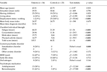
Characteristics of the study groups are presented in Table 1. All patients (with one exception) were unable to work because of chronic fatigue, whereas all control subjects had a working employment status. As expected, the scores on the different items of the Checklist Individual Strength were significantly higher in the patient group (Vercoulen et al. Reference Vercoulen, Alberts and Bleijenberg1999). None of the participants met the criteria for a current diagnosis of post-traumatic stress disorder (PTSD), although this was not an exclusion criterion. As expected, there was a higher frequency of other current anxiety disorders [specific phobia (n=1), social phobia (n=1), generalized anxiety disorder (n=3), panic disorder with or without agoraphobia (n=3), anxiety not otherwise specified (n=1)] and a higher frequency of a history of one or more major depressive episodes in the patient group (Afari & Buchwald, Reference Afari and Buchwald2003). None of the controls were diagnosed with generalized anxiety disorder or panic disorder and none of the participants had obsessive–compulsive disorder. The severity of the current anxiety disorders in both samples was mild or moderate; in three participants the anxiety disorder was in partial remission. The frequency of ELS was not different between the patients and the controls.
Characteristics of study groups

PTSD, Post-traumatic stress disorder; MDD, major depressive disorder.
Significant differences are indicated in bold.
In relation to medication, there were differences in the frequency of individuals who were taking oral oestrogens (all but one as oral contraceptive), antidepressants and benzodiazepines. Patients took the following antidepressants: amitriptyline (n=3, 25–75 mg/day); paroxetine (n=3, 20–30 mg/day); citalopram (n=2, 20 mg/day); escitalopram (n=1, 10 mg/day); fluoxetine (n=1, 20 mg/day); mirtazapine (n=1, 30 mg/day); melitracen/flupentixol (n=1, 10/0.5 mg/day) and trazodone (n=1, 50 mg/day). In addition, four patients were treated with trazodone as a second antidepressant for sleep disturbances (50–150 mg/day). Furthermore, 10 patients took ⩽1 mg/day clonazepam and one patient took 2 mg/day. The other benzodiazepines were: tetrazepam (n=3), lorazepam (n=2), lormetazepam (n=2), alprazolam (n=2) and prazepam (n=1) in patients, and lormetazepam (n=1) and tetrazepam (n=1) in controls.
Combined low-dose Dex/CRF test
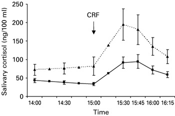
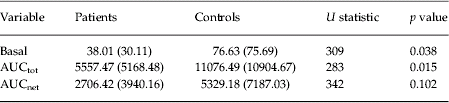
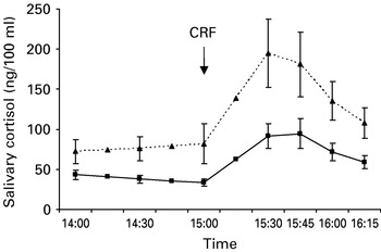
Fig. 1 presents the mean free salivary cortisol responses in CFS patients and controls in the combined Dex/CRF test. The mean cortisol responses before and after CRF were lower in the patient sample. Table 2 lists the mean vales of the three Dex/CRF variables (Basal, AUCtot and AUCnet) with the standard deviations. None of these variables showed a normal distribution in either sample (Kolmogorov–Smirnov, p<0.020). In the non-parametrical testing (Mann–Whitney, one-sided), patients showed significantly lower values of Basal and AUCtot and a trend towards lower values of AUCnet.
Mean salivary (free) cortisol values (ng/100 ml)±s.e.m. at seven time-points during the combined low-dose dexamethasone/corticotropin-releasing factor (CRF) test in CFS patients (■, n=34) and in control subjects (▲, n=25). Participants took 0.5 mg dexamethsone orally at 23:00 hours on the evening before neuroendocrine testing. Human CRF (100 μg) was administered 16 h later at 15:00 hours.
Mean values of the three computed variables in the combined low-dose dexamethsone/corticotropin-releasing factor test in patients and controls
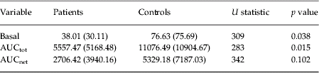
Basal, mean salivary cortisol concentration (ng/100 ml) over three time-points after dexamethasone but before CRF; AUCtot is the total area under the curve and represents the total cortisol output after CRF; AUCnet is identical to AUCtot, except for removal of the area between ground and Basal.
Standard deviations are presented within parentheses. In the non-parametrical testing (Mann–Whitney, one sided), patients (n=34) showed significantly lower values of Basal and AUCtot and a trend towards lower values of AUCnet compared to controls (n=25).
When comparing CFS patients with (n=12) and without ELS (n=22) (ELS+/ELS−), the cortisol responses before and after CRF were significantly lower in the ELS− group (Mann–Whitney, two-sided: Basal, p=0.016; AUCtot, p=0.008; AUCnet, p=0.012). Regarding the patient characteristics, there was a higher frequency of patients who experienced one or more major depressive episodes in the past in the patient group with ELS (75%) compared to patients without ELS (37%) (p=0.038), which is consistent with findings in other studies (Chapman et al. Reference Chapman, Whitfield, Felitti, Dube, Edwards and Anda2004). With the exception of a higher level of subjective complaints of concentration (as measured with Checklist Individual Strength) in the ELS+ group, there were no other significant differences in characteristics between both patient groups. Within the control group, subjects with ELS (n=10) showed lower cortisol responses before and after CRF than subjects without ELS (n=15), inversely to the findings in the patient group. However, the differences between the two groups were not statistically significant (Mann–Whitney, two-sided: Basal, p=0.375; AUCtot, p=0.405; AUCnet, p=1).
There were no significant differences in the distributions of any of the three Dex/CRF variables between patients with and without a history of one or more major depressive episodes in the combined Dex/CRF test (p>0.45). ELS and MDD were correlated, but within the CFS group with a history of one or more major depressive episodes (n=16), cortisol responses remained significantly different between ELS− (n=7) and the ELS+ (n=9) patients (Mann–Whitney, two-sided: Basal, p=0.017; AUCtot, p=0.005; AUCnet, p=0.003). Furthermore, there were no significant differences in the distributions of the three Dex/CRF variables when dividing the patient sample on the basis of benzodiazepine use (p>0.22, benzodiazepines higher mean rank) or antidepressants (p>0.38, antidepressants higher mean rank, except for Basal).
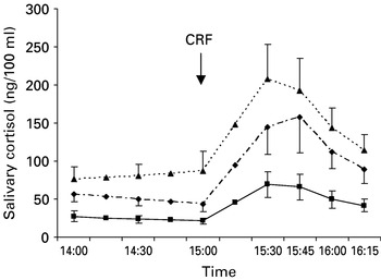
Patients who took an oral oestrogen (n=8) showed significantly lower cortisol responses in the combined Dex/CRF test than patients who did not take an oral oestrogen (n=26), as measured by AUCnet (p=0.023). The values of AUCtot were also lower in the oestrogen group, whereas the values of Basal were higher. However, the differences for these two variables were not statistically significant between the two groups (Mann–Whitney, two-sided: Basal, p=0.350; AUCtot, p=0.417). Since there was a higher frequency of subjects taking an oral oestrogen in the patient group compared with the control group (see Table 1), oral oestrogens could have represented a confounding factor in the previous analyses. Therefore, we performed an additional analysis after removal of the eight patients and two control subjects taking an oral oestrogen. Fig. 2 presents the mean free salivary cortisol responses in CFS patients with and without ELS and controls in the combined Dex/CRF test after removal of subjects taking an oral oestrogen. The cortisol responses before and after CRF were clearly lower in patients without ELS than in the other two groups. Table 3 lists the mean values of the three Dex/CRF variables with the standard deviations. The differences in distributions of Basal and AUCtot were significant in the (one-sided) analysis between two groups (patients and controls) and in the analysis between three groups (patients with and without ELS and controls), but not the differences in distributions of AUCnet. The Dunn test for post-hoc analysis revealed significant differences for Basal and AUCtot between ELS− patients and controls, and between the ELS− and ELS+ patients. The differences between the ELS+ patients and controls were not significant for any of the variables. Comparable results were obtained when excluding controls with a past history of ELS: patients (n=26) versus controls ELS− (n=14): Mann–Whitney, two-sided: Basal, p=0.050; AUCtot, p=0.033; AUCnet, p=0.427. Patients ELS+ (n=15) versus patients ELS− (n=11) versus controls ELS− (n=14): Kruskal–Wallis: Basal, p=0.004; AUCtot, p=0.008; AUCnet, p=0.223 (Dunn: significant differences for Basal and AUCtot between ELS− patients and controls, and between ELS− and ELS+ patients).
Mean salivary (free) cortisol values (ng/100 ml)±s.e.m. at seven time-points during the combined low-dose dexamethasone/corticotropin-releasing factor (CRF) test in CFS patients with (ELS+, ◆, n=11) and without (ELS−, ■, n=15) early-life stress and control subjects (▲, n=23) after removal of the eight patients and two controls who were taking an oral oestrogen.

Mean values of the three computed variables in the combined low-dose dexamethsone/corticotropin-releasing factor test when differentiating patients on the basis of early-life stress (ELS) and after removal of eight patients and two control subjects who were taking an oral oestrogen

For an explanation of the variables see Table 2. Standard deviations are presented within parentheses. When repeating the statistical analyses without the eight patients and two control subjectss who were taking an oral oestrogen, the differences in distributions of Basal and AUCtot were significant in the (one-sided) analysis between patients (total sample, n=26) and controls (n=23) [1] and in the analysis between patients without ELS (ELS−) (n=15) and with ELS (ELS+) (n=11) and controls [2], but not the differences in distributions of AUCnet. The Dunn test for post-hoc analysis revealed significant differences for Basal and AUCtot between the ELS− patients and controls and between ELS− and ELS+ patients. The differences between the ELS+ patients and controls were not significant for any of the variables.
Discussion
This is the first study to investigate HPA axis function in patients with CFS by means of the combined Dex/CRF test and also to examine the influence of a history of ELS on the outcome in this test. We intentionally administered a low dose of Dex (0.5 mg) because of the a priori hypothesis that CFS would be associated with an enhanced glucocorticoid feedback and/or a reduced central drive of the HPA axis. In accordance with this hypothesis, the salivary free cortisol responses in the combined Dex/CRF test were lower in CFS patients than in controls.
The total free cortisol responses before (Basal) and after CRF (AUCtot) were significantly different between the two groups. The differences in absolute free cortisol output after CRF (AUCnet) did not reach statistical significance, although CFS patients showed lower values than controls. A larger sample may be required to examine AUCnet in CFS.
In MDD, the high-dose (1.5 mg) Dex/CRF test has been shown to be more sensitive than the Dex suppression test (Heuser et al. 1994 a). The results of the current study do not point to the same conclusion with regard to the low-dose Dex/CRF test in CFS. More precisely, comparable p values were obtained for Basal and AUCtot (see Table 3). The less invasive low-dose Dex suppression test may be sensitive enough to examine the glucocorticoid feedback function in CFS, but further studies on the validity of these tests in CFS are required.
The current findings are consistent with the findings from Gaab et al. (Reference Gaab, Hustern, Peisen, Engert, Schad, Schurmeyer and Ehlert2002b) . These authors demonstrated enhanced suppression of salivary free cortisol after the administration of a low-dose of Dex (0.5 mg) in CFS. Recently, Jerjes et al. (Reference Jerjes, Taylor, Wood and Cleare2007) reported a higher level of cortisol suppression after predisnolone in CFS, providing further evidence for enhanced negative feedback sensitivity in CFS. Next to the glucocorticoid negative feedback mechanism, the outcome in the combined Dex/CRF test may also influenced by the central drive of the HPA axis (Holsboer, Reference Holsboer2000). Regarding to this, several studies have pointed to a reduced hypothalamic central secretion of CRF and arginine vasopressin or to a reduced supra-hypothalamic stimulus to the HPA axis in CFS (Demitrack et al. Reference Demitrack, Dale, Straus, Laue, Listwak, Kruesi, Chrousos and Gold1991; Scott et al. Reference Scott, Medbak and Dinan1998a, Reference Scott, Medbak and Dinan1999; Altemus et al. Reference Altemus, Dale, Michelson, Demitrack, Gold and Straus2001; Gaab et al. Reference Gaab, Huster, Peisen, Engert, Heitz, Schad, Schurmeyer and Ehlert2002a), although there have also been negative reports (Moorkens et al. Reference Moorkens, Berwaerts, Wynants and Abs2000; Cleare et al. Reference Cleare, Miell, Heap, Sookdeo, Young, Malhi and O'Keane2001; Inder et al. Reference Inder, Prickett and Mulder2005). Alternatively, desensitization of target receptors on anterior pituitary corticotropes may also explain the reduced cortisol output in CFS, although this is inconsistent with hypothalamic deficiency (Scott et al. Reference Scott, Medbak and Dinan1998a, Reference Scott, Medbak and Dinan1999). Furthermore, other neurobiological pathways may also be involved primarily or secondarily in HPA axis dysfunction. For instance, studies measuring cortisol and prolactin responses to a serotonin agonist have provided evidence for a disturbed relationship between the serotonergic system and the HPA axis in CFS (Parker et al. Reference Parker, Wessely and Cleare2001).
Further analysis revealed an influence of ELS and oestrogen intake on the cortisol responses. Interestingly, patients without a history of ELS showed significantly lower cortisol responses than patients with ELS and controls. CFS patients with ELS did not show a significantly enhanced glucocorticoid negative feedback. This is not surprising when considering the findings of Rinne et al. (Reference Rinne, de Kloet, Wouters, Goekoop, DeRijk and van den Brink2002), who demonstrated that ELS was associated with an enhanced ACTH and cortisol response in the combined high-dose Dex/CRF test in women with borderline personality disorder, pointing to an enhanced central CRF drive and/or a reduced glucocorticoid negative feedback, rather than the opposite. In contrast, there have also been reports of an enhanced glucocorticoid negative feedback in patients who experienced ELS (Stein et al. Reference Stein, Yehuda, Koverola and Hanna1997; Newport et al. Reference Newport, Heim, Bonsall, Miller and Nemeroff2004). However, in both studies, patients with ELS showed a high co-morbidity of current PTSD (94% and 68% respectively), a syndrome that has been associated with enhanced glucocorticoid negative feedback (Yehuda et al. Reference Yehuda, Southwick, Krystal, Bremner, Charney and Mason1993). Moreover, in the previously mentioned study by Rinne et al. (Reference Rinne, de Kloet, Wouters, Goekoop, DeRijk and van den Brink2002), co-morbid PTSD was shown to attenuate the ACTH response in the combined Dex/CRF test. Furthermore, in the studies by Newport et al. (Reference Newport, Heim, Bonsall, Miller and Nemeroff2004) and Yehuda et al. (Reference Yehuda, Halligan, Golier, Grossman and Bierer2004), ELS was only associated with Dex hypersuppression in patients with co-morbid MDD or PTSD, respectively. This suggests that enhanced glucocorticoid feedback is not an invariable consequence of ELS, but is more related to the resultant psychiatric illness in traumatized individuals. In the current study, PTSD or MDD were not confounding factors, since none of the participants were diagnosed with current PTSD or MDD. Further research on the neuroendocrine characteristics of ELS is required.
Some limitations need to be considered when interpreting the results. All patients were female and they were all attending a tertiary care centre. Consequently, we cannot directly extrapolate these results to male CFS patients and to CFS patients in primary or secondary care. The validity of retrospective reports of ELS has been critically discussed (Widom et al. Reference Widom, Raphael and DuMont2004). Problems include: accuracy, recall bias, sampling bias and examination of causal relationships. However, the conclusion of a review on this topic was that retrospective reports of ELS are sufficiently valid, if they rely on reasonable operationalization (Hardt & Rutter, Reference Hardt and Rutter2004). Although there are no clear-cut guidelines for severity rating of child abuse, we focused on behaviors that unambiguously point to physical or sexual abuse. When applying the operational criteria, ELS could clearly be rated for each subject. Furthermore, the frequency of abuse has previously been found to be a reliable predictor of neuroendocrine sequels of ELS (Rinne et al. Reference Rinne, Westenberg, den Boer and van den Brink2000, Reference Rinne, de Kloet, Wouters, Goekoop, DeRijk and van den Brink2002). According to Hardt & Rutter (Reference Hardt and Rutter2004), retrospective reports are likely to provide underestimates of the incidence of abuse or neglect, rather than overestimates. Regarding this, the use of a structured interview is likely to be more valid than a questionnaire. The rates for ELS in the control subjects (40%) were slightly higher than the ones reported in a large study with 8667 participants (35%) (McCauley et al. Reference McCauley, Kern, Kolodner, Dill, Schroeder, DeChant, Ryden, Derogatis and Bass1997). However, the high prevalence of ELS did not affect the results of this study. More precisely, comparable results were obtained after excluding controls with a history of ELS. In contrast with the studies by Van Houdenhove et al. (Reference Van Houdenhove, Neerinckx, Lysens, Vertommen, Van Houdenhove, Onghena, Westhovens and D'Hooghe2001) and Heim et al. (Reference Heim, Wagner, Maloney, Papanicolaou, Solomon, Jones, Unger and Reeves2006), there was no higher rate of ELS in CFS. This may be due to the fact that in the current study, emotional neglect and adult victimization were not assessed.
Furthermore, concentrations of ACTH were not measured in the combined Dex/CRF test. Therefore, it cannot be excluded that the observed lower cortisol output in CFS patients is due to adrenal insufficiency. However, in two challenge studies with ⩾1 μg ACTH, there were no differences in cortisol responses between CFS patients and healthy subjects (Hudson & Cleare, Reference Hudson and Cleare1999; Gaab et al. Reference Gaab, Huster, Peisen, Engert, Heitz, Schad, Schurmeyer and Ehlert2003b), suggesting that adrenal insufficiency is unlikely to play a significant role in CFS. Scott et al. (Reference Scott, Medbak and Dinan1998c) and Demitrack et al. (Reference Demitrack, Dale, Straus, Laue, Listwak, Kruesi, Chrousos and Gold1991) reported reduced cortisol responses in the ACTH test, but taking the findings of challenge studies into account, both research groups interpreted these results as a diminished adrenocortical reserve in CFS, secondary to a reduced stimulation from pituitary ACTH. Another limitation is the absence of a baseline collection of salivary cortisol at the same time of day without Dex. Therefore, it cannot be excluded that patients were hypocortisolaemic at baseline, with a subsequent reduced response to both Dex and CRF. However, in every analysis, the values of the absolute cortisol increase after CRF (AUCnet) were lower in CFS patients compared to controls (see Tables 2 and 3). Based on this finding and on the results of the previously mentioned study by Gaab et al. (Reference Gaab, Hustern, Peisen, Engert, Schad, Schurmeyer and Ehlert2002b) (in which there was no significant baseline difference between patients and controls), it seems unlikely that the differences in total free salivary cortisol concentrations between CFS patients and controls after Dex and after CRF were only due to a possible baseline hypocortisolism in CFS. An additional limitation is that it cannot be excluded that there was no suppression in the control group, resulting in a CRF test in this group, rather than a Dex/CRF test. However, the values of salivary cortisol after 0.5 Dex (CFS patients: 38.01 ng/100 ml=1.05 nmol/l; controls: 76.63 ng/100 ml=2.11 nmol/l; see Table 2) were very low in both groups, pointing to Dex suppression. Moreover, these salivary cortisol values were comparable with the values at the 15:00 hours time-point after 0.5 mg Dex in the study by Gaab et al. (Reference Gaab, Huster, Peisen, Engert, Heitz, Schad, Schurmeyer and Ehlert2002b). These authors did measure baseline salivary cortisol and reported significantly lower values after 0.5 mg Dex in both CFS patients and controls (although a stronger decrease in CFS patients). Taking this evidence into account, it is unlikely that there was no suppression in the control group in our study.
Psychotropic drugs may have an influence on HPA axis function, but there was no significant influence of antidepressants or benzodiazepines in this study (and the CRF response was even higher in patients taking those medications). In accordance with the findings of Kirschbaum et al. (Reference Kirschbaum, Kudielka, Gaab, Schommer and Hellhammer1999), oestrogen intake affected the absolute free cortisol concentrations after CRF (AUCnet). Oestrogens induce an increased production of corticosteroid-binding globulin, resulting in a reduced fraction of free cortisol (Kirschbaum et al. Reference Kirschbaum, Kudielka, Gaab, Schommer and Hellhammer1999). The frequency of oestrogen intake was higher in the patient group, but the main findings of this study remained significant after correcting for this confounding factor (Table 3). However, we did not control for menstrual cycle or menopause status, nor did we control for Dex plasma levels.
In conclusion, CFS was associated with reduced cortisol responses in the combined low-dose Dex/CRF test, but this effect was only significant in CFS patients without a history of ELS. This study provides further support for an enhanced glucocorticoid negative feedback and/or a reduced central HPA axis drive in CFS. Further validation of the low-dose Dex suppression test and Dex/CRF test in CFS is required. Furthermore, CFS with and without a history of ELS are possibly different from a neurobiological point of view, and future studies on the pathogenesis and treatment of the disorder should take this into account.
Acknowledgements
This study has been supported by the Special Research Fund of the University of Antwerp, the Fund for Scientific Research Flanders (FWO-F) and the Inter-university Attraction Poles (IUAP) program P5/19 of the Belgian Science Policy Office. S. J. Claes is a senior Clinical Researcher of the FWO-F. We thank N. Draijer for her permission to use the Dutch version of the Structured Trauma Interview. We acknowledge M. Van Ham and K. Peeters for the technical support when performing the Dex/CRF test and J. Sulon of the Department of Reproduction Physiology of the University of Liège in Belgium for the analyses of salivary cortisol. We thank J. Timms for revising the English text. We are grateful to the patients and control subjects for their participation.
Declaration of Interest
None.