Introduction
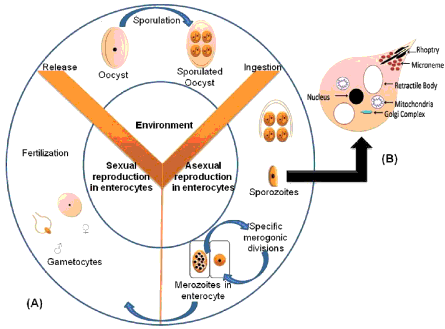
Coccidiosis is a complex intestinal disease of major economic importance in chickens caused by multiple species of the protozoan Eimeria. Infection by coccidial parasites has a huge impact on worldwide poultry production due to the morbidity, mortality, and reduced body weight gain (Del-Cacho et al., Reference DEL-CACHO, MARGARITA, HYUN, JOAQUIN, ERIK and CARIDAD2012; Kaboudi et al., Reference KABOUDI, UMAR and MUNIR2016). The life cycle of Eimeria spp. involves ingestion of the sporulated oocysts that excyst in the intestine and each releases eight sporozoites. Once free within the intestine, the sporozoites penetrate the host's enterocyte in very short time, encapsulate themselves safely within a parasitophorous vacuole, and replicate (Shah, Reference SHAH2013). Merozoites are then released in tremendous numbers; they rupture the host cell, and invade new ones. It has been proven that each Eimeria spp. is programmed genetically for a specific number of merogonic divisions. As much the parasite reinvades the cell, as much it damages the intestine and causes severe lesions (Renault, Reference RENAULT2007; Idris et al., Reference IDRIS, ABBAS, MASOOD, REHMAN, FAROOQ, BABAR, HUSSAIN, RAZA and RIAZ2016; Ahmad et al., Reference AHMAD, EL-SAYED and EL-SAYED2016). After the last species specific merogonic generation, merozoites invade the enterocytes to differentiate into male and female gametocytes. After fertilisation, the oocyst ruptures from the host's enterocytes and leaves the host with faeces ( Figure 1 ).
(A) Diagrammatic life cycle of Eimeria and (B) simplified structure of sporozoites (Adapted from Ahmad et al., Reference AHMAD, EL-SAYED and EL-SAYED2016).

The annual cost of losses to the global poultry industry caused by coccidiosis has been predicted to exceed US $3 billion (Shah et al., Reference SHAH, XU, YAN, SONG and LI2010a). Economic losses due to coccidiosis include mortality, malabsorption, inefficient feed utilisation, impaired growth rate in broilers, and reduction of egg production in layers (Chapman et al., Reference CHAPMAN, CHERRY, DANFORTH, RICHARDS, SHIRLEY and WILLIAMS2002). Moreover, coccidiosis may also predispose to necrotic enteritis in poultry (Abid et al., Reference ABID, AZEEM and UMAR2016).
A variety of strategies are used to control coccidiosis, including medication with anti-coccidial drugs and immunisation of chickens with live vaccines (Shah et al., Reference SHAH, UMAR, IQBAL, REHMAN, QADRI and HE2014; Idris et al., Reference IDRIS, ABBAS, MASOOD, REHMAN, FAROOQ, BABAR, HUSSAIN, RAZA and RIAZ2016). Currently, although chemoprophylaxis is the predominant method used to control coccidiosis, alternate options are needed due to the emergence of drug resistance of Eimeria spp., drug residue in animal food products, and high costs for development of new drugs (Dexing et al., Reference DEXING, CHUNLI, GAO, LI, NIU and XIADON2012). Live vaccines and attenuated vaccines available commercially could provide partial immune protection against reinfection, but pose the risk of unintended infection under the immunosuppressive conditions. It has been shown that host immunity to avian coccidiosis is largely dependent on immunity (Dalloul et al., 2005), thus generating interest in developing recombinant vaccines and adjuvants that preferentially elicit cellular immune responses. Many secreted or membrane-bound proteins involved in the interaction with the host immune system have been considered as targets for immunological interventions. Some antigens located on the surface of sporozoites and schizonts have been identified as potential immune regulator and effector genes that could influence host-parasite interaction at the molecular and cellular levels (Dexing et al., Reference DEXING, CHUNLI, GAO, LI, NIU and XIADON2012). A practical vaccine against coccidiosis should confer stable protection between different bathes as well as long-lasting protection covering the production cycle of chickens. Cell-mediated immune response plays a major role in host protective immunity to coccidia. The antibody titres in serum and intestine did not correlate with the level of protection after oral infection with coccidia (Xu et al., Reference XU, SONG, XU, YAN, SHAH and LI2008). Immunity against Eimeria spp. depends on the cellular arm of the immune response including CD4+ and CD8+ T cells with antibodies playing only a minor role in protection. Thereby, DNA immunisation has been widely studied as a novel strategy to elicit protection against coccidiosis for the ability to stimulate cellular immune response (Shah, Reference SHAH2013). DNA vaccines or recombinant antigen can provoke both humoral and cell-mediated immune responses and co-delivery of cytokines as adjuvants could enhance the potential for DNA vaccines or recombinant antigen to induce broad and long-lasting humoral and cellular immunity (Shah et al., Reference SHAH, XU, YAN, SONG and LI2010b). From the current literature, a review article about role of DC in immunity against avian coccidiosis is needed. This review briefly summarises the current state of knowledge of DC and their specific functions in immunity against avian coccidiosis.
DC as a regulator of immune response
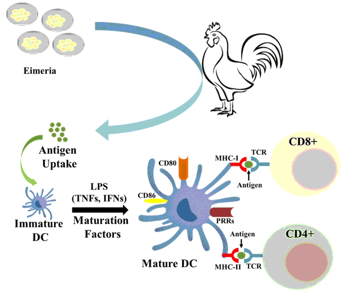
DCs are antigen-presenting cells (APCs) with the unique ability to induce both innate immune responses and a highly specific acquired immunity. DC are crucial to induce immunity and their maturation and functions are influenced by microbial and environmental stimuli. Chicken DC are composed of several subtypes including bursal secretory DC (BSDCs), follicular DC (FDCs), and thymic DC (TDCs). DC maturation depends on the nature of the perturbation and permits unique and efficient immune responses for each pathogen. They originate from haematopoietic stem cells in bone marrow. These cells are derived from myeloblast along with other granulocytes. At this stage, DC are referred to as immature, and have high endocytic activity and low potential of activating T cells. Immature DC survey the extra cellular environment for pathogens ( Figure 2 ). Once DC phagocytose an antigen, they become mature DC and migrate to regional lymph nodes where they present antigen to T lymphocytes (Liu and Nussenzweig, Reference LIU and NUSSENZWEIG2010; Maverakis et al., Reference MAVERAKIS, KIM, SHIMODA, GERSHWIN, PATEL, WILKEN, RAYCHAUDHURI, RUHAAK and LEBRILLA2015).
DC are a heterogeneous group of cells which mostly reside in peripheral tissues, especially in areas where body comes in contact with external environment. Under normal condition DC constantly survey the blood, lymph, peripheral tissues and secondary lymphoid organs (Guermonprez et al., Reference GUERMONPREZ, VALLADEAU, ZITVOGEL, THÉRY and AMIGORENA2002). DC present antigen on their surface along with co-stimulation molecules after capturing and processing the antigen. They play role in maintenance of B cells and are important in process of immunological memory (Wieder, Reference WIEDER2003). DC are unique APCs and have been referred to as ‘professional’ APCs, since their principal function is to present antigens, because only they have the ability to induce a primary immune response in resting naive T lymphocytes (Ohgimoto et al., Reference OHGIMOTO, OHGIMOTO, IHARA, MIZUTA, ISHIDO, AYATA, OGURA and HOTTA2007; Yasmin et al., Reference YASMIN, YEAP, TAN, HAIR-BEJO, FAKURAZI, KAISER and OMAR2015). DC have three main functions each of which includes antigen presentation. DC not only present antigen and activate T cells but also induce and maintain immune tolerance. Follicular DC are thought to be involved in establishing immunological memory in tandem with B cells (Wieder, Reference WIEDER2003).
Dendritic cell maturation and activation of CD4+ and CD8+ T cells in response to pathogens. Schematic drawing depicting the differentiation of immature DC into mature DC by up-taking antigen. This process involved in morphological changes in mature DC as well as development of strong co-stimulatory molecules for the activation of T cells as well as pattern recognition sites on the surface of DC (adapted from Rehman et al., Reference REHMAN, SAJID, CHUNCHUN, ZAFAR, FAROOQ, SAIF and CHAN2017).

Role of DC in immunity against coccidiosis
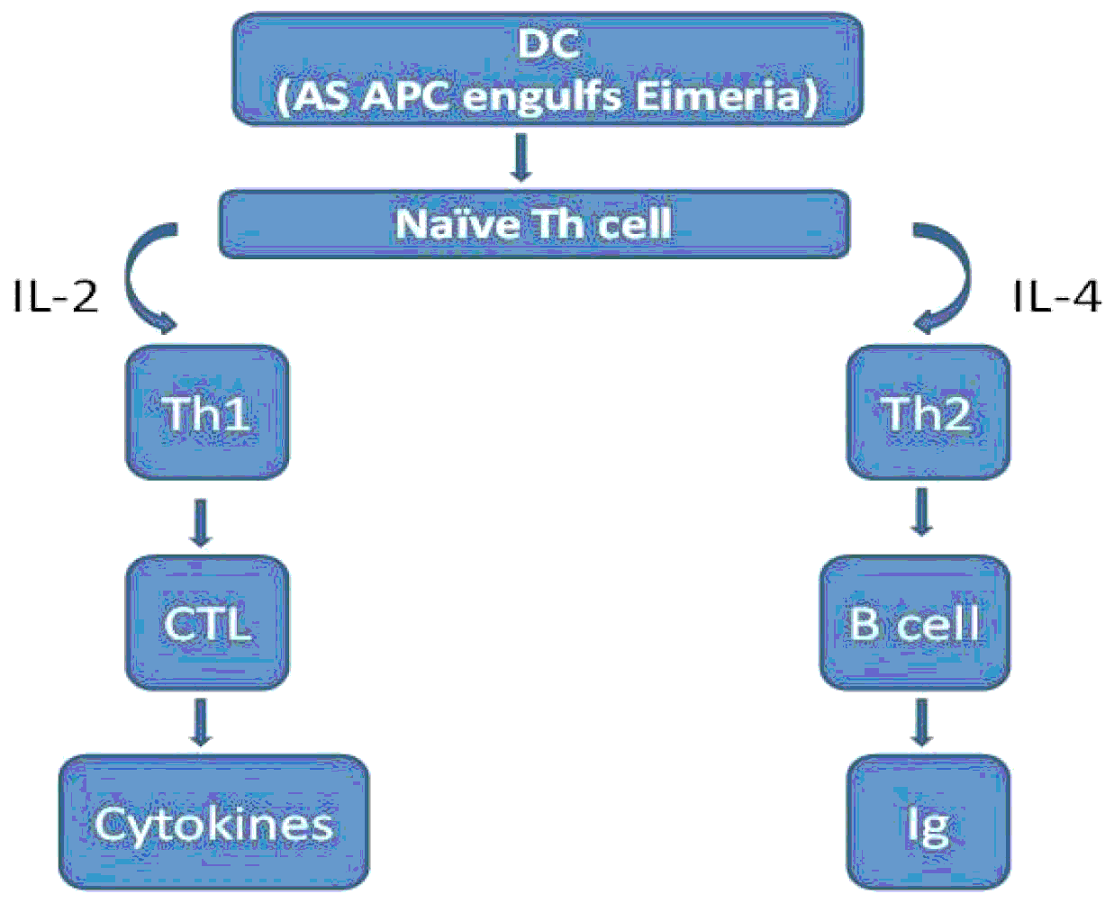
The main function of DC is to process antigen material and present it on the cell surface to the T cells of the immune system. They act as messengers between the innate and the adaptive immune systems. Owing to their properties, DC are often called ‘nature's adjuvants’ and thus have become the natural targets for antigen delivery. DC provide an essential link between the innate and the adaptive immune responses. DC are at the centre of the immune system owing to their ability to control both tolerance and immune responses ( Figure 3 ). These key properties of DC render them the central candidates for antigen delivery and vaccination (Karolina and Jacques, Reference KAROLINA and JACQUES2012). The cellular and molecular mechanisms leading to immune protection against coccidiosis in chickens are complex and include multiple aspects of innate and adaptive immunity. When a pathogen enters the bird, it is initially contained by physical barriers but if it is able to penetrate and successfully infect it, the innate immunity takes an active role. At this point in a primary infection, DC along with macrophages are important. They capture the pathogen, digest the proteins and present them. APC (macrophages and DC) will activate naïve T-cells. DC in fact are seen as the link between innate and adaptive immunity. Once the adaptive immunity is created, this will provide a specific response that will eliminate the pathogen (Lillehoj et al., Reference LILLEHOJ, LEE, JANG, KIM and LEE2011).
Role of DC in cell mediated immunity against Eimeria spp.

Current control strategy relies primarily on routine chemoprophylaxis using anticoccidial drugs. However, alternative methods are needed due to the rapid emergence of drug-resistant parasites and drug residues in chicken products (Shah et al., Reference SHAH, XU, YAN, SONG and LI2011; Clarke et al., Reference CLARKE, FODEY, CROOKS, MOLONEY, O'MAHONY, DELAHAUT, O'KENNEDY and DANAHER2014). Immunisation is a practical alternative to chemotherapy for the control of coccidiosis. However, most current commercial vaccines consisting of live oocysts of attenuated or non-attenuated coccidia are expensive and difficult for large-scale manufacturing. Furthermore, live vaccines have the risk of vaccinal pathogenicity, the potential reversion to virulence and coccidiosis breakout (Williams, Reference WILLIAMS2002; Del-Cacho et al., Reference DEL-CACHO, MARGARITA, HYUN, JOAQUIN, ERIK and CARIDAD2016 ). Therefore, it is urgent to develop safe and effective vaccine against avian coccidiosis. In case of DNA vaccination against coccidiosis, after in vivo generation, the antigenic peptides are processed and presented by professional antigen presenting cells (APCs), like DC, by getting primed through direct or by obtaining proteins from myocytes (Shah, Reference SHAH2013).
Antigens can reach lymph nodes through two pathways: via lymphatics, where the antigen is captured by lymph node-resident DC, or via tissue-resident DCs. These immature DC capture antigens, and DC activation triggers their migration towards secondary lymphoid organs and their maturation. DC display antigens in the context of classical major histocompatibility (MHC) class I and MHC class II molecules or in the context of non-classical CD1 molecules, which allow the selection of rare antigen-specific T lymphocytes. Activated T cells drive DC towards their terminal maturation, which induces further expansion and differentiation of T lymphocytes into effector T cells. If DC do not receive maturation signals, they will remain immature and antigen presentation will lead to immune regulation and/or suppression (Karolina and Jacques, Reference KAROLINA and JACQUES2012).
Dendritic cell derived exosomes (vaccine candidates) against coccidiosis
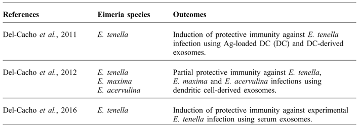
Exosomes are cell-derived, phospholipid-enclosed vesicles less than 100 nm in diameter released into the extracellular milieu that can be taken up by recipient cells at sites distal to their release. Exosomes from different cellular sources contain proteins specific for their cell type of origin, and these proteins, as well as those of their target cells, mediate exosome uptake (Del-Cacho et al., Reference DEL-CACHO, MARGARITA, HYUN, JOAQUIN, ERIK and CARIDAD2016). Exosomes secreted from antigen-presenting cells (APCs) play a functional role in mediating innate and adaptive immunity. DC derived exosomes expressing major histocompatibility complex class I (MHC-I) and MHC-II molecules induce and enhance antigen (Ag)-specific T cell responses in vitro and in vivo (Denzer et al., Reference DENZER, EIJK, KLEIJMEER, JAKOBSON, GROOT and GEUZE2000). DC-derived exosomes also express on their surface co-stimulatory molecules, such as CD80, necessary for Ag presentation (Viaud et al., Reference VIAUD, THÉRY, PLOIX, TURSZ, LAPIERRE, LANTZ, ZITVOGEL and CHAPUT2010). Current technologies, however, limit the practicality of isolating chicken DC exosomes on a scale sufficient for mass vaccination against avian coccidiosis in the field. Alternatively, exosomes from serum might constitute a more convenient source for such an application. It is well known that exosomes not only act locally, as it is the case in caecal tonsils during coccidiosis, but also circulate systemically through the vasculature and lymphoid systems (Caby et al., Reference CABY, LANKAR, VINCENDEAU-SCHERRER, RAPOSO and BONNEROT2005). One novel approach to vaccination against coccidiosis is to exploit DC (DCs) as an initial step in the development of a second-generation coccidiosis vaccine. DC are APC serving as immunologic sentinels that are critical for the induction of protective immunity against pathogens (Steinman, Reference STEINMAN2007). DC-mediated vaccination strategies rely on the technique of in vitro loading of DC with pathogen-derived Ag. One of the proposed mechanisms through which DC produce Ag-specific cellular and humoral immune responses involves the secretion of exosomes (Colino et al., 2006). DC-derived exosomes expressing major histocompatibility complex class I (MHC-I), MHC-II, and costimulatory molecules induce and enhance Ag-specific T cell responses in vitro and in vivo (Delcayre and Le-Pecq, Reference DELCAYRE and LE-PECQ2006). Because cytoplasmic vesicles are capable of becoming exosomes, among other fates, intracellular parasites, bacteria, and viruses that enter cells via an endocytotic pathway are prime candidates for DC-based exosome vaccines. Intracellular pathogens that are taken up by APCs are processed into MHC-II-binding peptide Ag, predominantly within endosomes. The Ag-loaded complexes are delivered to the cell surface through lipid bilayer fusion of the endosome and plasma membranes or, alternatively, are released extracellularly as intact exosomes ( Figure 4 ). Thus, MHC-II–Ag complexes are presented to naïve T cells either on the surface of APCs or through their exosomes. Isolated APC-derived exosomes offer the additional advantage of amplifying Ag-specific T cell activation through the synergistic effects of direct T cell activation as well as indirect transfer of MHC-II–Ag complexes to DC prior to antigen presentation. Remarkably, MHC barriers do not restrict exosome efficacy since syngeneic exosomes do not appear to work significantly better than allogeneic exosomes. Because protective immunity against Eimeria infection is T cell dependent, coccidiosis vaccines based on DC exosomes are promising vehicles for activating parasite-specific T cells (Del-Cacho et al., Reference DEL-CACHO, MARGARITA, HYUN, JOAQUIN, ERIK and CARIDAD2012) ( Table 1 ).
DC derived immune responses against Emeria species.

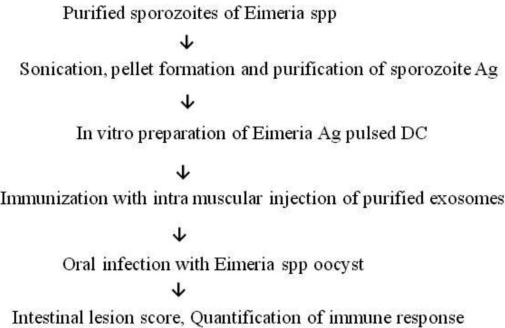
Loading of dendritic cells with Eimeria derived antigen.

Eimeria tenella can be controlled by using parasite Ag-loaded DC, or their derived exosomes, in the absence of free Ag CD45 (+) intestinal DC were isolated from E. tenella-infected chickens and loaded ex vivo with an extract of sporozoites as parasite Ag. Extracellular vesicles purified from the Ag-pulsed DC expressed surface proteins associated with DC-derived exosomes, including major histocompatibility complex proteins (MHC I and MHC II), CD80, flotillin, and heat shock protein (HSP70). Following intramuscular immunisation of chickens with Ag-pulsed DC or Ag-pulsed DC-derived exosomes, Ag-containing cells were observed diffusely localised in the lymphoid tissue and concentrated in germinal centres of caecal tonsils, and restricted to germinal centres (GC) in the spleen. Chickens immunised with pulsed DC or exosomes exhibited higher numbers of caecal tonsil and spleen cells expressing IgG and/or IgA antibodies that were reactive with E. tenella Ag, greater numbers of IL-2-, IL-16-, and IFN-γ-producing cells, and higher E. tenella Ag-driven cell proliferation, compared with chickens immunised with Ag in the absence of DC or exosomes. Chickens immunised with Ag-pulsed DC or Ag-pulsed DC-derived exosomes and subsequently given a live E. tenella challenge infection at 10d post-immunisation displayed increased body weight gains, decreased feed conversion ratios, reduced faecal oocyst shedding, diminished intestinal lesions, and lower mortality, compared with animals given Ag alone (Del-Cacho et al., Reference DEL-CACHO, GALLEGO, LEE, LILLEHOJ, QUILEZ, LILLEHOJ and SÁNCHEZ-ACEDO2011).
Strategies for vaccination against avian coccidiosis should be based on stimulation of these gut-associated lymphoid organs. Because exosomes are capable of migrating to the intestinal mucosa and present Ags to immunocompetent cells, it seems possible that administered Ag-pulsed DC exosomes similarly interact with T and B lymphocytes within the gut CTs and PPs to stimulate cellular and humoral immunity (Aline et al., Reference ALINE, BOUT, AMIGORENA, ROINGEARD and DIMIER2004). The use of exosomes in chicken immunisation against Eimeria spp. could not, to date, compete on price with the use either anticoccidial drugs or live parasite vaccines. Nevertheless, a single immunisation of chickens with DC-derived exosomes induced protective immunity against E. tenella, E. maxima, and E. acervulina infection (Del-Cacho et al., Reference DEL-CACHO, MARGARITA, HYUN, JOAQUIN, ERIK and CARIDAD2012). There is need for further studies regarding this mode of action (Masood et al., Reference MASOOD, WANG, YAO, GU, ANSARI, ZHANG, LIU and MASOOD2017; Monica et al., 2017; Simon et al., 2016; Atiba et al., Reference ATIBA, TEMU, LADO and SAYRE2016).
Conclusions
DC provide an essential link between the innate and adaptive immune responses. In recent years, with the emergence of new chicken DC markers, it has become increasingly possible to culture chicken DC in vitro. DC are an attractive target for therapeutic manipulation of the immune system, to enhance insufficient immune responses against infectious diseases. Immunisation with DC derived exosomes is effective against a combined infection with Eimeria spp.. Immunisation with exosomes could be an alternative strategy to the conventional disease control methods in case the regulation of anticoccidial drugs is tightened. Vaccines consisting of exosomes will both preserve all the positive aspects of live parasite vaccines and avoid their inherent risks. In addition to activating both arms of the immune system, exosome vaccines will never be able to cause infection, because they lack live parasite. Furthermore, recent investigations have proved that DC exosomes are a useful tool in the development of vaccines against other pathogens. Therefore, one may assume that development of polyvalent vaccines consisting of DC exosomes loaded with Ags from a variety of pathogens such as viruses, bacteria, and parasites may be feasible.
Conflict of interest
None of the authors of this paper has a financial or personal relationship with other people or organisations that could inappropriately influence or bias the content of the paper.
Acknowledgements
This study was supported by NSFC-PSF/13 project entitled ‘Determination of DC stimulation antigen of chicken coccidian and their application in the development of coccidiosis vaccine in chicken’. We are thankful to Dr Zaib-ur-Rehman (Shanghai Veterinary Research Institute, Shanghai, China) for helpful discussion and scientific advice on the preparation of this manuscript.