Introduction
Pseudephebe M. Choisy is a genus of lichenized fungi in the alectorioid clade of Parmeliaceae (Divakar et al. Reference Divakar, Crespo, Wedin, Leavitt, Hawksworth, Myllys, McCune, Randlane, Bjerke and Ohmura2015). It is distinguished from the two other genera in the clade with non-septate colourless ascospores, Bryoria Brodo & D. Hawksw. and Nodobryoria Common & Brodo, primarily by a distinct superficial layer of cells on the cortex (Brodo & Hawksworth Reference Brodo and Hawksworth1977). Pseudephebe species form small, fruticose to subcrustose cushion-like thalli on hard siliceous rock surfaces and have never been described as producing extrolites (i.e. secondary metabolites). The genus is known from both hemispheres, is circumpolar, and is found in mountains with arctic-alpine conditions, from Europe, North America and southern South America (Øvstedal & Smith Reference Øvstedal and Smith2001). It is, however, not known from Africa and is uncommon in Asia and Australia (Kantvilas Reference Kantvilas1994; Wang & McCune Reference Wang and McCune2010). The genus traditionally comprised two morphologically similar species (Hillmann Reference Hillmann1936; Lamb Reference Lamb1964; Brodo & Hawksworth Reference Brodo and Hawksworth1977): 1) P. minuscula (Arnold) Brodo & D. Hawksw., with strongly appressed and tending to be flattened branches, the tips becoming adnate, and with internodes to 1 mm in length; and 2) P. pubescens (L.) M. Choisy, with terete and never strongly appressed branches, the tips generally free, and internodes 1−3 mm in length. The known distributions of the two species overlap, but P. minuscula is generally reported from more extreme arctic-alpine habitats than P. pubescens, which is usually more common in moister areas (Myllys et al. Reference Myllys, Velmala and Holien2011). Both species have a variable morphology, which has led to the description of many infraspecific taxa, especially forms. These forms have been delimited mainly based on branching and appression degree and the thalli become almost subcrustose in part when growing in the most severe environments. Intermediate morphs occur, particularly in extreme habitats, making morphological species delimitation difficult (Imshaug Reference Imshaug1957; Brodo & Hawksworth Reference Brodo and Hawksworth1977).
Bryoria mariensis, recently described from the Falkland Islands (Fryday & Øvstedal Reference Fryday and Øvstedal2012), morphologically resembles large specimens of P. pubescens. It was described as containing lichenan and having a morphologically similar cortex to Pseudephebe; a prosoplechtenchymatous hyphal inner layer and a pseudoparenchymatous surface with knobby outermost cells. The presence of lichenan excludes a placement in Nodobryoria (Common Reference Common1991; Common & Brodo Reference Common and Brodo1995). Furthermore, species of Nodobryoria also have matt rather than shiny thalli and a knobbly cortex with a jigsaw pattern in surface view. The species was described in Bryoria, despite the similarities in cortical structure to Pseudephebe species, because of the presence of pseudocyphellae, production of norstictic acid and what were referred to as soralia; all characters not previously reported in Pseudephebe (Fryday & Øvstedal Reference Fryday and Øvstedal2012). Given the similarities between B. mariensis and Pseudephebe, we also included this species in our study.
For this study we performed morphological, microchemical, and three-locus phylogenetic analyses of a range of Pseudephebe specimens, and two recently collected B. mariensis samples from the type locality.
Materials and Methods
Materials
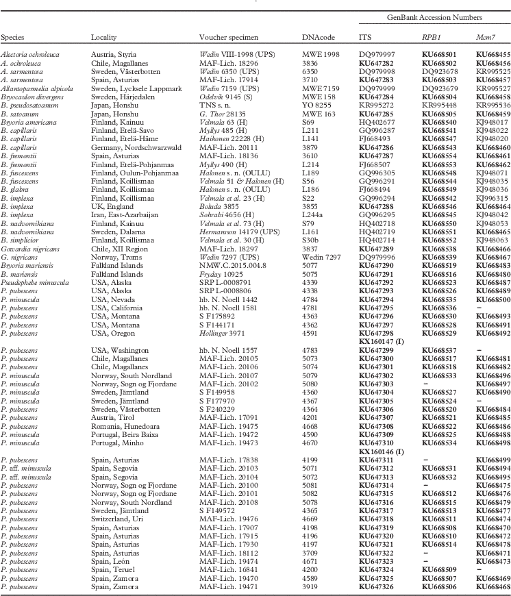
We studied over 120 Pseudephebe specimens from various institutional collections (AAS, BM, K, MAF, MSC, NMW, UBC, and ZT), and the private collection of Nastassja Noell (Reno, Nevada, USA; as hb. N. Noell). Thirty-seven of these were used for phylogenetic reconstruction, with the addition of 25 outgroup specimens (Table 1). Material that was morphologically and microchemically studied, but not molecularly analyzed, is listed according to the names used in the original identification, in Supplementary Material S1 (available online).
GenBank accession numbers and species data for specimens used for the phylogenetic tree shown in Fig. 1. Newly obtained sequences are in bold. DNAcode refers to the genetic material extracting code, newly obtained extractions preserved in MAF Herbarium DNA-bank. Pseudephebe species names are according to the original identifications. Specimens 4591 and 4670 have ITS intrathalline variability, and the alleles with an intron are indicated with (I)

Morphology and chemistry
Traditional and additional characters used to distinguish Pseudephebe pubescens and P. minuscula were examined in all material studied (Table 1, cited under Taxonomy, and in Supplementary Material S1 & S2, available online). For loaned specimens the envelope identification was maintained, while new collections were identified using Brodo & Hawksworth (Reference Brodo and Hawksworth1977), Myllys et al. (Reference Myllys, Velmala and Holien2011) and Smith et al. (Reference Smith, Aptroot, Coppins, Fletcher, Gilbert, James and Wolseley2009). Specimens were examined morphologically under a Nikon SMZ-1000 stereomicroscope, and hand-cut sections were studied using a Nikon Eclipse-80i microscope. Photographs were taken with a Nikon 105 mm f/2.8D AF Micro-Nikkor lens connected to a Nikon D90 camera. Spot tests (K, C, and PD) and thin-layer chromatography (TLC) were carried out following Orange et al. (Reference Orange, James and White2010). We used TLC solvent system C (200 ml toluene / 30 ml acetic acid), with concentrated acetone extracts at 50°C spotted onto silica gel 60 F254 aluminium sheets (Merck, Darmstadt). The aluminium sheets were dried for 10 min in an acetic acid atmosphere to maximize resolution. The same lichen fragment used for TLC was used for DNA extraction.
Molecular techniques
DNA was extracted from a single, clean (under a dissecting microscope) lichen branch using the DNeasy Plant Mini Kit (Qiagen, Barcelona) with a slight modification to the manufacturer’s instructions (Crespo et al. Reference Crespo, Blanco and Hawksworth2001). The fungal nuclear internal transcribed spacer (ITS) rDNA, and a partial sequence of the low copy protein coding genes RNA polymerase II largest subunit (RPB1), and minichromosome maintenance complex component 7 (Mcm7) were amplified using, respectively, the primers ITS1FKYO2 (5'-TAG AGG AAG TAA AAG TCG TAA-3') and ITS4KYO2 (5'-RBT TTC TTT TCC TCC GCT-3') (Toju et al. Reference Toju, Tanabe, Yamamoto and Sato2012), RPB1 MH F (5'-ACGTCGCCGAGACCCHAARA-3'; Leavitt et al. Reference Leavitt, Esslinger, Divakar and Lumbsch2012) and fRPB1-C rev (5'-CCNGCDATNTCRTTRTCCATRTA-3'; Matheny et al. Reference Matheny, Liu, Ammirati and Hall2002), and Xmcm7 F1 (5'-CGTACACYTGTGATCGATGTG-3'; Leavitt et al. Reference Leavitt, Johnson and St. Clair2011) and Mcm7-1348rev (5'-GAYTTDGCIACICCIGGRTCWCCCAT-3'; Schmitt et al. Reference Schmitt, Crespo, Divakar, Fankhauser, Herman-Sackett, Kalb, Nelsen, Nelson, Rivas-Plata and Shimp2009). For amplification, we used a reaction mixture of c. 25 µl, containing 18 µl of sterile water, 2·5 µl of 10× buffer with 2 mM MgCl2, 0·5 µl dNTPs (10 mM of each base), 1·25 µl of each primer at 10 µM, 0·625 µl of DNA polymerase (1U µl−1), and 0·5–2 µl of DNA elution 2 template. For any failed samples the PCR was repeated using PuReTaq Ready-To-Go PCR Beads (2·5 U of PuReTaq DNA Polymerase, 200 µM of each dNTP, BSA, buffer reaction and stabilizers: 10 mM Tris-HCl pH 9·0, 50 mM KCl, 1·5 mM MgCl2; GE Healthcare, Little Chalfont, UK), adding to the lyophilized bead 20 µl of sterile water, 1 µl of each primer at 10 µM and 2 µl of elution 1 DNA template.
Amplifications were run in an automatic thermocycler (XP Cycler, Bioer, Hangzhou, China) using the following parameters: initial denaturation of 5 min at 95°C, then 35 cycles of 60 s at 95°C, 60 s at 56°C, 90 s at 72°C, and a final extension of 10 min at 72°C for ITS; initial denaturation of 10 min at 94°C, followed by 4 cycles of 45 s at 94°C, 50 s at 56°C, 1 min at 72°C, 4 cycles of 45 s at 94°C, 50 s at 54°C, 1 min at 72°C, 36 cycles of 45 s at 94°C, 50 s at 52°C, 1 min at 72°C and a final extension of 8 min at 72°C for RPB1 and Mcm7. Double bands in an electrophoresis gel of PCR products were isolated using the FavorPrepTM MicroElute Gel / PCR Purification Kit (Favorgen® Biotech, Vienna, Austria) following the manufacturer’s instructions. PCR products were cleaned using IllustraTM ExoProStar (GE Healthcare, Little Chalfont, UK), according to the manufacturer’s instructions. Sequencing was performed by the Unidad de Genómica (Parque Científico de Madrid). The DNA sequences obtained were manually adjusted using SeqMan version 7.0 (DNAstar, Madison, USA) and MEGA5 (Tamura et al. Reference Tamura, Peterson, Peterson, Stecher, Nei and Kumar2011).
Phylogenetic analyses
Alignments for each locus were performed using MAFFT v7 (http://mafft.cbrc.jp/alignment/server/; Katoh & Standley Reference Katoh and Standley2013) with the G-INS-i alignment algorithm and ‘1PAM/K=2’ scoring matrix, with an offset value of 0·1, and the remaining parameters set as default. Some Pseudephebe specimens contained an intron at the 3' end of SSU rDNA, which was removed for the analysis at this level. While alignments for RPB1 and Mcm7 were straightforward and did not require manual corrections, exploratory analyses revealed several ambiguous regions in the ITS alignment. Therefore, we used the program Gblocks v0.91b (Talavera & Castresana Reference Talavera and Castresana2007) to delimit and remove ambiguous alignment nucleotide positions from the final ITS alignment using the online web server (http://molevol.cmima.csic.es/castresana/Gblocks_server.html) and implementing the options for a less stringent selection of ambiguous nucleotide positions, including the ‘Allow smaller final blocks’, ‘Allow gap positions within the final blocks’, and ‘Allow less strict flanking positions’ options. The single and concatenated datasets were analyzed using maximum likelihood (ML) and Bayesian (B/MCMC) approaches. To detect topological conflicts among loci, the CADM test (Legendre & Lapointe Reference Legendre and Lapointe2004; Campbell et al. Reference Campbell, Legendre and Lapointe2011) was performed using the function ‘CADM.global’ implemented in the library ‘ape’ of R (Paradis et al. Reference Paradis, Claude and Strimmer2004). The analysis resulted in a W of 0·83, which means there is no evidence of well-supported topological conflict. For the maximum likelihood (ML) tree reconstructions, the program RAxML v7.2.8 (Stamatakis Reference Stamatakis2006) implemented on the Cipres Science Gateway (http://qball2.sdsc.edu:7070/portal2/home.action; Miller et al. Reference Miller, Pfeiffer and Schwartz2010) was used with the GTRGAMMA model (Stamatakis Reference Stamatakis2006; Stamatakis et al. Reference Stamatakis, Hoover and Rougemont2008). Support values were assessed using the ‘rapid bootstrapping’ option with 1000 replicates. For the concatenated dataset, the ITS regions ITS1, 5.8S and ITS2 were partitioned and for the protein-coding markers we used a three-partition approach, with the first, second, and third codon positions as separate model partitions. For the Bayesian reconstruction, MrBayes v3.2.1 (Ronquist & Huelsenbeck Reference Ronquist and Huelsenbeck2003) was used. Models of DNA sequence evolution for each locus were selected with the program jModeltest2.0 (Darriba et al. Reference Darriba, Taboada, Doallo and Posada2012), using the Akaike Information Criterion (AIC; Akaike Reference Akaike1974). The best-fit model of evolution was as follows: ITS, ITS1=TIM2+G, 5.8S=K80, ITS2=TIM3ef+G; RPB1; 1st position, TIM2+I, 2nd position, TrN, 3rd position, TIM1ef+G; Mcm7; 1st position, TPM3, 2nd position, F81, 3rd position, TrNef+G. A run with 10 million generations, starting with a random tree and employing 12 simultaneous chains, was executed. Every 500th tree was saved to a file. We plotted the log-likelihood scores of sample points against generations using Tracer v1.5 (Rambaut et al. Reference Rambaut, Suchard, Xie and Drummond2014) and determined that stationarity had been achieved when the log-likelihood values of the sample points reached an equilibrium and ESS values exceeded 200 (Huelsenbeck & Ronquist Reference Huelsenbeck and Ronquist2001). Preliminary analysis resulted in an overestimation of branch lengths and to correct this we used the uniform compound Dirichlet prior ‘brlenspr=unconstrained:gammadir (1,1,1,1)’ (Zamora et al. Reference Zamora, Calonge and Martín2015), obtaining rather reasonable branch length estimates. Posterior probabilities (PPs) were obtained from the 50% majority rule consensus of sampled trees after excluding the initial 25% as burn-in. The phylogenetic tree was drawn using FigTree v1.4 (Rambaut Reference Rambaut2009).
Species delimitation
In order to establish species limits in the phylogenetic tree, three computational approaches were used: 1) Automatic Barcode Gap Discovery ABGD (Puillandre et al. Reference Puillandre, Lambert, Brouillet and Achaz2011) based on barcode gaps using genetic distances; 2) Poison Tree Processes PTP (Zhang et al. Reference Zhang, Kapli, Pavlidis and Stamatakis2013), based on gene trees; and 3) Bayesian Phylogenetics and Phylogeography BP&P (Yang & Rannala Reference Yang and Rannala2010), based on a multispecies coalescent model for species validation.
Results
Molecular analysis
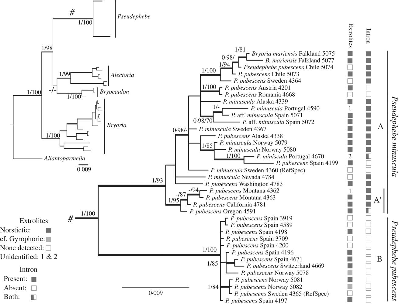
The GenBank accession numbers of 146 newly obtained sequences are included in Table 1. The final concatenated matrix used as input for the phylogenetic reconstruction contained 1448 unambiguously aligned base pairs (bp) (496 bp for RPB1, 458 for Mcm7 and 494 for ITS), with no topological incongruences among loci. Alignments are available in TreeBASE (TB2:S15972). The tree reconstruction (Fig. 1) included all genera belonging to the alectorioid clade (Divakar et al. Reference Divakar, Crespo, Wedin, Leavitt, Hawksworth, Myllys, McCune, Randlane, Bjerke and Ohmura2015), with the exception of Nodobryoria as no DNA-fresh material could be obtained, and using Allantoparmelia (Vain.) Essl. as outgroup. The genera Alectoria Ach. (including Gowardia Halonen et al.; see Lumbsch & Huhndorf Reference Lumbsch and Huhndorf2010), Bryocaulon Kärnefelt, and Bryoria were included to check for any possible alternative affinities for B. mariensis. Several species for each genus were included to facilitate the comparison of species branch lengths with the ones in Pseudephebe. Pseudephebe was shown to be monophyletic, including B. mariensis within it. Two isolated clades were recovered within the genus (A and B; Fig. 1). Clade A is a variable group containing two major clades, one of which is well-supported so we distinguish it as clade A’.
Phylogenetic consensus tree based on ITS, RPB1 and MCM7 markers analyzed as a concatenated data matrix (Table 1). Whole tree in the upper left corner using Allantoparmelia as outgroup, with the Pseudephebe clade shown in detail. Tree topology depicts the results of Bayesian inference, showing significant posterior probabilities ≥0·95 and bootstrap values ≥70% obtained in the maximum likelihood analysis. Intron presence and extrolite composition are indicated on the lower left corner. RefSpec=sequenced reference specimen.
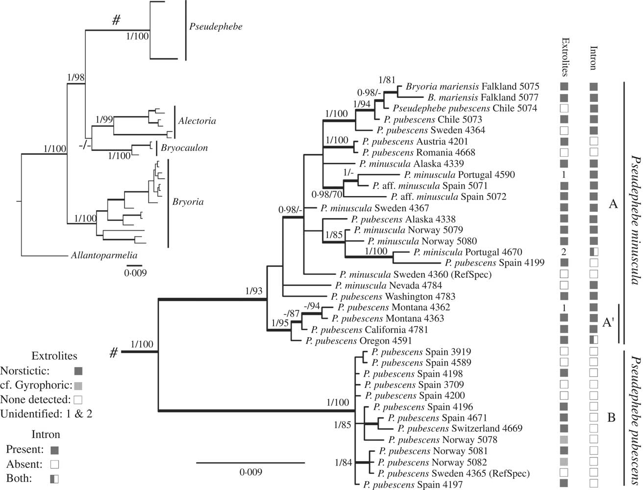
The region amplified with the primers ITS1FKYO2 and ITS4KYO2 comprises the ITS (ITS1, 5.8 S and ITS2) and a small portion of the 3' end of the SSU gene, which included in some specimens a group I intron of 233 bp inserted in site 1516 relative to the SSU rDNA sequence of Escherichia coli (Gutiérrez et al. Reference Gutiérrez, Blanco, Divakar, Lumbsch and Crespo2007) or site 1777 of Saccharomyces cerevisiae. This intron was detected in many but not all samples in clade A (Fig. 1), but was absent from all samples in clade B. In the electrophoresis gel of two samples, two bands corresponding to sequence lengths with and without the intron were distinguished; we checked that result by sequencing each band separately. ITS and SSU are multicopy loci and as DNA was extracted from a single branch, this result suggests that some copies could contain the intron while others would not.
Morphological and chemical analyses
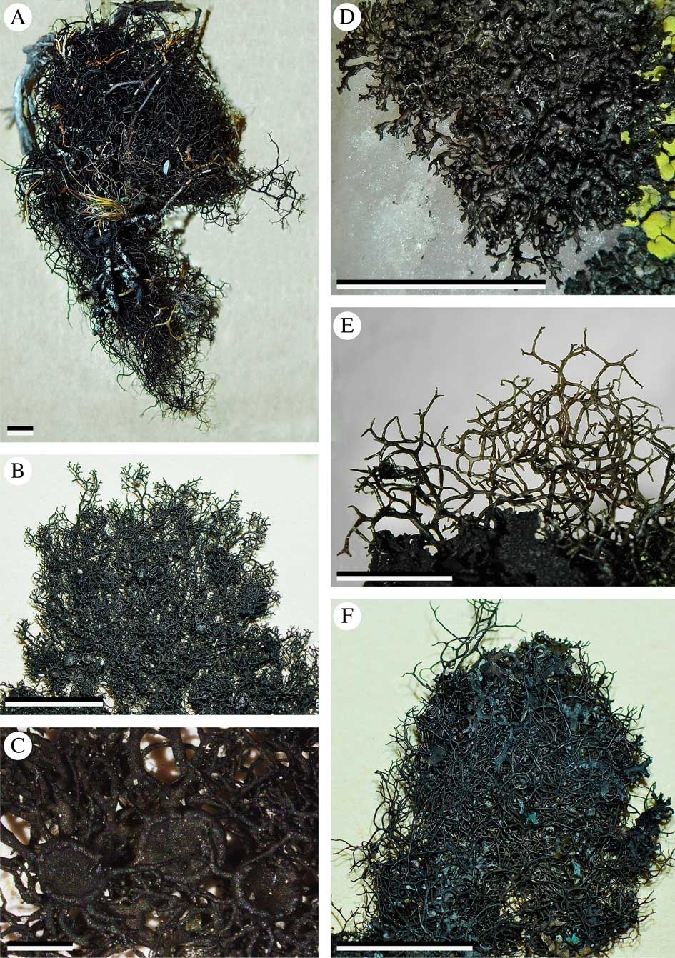
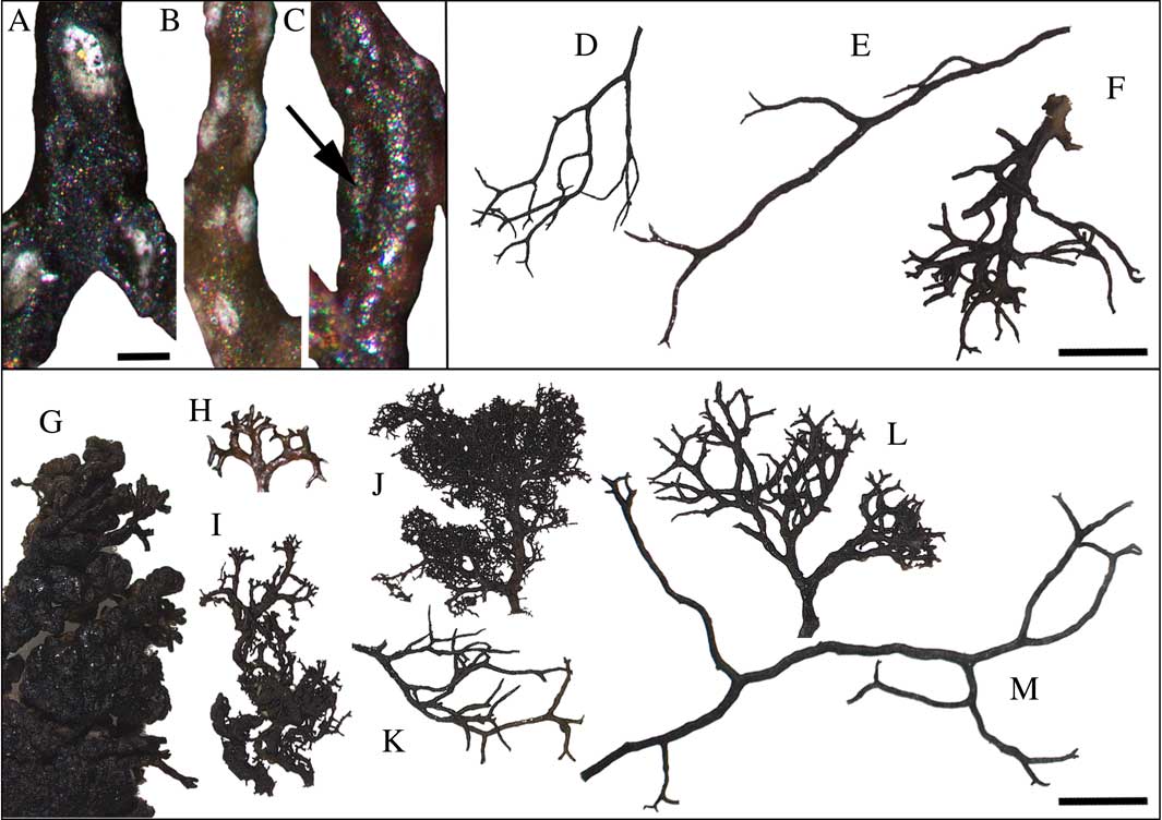
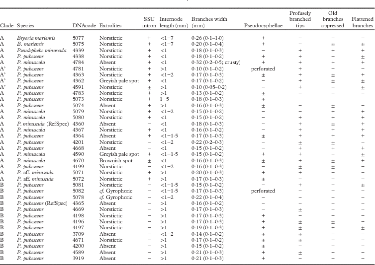
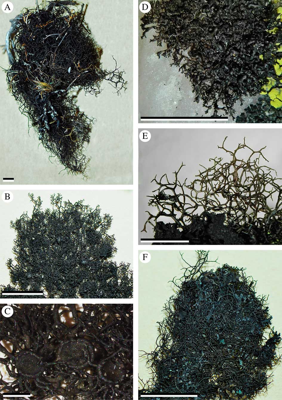
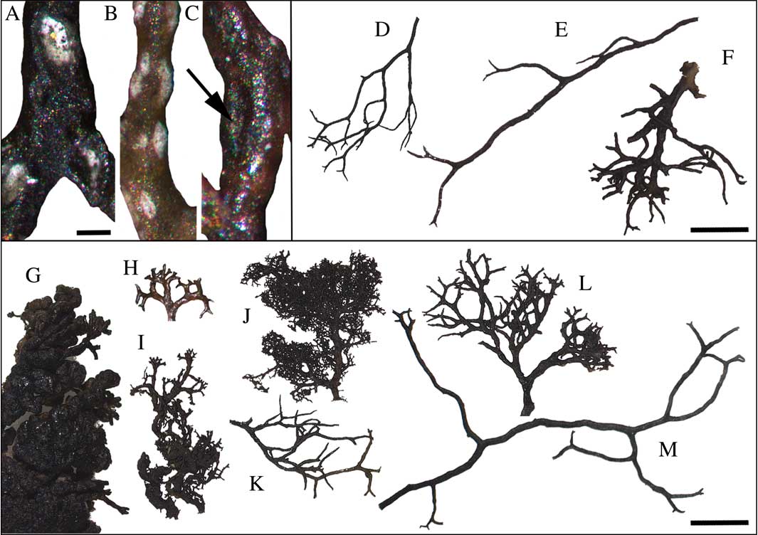
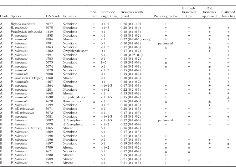
Morphological and chemical characters of the specimens studied in the phylogenetic analysis are presented in Table 2. The characters traditionally used to distinguish Pseudephebe minuscula from P. pubescens (i.e. loose or dense appearance, evenly/unevenly thickened branches, branch diameter, internode branch length, and presence of flattened branches) were very variable. The gradient of values found made unequivocal separation into two groups difficult. However, some of the characters studied were taxonomically informative. The presence of a flattened branching pattern, which is related to the presence of dorsoventrally flattened branches, had fewer intermediate states than other characters. The profusion of new ramifications on branch tips (Figs 2 & 3) was also informative when used together with other characters. Characters related to apothecia and pycnidia, such as spore size and the presence of marginal cilia, however, could not be adequately assessed as only four of the Pseudephebe specimens analyzed molecularly had apothecia (see Supplementary Material S2, available online). On those specimens, ciliate apothecia were observed in both clades, and spore measurements were not discriminatory.
Variability in Pseudephebe species. A–D, P. minuscula; A, long branched fruticose habit (NMW.C.2015.004.8); B, short to intermediate, more typical, branched habit (S F149958, sequenced reference specimen); C, specimen with apothecia in detail (S F149958, sequenced reference specimen); D, extreme subcrustose habit (hb. N. Noell 1561). E & F, P. pubescens; E, long branched fruticose habit (MAF-Lich. 20100); F, typical habit (S F149572, sequenced reference specimen). Scales: A, B, D−F=5 mm; C=1 mm.
Pseudocyphellae and branching variability in Pseudephebe. A–C, pseudocyphellae in P. minuscula: A, perforated (MAF-Lich. 20100); B, superficial (MAF-Lich. 19472); C, inconspicuous (MAF-Lich. 20103). D–F, P. pubescens branching variability; D, S F149572; E, MAF-Lich. 19474; F, MAF-Lich. 20101. G–M, P. minuscula branching variability; G, hb. N. Noell 1442; H, MAF-Lich. 20103; I, hb. N. Noell 1561; J, S F177970; K, MAF-Lich. 19475; L, MAF-Lich. 19472; M, NMW.C.2015.004.8. Scales: A–C=0·1 mm; D–M=2 mm.
Main characters used to distinguish Pseudephebe species in specimens used for the phylogenetic trees shown in Fig. 1. Pseudephebe species names are according to the original identifications. Internode length and branch width measurements, n = 30, are given with extreme values in brackets. RefSpec = sequenced reference specimen;±= ambiguous
Pseudocyphellae, reported here for the first time in the genus, were observed in 23 of the genetically analyzed samples (62%). They varied from inconspicuous to very conspicuous (Fig. 3), and in two samples were perforated. The presence or absence of pseudocyphellae is a taxonomically informative character at the generic level in different clades of Parmeliaceae (Elix Reference Elix1993; Crespo et al. Reference Crespo, Kauff, Divakar, del Prado, Pérez-Ortega, Amo de Paz, Ferencova, Blanco, Roca-Valiente and Núñez-Zapata2010). Although reported in the original species description, no true soralia were observed in Bryoria mariensis, neither in the type material nor in the additional specimens studied, but frequent short side-branch hapters with globose ends were present.
Pseudephebe is described in all previous literature as lacking extrolites detectable by spot tests or TLC (Brodo & Hawksworth Reference Brodo and Hawksworth1977; Smith et al. Reference Smith, Aptroot, Coppins, Fletcher, Gilbert, James and Wolseley2009; Myllys et al. Reference Myllys, Velmala and Holien2011). However, c. 60% of the studied genetically samples contained norstictic acid, generally in small quantities or in traces, but in some as a major substance. In some specimens of both P. pubescens and P. minuscula, we observed the characteristic needle-like red crystals of norstictic acid after the application of K. The presence of norstictic acid was not correlated with geographical distribution or any morphological character studied. In addition, a similar spot (with TLC) to gyrophoric acid, and two unidentified substances (substances 1 and 2), were also detected in some samples.
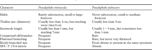
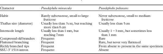
A selection of the most informative characters that can help to distinguish the two Pseudephebe species recognized here (i.e. clades A and B) is provided in Table 3.
Characters giving the most discrimination for the separation of Pseudephebe species, based on genetically analyzed material. An unequivocal identification to species is only possible using DNA barcoding
Species delimitation
Pseudephebe specimens fall into two genetically well-isolated clades (clades A and B) separated by an unexpectedly long branch (Fig. 1). The morphological characters traditionally used to separate these species were not, however, fully congruent with the two clades.
Clade B included specimens of P. pubescens conforming to the traditional concept of the species (Brodo & Hawksworth Reference Brodo and Hawksworth1977), but also a small number which were similar morphologically to P. minuscula. In contrast, 42% of the specimens from clade A were originally identified as P. minuscula, and the remaining 58% had been named P. pubescens or Bryoria mariensis.
Clade A (Fig. 1) contained a much greater genetic variability than clade B (here assigned to P. pubescens), with branch lengths and a topology similar to other alectorioid genera with multiple species (Velmala et al. Reference Velmala, Myllys, Goward, Holien and Halonen2014). Subclade A' (Fig. 1) is well supported and comprised specimens from western North America. That material has thin elongated terminal branches that are themselves minutely branched at the end; we initially speculated that this might merit recognition as a separate taxon, but discovered that this character was also evident in other clades.
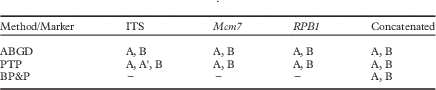
Species delimitation approaches (Table 4) confirmed the existence of two species as the most probable scenario for the genus since no genetic gap was detected within clade A (Leavitt et al. Reference Leavitt, Moreau and Lumbsch2015). We therefore assign clade A to Pseudephebe minuscula and clade B to P. pubescens. Although the two species overlap morphologically, an assay with a node dating analysis established that they could have split from a common ancestor around 9·5 to 11·6 mya (see Supplementary Material S3, available online).
Species delimitation prediction according to each method and marker
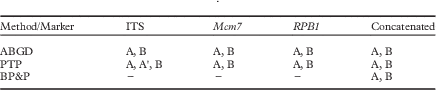
A, A' and B refers to clades named in Fig.1. ABGD=Automatic Barcode Gap Discovery; PTP=Poison Tree Processes; BP&P=Bayesian Phylogenetics and Phylogeography
Taxonomy
Where morphological identification cannot be made with confidence, species-level identification in Pseudephebe should be carried out by molecular methods. In cases where this is not possible, we suggest that collections should be referred to as either P. pubescens s. lat. or P. pubescens aggr., as discussed by Crespo & Lumbsch (Reference Crespo and Lumbsch2010). We prefer ‘aggr.’, as that term is used for plants, particularly in cases covering two or more named species.
In order to leave no doubt in the application of names to the two major clades distinguished here, and for assigning sequences obtained from fresh collections, we designated a sequenced reference specimen for both Pseudephebe species.
We have not reinvestigated species names other than those listed below, viz. Alectoria biformis (Vain.) Dodge, A. congesta (Zahlbr.) Dodge, A. intricata Hue, and A. nigerrima Hue, nor the application of numerous infraspecific names employed in the group. Information on these is provided in Hillmann (Reference Hillmann1936), Lamb (Reference Lamb1948, Reference Lamb1964), Hawksworth (Reference Hawksworth1972), Brodo & Hawksworth (Reference Brodo and Hawksworth1977), and Myllys et al. (Reference Myllys, Velmala and Holien2011). Many of these names are based on material from Antarctica, and are likely to represent P. minuscula as circumscribed here because of the morphology, but ideally they should be re-collected and sequenced from the type localities to verify their status.
Pseudephebe minuscula (Nyl. ex Arnold) Brodo & D. Hawksw.
Opera Bot. 42: 140 (1977).—Imbricaria lanata var. minuscula Nyl. ex Arnold, Verh. Zool.-Bot. Ges. Wien 28: 293 (1878).—Parmelia minuscula (Nyl. ex Arnold) Nyl., Bull. Soc. Linn. Normandie, sér. 4 1: 205 (1887); type: Finland, Lapponia enontekiensis, Enontekio, in alpe Pietsovaara prope Kilpisjärvi, 1867, J. P. Norrlin (H-NYL 34355—lectotype ?, designated by Brodo & Hawksworth Reference Brodo and Hawksworth1977: 141 [as “34255”] but see below); Sweden, Jämtland, Undersåker sn, Välliste, topplatån, 1300 m VSV om Norra stugan, 63º16'07''N, 13°08'16·1''E, fjällhed, klippor, på stenblock, 22 July 2009, G. Odelvik 9152 (S F149958—reference specimen selected here, DNAcode 4360; GenBank accession numbers: ITS KU647304, RPB1 KU668527, Mcm7 KU668490).
Alectoria pacifica Stizenb., Proc. Calif. Acad. Sci., ser. 2 5(2): 537 (1895): type: [Mexico], Insulae californica Guadalupe, supra terram humosam, 1875, E. Palmer (ZT Myc55359—holotype; US—isotype n. v.).
Bryoria mariensis Øvstedal et al., Lichenologist 44: 487 (2012); type: Falkland Islands, West Falkland, Port Howard, on summit of Mt. María, UTM 21F UC 2079, 2158 ft [658 m], feldmark, 28 January 1968, H. A. Imshaug 41327 & R. C. Harris (MSC 80870—holotype).
Myllys et al. (Reference Myllys, Velmala and Holien2011: 140) designated a different specimen from that selected by Brodo & Hawksworth (Reference Brodo and Hawksworth1977: 141) as lectotype for Nylander’s “minuscula”. They cited an undated specimen from Austria collected by Arnold (H-NYL 34356), without discussion, but presumably as they considered that a lectotype had to be chosen only from material studied by the validating author, in preference to one from Finland collected in 1867 and studied by Nylander (H-NYL 34355), which was selected by Brodo & Hawksworth. This case is, however, complex as Arnold (Reference Arnold1878: 293) also referred to earlier usages of the name by Nylander, Stizenberger, and himself. There is no indication that Arnold was intending to do anything but cite an identification in a floristic list of species in a region of Austria. Under Art. 9.3, original material in validating descriptions can include materials not seen by the validating author. Also mentioned by Arnold is “Nyl. Lapp. Or. 120”, which is a direct reference to Nylander material, and it seems preferable to have a Finnish specimen as lectotype as Arnold cited Nylander as the author. “Lapp. Or. 120” would seem to be a cryptic reference to an exsiccate of Fellman issued in 1865 which, however, has “Parmelia lanata f. minuscula” as no. 83, while no. 120 is “Lecanora dicksonii” (Lynge 1915–Reference Lynge16). However, there is a Fellman specimen in H-NYL from “Karelia pomorica orientalis, Suma” dated 1863 (H-NYL 34357) and examined by DLH in 1968 which could therefore be a better candidate. The issue merits further study and a resolution of this typification is beyond the scope of the present work. Rather than complicate matters further here, we have indicated a reference specimen (RefSpec) to serve as a proxy sequenced type, following the proposal of Ariyawansa et al. (Reference Ariyawansa, Hawksworth, Hyde, Jones, Maharachchikumbura, Manamgoda, Thambugala, Udayanga, Camporesi and Daranagama2014), as an interim solution to fixing the molecular application of the epithet minuscula in our sense.
The name Alectoria antarctica Dodge & Baker is excluded here as it was based on a specimen of Pseudephebe minuscula infected by a lichenicolous fungus. The lichenicolous fungal element was selected as lectotype for the name by Hawksworth & Iturriaga (Reference Hawksworth and Iturriaga2006: 202), who transferred it into Carbonea.
Specimens referred to Pseudephebe pubescens f. subciliata (Nyl.) D. Hawksw. forming appressed rosettes and with short internodes appear to belong here. They were retained under P. pubescens by Hawksworth (Reference Hawksworth1972) as the lobes were terete rather than dorsoventrally compressed, but from the data presented here that character does not appear to be taxonomically informative and environmentally induced. Of the total samples analyzed molecularly, specimens from Austria, Chile, the Falkland Islands (Islas Malvinas), Norway, Portugal, Romania, Spain, Sweden, and the USA were clustered in the Pseudephebe minuscula clade.
Additional specimens examined (DNA, morphology, and chemistry examined). Austria: Tirol: Otzaler Alpen, SE of Martin Busch Haus, 46º47'57''N, 10º52'47''E, 2580 m, on gneissic boulder, 2011, R. Türk 49630 [Obermayer, Lichenoth. Graec. No. 379] (MAF-Lich. 17091, as Pseudephebe pubescens, DNAcode 4201).—Chile: Magallanes y Antártida Chilena (XII Región): Navarino Island, Cabo de Hornos Commune, Bandera Hill, 15 iii 2012, C. Laguna Defior (MAF-Lich. 20105, DNAcode 5073, TLC: norstictic acid; and MAF-Lich. 20106 (DNAcode 5074, TLC: no substances detected), 20110).—Falkland Islands (Islas Malvinas): West Falkland (Gran Malvina): Port Howard (Puerto Mitre), Mt. María, 51·60768°S, 59·58654°W, 580 m, on sandy soil in feldmark, 2015, A. Orange 22484 (NMW s. n., as Bryoria mariensis, DNAcode 5077); Mt María summit, D. Crabtree [A. Fryday 10925] (MSC, as Bryoria sp., DNAcode 5075).—Norway: Sogn og Fjordane: Sogn, c. Luster, 61º31'58''N, 07º50'49''E, 1316 m, on rocky soil with moss and lichens, 12 viii 2015, C. G. Boluda & N. Calpena (MAF-Lich. 20101, DNAcode 5082, TLC: cf. gyrophoric acid). Nordaland: E6 road, contact with the Arctic Circle, 66º34'36''N, 15º20'57''E, 679 m, on soil with mosses and lichens, 14 viii 2015, C. G. Boluda & N. Calpena (MAF-Lich. 20107, DNAcode 5079, TLC: norstictic acid).—Portugal: Beira Baixa: Covilha, Serra da Estrela, Torre, 40°19′N, 07°36'W, 1966 m, on granitic boulders, 11 vi 2014, V. J. Rico (MAF-Lich. 19472, DNAcode 4590). Minho: Peneda-Gerés Natural Park, c. Castro Laboreiro, 42°01'N, 08°08'W, 1028 m, 9 ix 2014, C. G. Boluda & V. J. Rico (MAF-Lich. 19473, DNAcode 4670).—Romania: Hunedoara: Vrdele Pass, 45°20'40''N, 23°39'31''E, 2115 m, 12 x 2014, on siliceous rock, C. G. Boluda & E. Araujo (MAF-Lich. 19475, as P. pubescens, DNAcode 4668).— Spain: Asturias: Caso, Campo de Caso, Redes Natural Park, Valdebezón, from Monasterio River to peña’l Vientu, 43º05′21′′N, 05°19′33′′W, 1553 m, S slope, on quartzite, 2012, V. J. Rico 4423 (MAF-Lich. 17838, as P. pubescens, DNAcode 4199). Segovia: La Granja de San Ildefonso, c. Puerto de Navacerrada to Puerto de Cotos road, brook of Los Puentes, towards the Loma del Noruego, 40º47'36''N, 03º57'12''W, 1900 m, on granite, 2014, V. J. Rico 4614 (MAF-Lich. 20103, DNAcode 5071, TLC: norstictic acid; MAF-Lich. 20104, DNAcode 5072, TLC: norstictic acid).—Sweden: Jämtland: Kall par., Skäckerfjällen Nature Reserve, Rutsdalen, SW slope of Dörsvalen, 63·80775ºN, 12·84044°E, 700 m, on siliceous rock in boulder area, 2010, M. Westberg 10-078 (S F177970, DNAcode 4367). Lycksele Lappmark: Tärna sn, Atoklintens, 65°40·904'N, 14°38·310'E, on rock, 2012, G. Odelvik 12609, M. Hamnede & L. Hedenäs (S F240229, DNAcode 4364).—USA: Alaska: Fairbanks North Star Co., Steese Highway, Eagle Summit, 65·4867, −145·4153, 1100 m, alpine tundra, on rocks, 2011, R. Rosentreter 17334, 17292 et al. (SRP L8791, L8806, as P. pubescens, DNAcode 4339, 4332). California: Placer Co., Tahoe Rim Trail just north of Blockway Summit Trailhead, 39·2646°N, 120·0607°W, 2361 m, on granite, 2015, N. Noell 1581 & J. Hollinger (hb. N. Noell, as P. pubescens, DNAcode 4781). Montana: Deerlodge Co., Beaverhead-Deerlodge National Forest, c. Four Mile Basin, 46º05·680'N, 113º13·918'W, 2438 m, on granite boulders, 2009, St. Clair 16685 [St. Clair et al. Lich. W. N. Amer. no. 145] (S F175892, as P. pubescens, DNAcode 4363); Jefferson Co., just E of Homestake Pass on I90, 45º55'01''N, 112º22'33''W, 6200 ft [1890 m], on boulders, 2006, C. M. Wetmore 94730 (S F144171, as P. pubescens, DNAcode 4362). Nevada: White Pine Co., Great Basin National Park, Mount Wheeler, 38·9859°N, 114·3148°W, 3981 m, rocky summit, on quartzite, 2014, N. Noell (1442) & J. Hollinger (hb. N. Noell, DNAcode 4784). Washington: Spokane Co., Turnbull National Wildlife Refuge, Kepple Lake overlook, 47·441°N, 117·528°W, 700 m, on top of basalt on forest floor, 2015, N. Noell 1557, J. Allen & R. O’Quinn (hb. N. Noell, as P. pubescens, DNAcode 4783).
Pseudephebe pubescens (L.) M. Choisy
Icon. Lich. Univ., ser. 2 1: sine pag. (1930).—Lichen pubescens L., Sp. Pl. 2: 1155 (1753); type: Dillenius, Hist. Musc.: pl. 13, fig. 9 (1742) (lectotype designated by Jørgensen et al. Reference Jørgensen, James and Jarvis1994: 343); sine loc. (LINN 1273.286—epitype designated by Jørgensen et al. Reference Jørgensen, James and Jarvis1994: 343); Sweden, Jämtland, Undersåker sn, Välliste, topplatån, 1300 m VSV om Norra stugan, 63º16'07''N, 13º08'16·1''E, fjällhed, klippor, på stenblock, 22 July 2009, G. Oldevik 9115 (S F149572—reference specimen selected here, DNAcode 4365; GenBank accession numbers: ITS KU647317, RPB1 KU668513, Mcm7 KU668477).
Jørgensen et al. (Reference Jørgensen, James and Jarvis1994: 343) pointed out that the specimen in the Linnean collections under the name Lichen pubescens (LINN 1273.286), which had been cited as lectotype by Hawksworth (Reference Hawksworth1972: 235), was annotated in the handwriting of Linnaeus’ son and not Linnaeus himself, something also pointed out by Howe (Reference Howe1912: 201). In the absence of evidence that the specimen was part of the original material studied by Linnaeus prior to publication of the name, it is not eligible as a lectotype and the Dillenian plate has to be used. Jørgensen et al. (Reference Jørgensen, James and Jarvis1994: 343) consequently designated LINN 1273.286 as epitype. As there appears to be no mechanism to change an epitype once selected, without going through the formal conservation process, we have indicated a reference specimen (RefSpec) to serve as a proxy sequenced type to fix our application of the epithet pubescens, as for minuscula (see above).
We selected a sequenced reference specimen from northern Sweden, as Linnaeus (Reference Linnaeus1753: 1155) gave the habitat information as “Habitat in Europa septentrionali, Lapponia, Suecia”. Dillenius (Reference Dillenius1742: pl. 13, fig. 9) gives the locality as “In rupibus Groenlandie leóta & ab me delata fuit a Franc. Soldano, Chirurgo.” (loc. cit.: 66), so from Greenland where it was evidently collected by Francesco (or perhaps Franco) Soldano (fl. 1700–1740), a surgeon and friend of Dillenius known to have collected at several sites in Greenland as far as 81°N. Some specimens of Soldano are evidently still present in the Sherard Herbarium in OXF (Clokie Reference Clokie1964). Specimens in that herbarium are not available for loan and have not been studied by us. Although potentially of historical interest, they are not now pertinent to the typification of the Linnean name as an epitype has already been designated by Jørgensen et al. (Reference Jørgensen, James and Jarvis1994).
Of the samples analyzed molecularly, specimens from Norway, Spain, Sweden, and regions of Switzerland clustered in the Pseudephebe pubescens clade.
Additional specimens examined (DNA, morphology, and chemistry examined). Norway: Sogn og Fjordane: Sogn, c. Luster, 61º31'58''N, 07º50'49''E, 1316 m, on rocky soil with mosses and lichens, 12 viii 2015, C. G. Boluda & N. Calpena (MAF-Lich. 20100, 20102, DNAcode 5081, 5080, TLC: norstictic acid). Nordaland: E6 road, contact with the Arctic Circle, 66°34'36''N, 15°20'57''E, 679 m, on soil with mosses and lichens, 14 viii 2015, C. G. Boluda & N. Calpena (MAF-Lich. 20108, DNAcode 5078, TLC: cf. gyrophoric acid).—Spain: Asturias: Caso, Campo de Caso, Redes Natural Park, surroundings of Ubales Lake, N slope of Cascayón pike, 43°06'05''N, 05°21'16''W, 1750 m, on quartzite, quartzite vertical wall, on sedges, other lichens and bryophytes, 2012, V. J. Rico 4490, 4498, 4513 (MAF-Lich. 17907, 17915, 17930, DNAcode 4198 (TLC: norstictic acid), 4196, 4197); 43º06'09''N, 05º21'11''W, 1750 m, on quartzite, 2012, C. G. Boluda 88 (MAF-Lich. 18112, DNAcode 3709). León: Riaño, Picos de Europa, close to the village, 43º07'47''N, 04°58'59''W, on siliceous conglomerates, 1 xi 2014, C. G. Boluda & N. Calpena (MAF-Lich. 19474, DNAcode 4671). Teruel: Orihuela del Tremedal, Nuestra Señora del Tremedal Sanctuary, 30TXK143874, 1725 m, Pinus sylvestris forest, on quartzite, 2010, V. J. Rico 4032 & M. Vivas (MAF-Lich. 16841, DNAcode 4200). Zamora: Sanabria Lake, close to the Laguna de los Peces, 42º09'50''N, 06º44'10''W, 1641 m, rocky areas among bushes, 28 vii 2013, C. G. Boluda & N. Calpena (MAF-Lich. 19470, 19471, DNAcode 4589, 3919).—Switzerland: Uri: c. Wassen, 46°45'08''N, 08°29'01''E, 2210 m, alpine siliceous rocky outcrops, 15 vi 2014, C. G. Boluda & Ch. Scheidegger (MAF-Lich. 19476, DNAcode 4669).
Discussion
Pseudephebe is confirmed as a separate genus in the alectorioid clade of Parmeliaceae, with two genetically isolated clades, recognized here as P. minuscula and P. pubescens. Pseudocyphellae (Fig. 3), norstictic acid, and other extrolites are common characters in the genus, although these have not previously been recognized. Pseudephebe pubescens and P. minuscula are not always morphologically distinguishable, as indicated by annotations of specimens examined by numerous lichenologists, and our molecular data. The morphological variability of P. pubescens overlaps with that of the more variable P. minuscula. Many specimens are distinguishable with confidence only by DNA analyses, especially of the ITS region, thus proving it to be cryptic. Nevertheless, specimens with extreme morphologies (subcrustose thalli, considerably flattened branching patterns or very compressed branches), as well as samples with an intron at the end of the SSU-3’ locus, are most likely to belong to P. minuscula.
In some cases, as in several Norwegian samples analyzed (MAF-Lich. 20108, 20107, 20102, 20100 and 20101), Pseudephebe minuscula and P. pubescens grow together with comparable morphology, suggesting that the final thallus form is environmentally determined, particularly in P. minuscula. Comparable situations in which thallus morphology appears to be related to environmental factors have been described in a range of other lichens (Hawksworth Reference Hawksworth1973) but many of those examples have yet to be examined by molecular methods. The underlying mechanisms of this process are unknown, and perhaps could be elucidated by gene-expression studies in due course. The possibility that some extra-chromosomal “evo-devo” processes (Sultan Reference Sultan2015) are involved may also merit investigation when technically feasible.
SSU-3’ 1516 introns are common in Parmeliaceae (Gutiérrez et al. Reference Gutiérrez, Blanco, Divakar, Lumbsch and Crespo2007), but a variable presence was not previously known to occur within a single species, as specimens with and without an intron generally belong to different cryptic species (Molina et al. 2011 Reference Molina, Del-Prado, Divakar and Crespoa , Reference Molina, Divakar, Millanes and Crespob ). In other lichen groups such as in Physcia, belonging to Physciaceae (Caliciales), similar introns in close-by regions (e.g. the LSU) are common and variable in a single species and even in the same specimen (Simon et al. Reference Simon, Hummel, Sheeley and Bhattacharya2005). PCR is a process in which the commonest or the most competitive locus allele can be fixed on the resulting sequence, masking the probability of detecting within-locus variability. Two of our samples were found to have intron presence and absence. As the SSU is a multi-copy locus, we cannot establish if this variability is among different regions of the branches, different fungal partners growing in a single thallus or inside a single nucleus. This could indicate that SSU-3' 1516 intron is an unstable element that can be deleted from the genome and regained thorough ‘homing’ (homing endonuclease) or reverse splicing mechanisms (Haugen et al. Reference Haugen, Reeb, Lutzoni and Bhattacharya2004; Bhattacharya et al. Reference Bhattacharya, Reeb, Simon and Lutzoni2005; Reeb et al. Reference Reeb, Haugen, Bhattacharya and Lutzoni2007). This intron seems present as an ancestral character in clade A and then secondarily lost independently in some clades, individuals, thallus regions, or even in some of the copies in a single fungal nucleus.
This contribution is dedicated to Irwin M. Brodo in celebration of his 80th birthday in 2015. We gratefully acknowledge Trevor Goward (UBC) and Leena Myllys (H) for sending samples, Alan M. Fryday (MSC), Alan Orange (NMW) and support from the UK Government Darwin Initiative for collections from the Falkland Islands, and Juan Carlos Zamora for his help with the Bayesian analysis. Thanks also to Angela Bond and Holger Thüs for providing access to material in the collections of the Royal Botanic Gardens Kew (K) and The Natural History Museum London (BM), respectively. Charlie E. Jarvis kindly assisted in clarifying the specimen details in Dillenius (Reference Dillenius1742). We also express appreciation to the editors and reviewers for thoughtful comments that helped improve this manuscript. This contribution was prepared with support from the Spanish Ministerio de Economía y Competitividad projects CGL2011-25003 and CGL2013-42498-P.
SUPPLEMENTARY MATERIAL
For supplementary material accompanying this paper visit http://dx.doi.org/10.1017/S0024282916000426. This includes: 1) a list of additional specimens studied morphologically and chemically, and arranged according to the original identifications; 2) data on apothecial morphological characters in Pseudephebe specimens studied; and 3) an assay analysis to node ages estimation.