Introduction
Pestalotiopsis leaf spot (or grey leaf blight) is caused by the fungal complex of infectious Colletotrichum, Curvularia, Gloeosporium Helminthosporium and Pestalotia (P. glandicula and P. palmarum) genera on the leaves of Elaeis guineensis Jacquin (Arecaceae) (oil palm) (Jiménez and Reyes, Reference Jiménez and Reyes1977; Escalante et al., Reference Escalante, Damas, Márquez, Gelvez, Chacón, Díaz and Moreno2010). In America, the disease is now prevalent in Colombia, Ecuador, Honduras, Peru and Venezuela (Genty et al., Reference Genty, Garzon and Garcia1983; Howard et al., Reference Howard, Moore, Giblin-Davis and Abad2001). Early symptoms of the disease are the appearance of brown spots with yellowish haloes on leaves that quickly turn brown, and necrosis spreads over the leaf parenchyma. Increased spots are irregularly shaped and concentric with desiccation and total leaf loss (Jiménez and Reyes, Reference Jiménez and Reyes1977; Genty et al., Reference Genty, Desmier De Chenon and Morin1978; Turner, Reference Turner1981). Pestalotiopsis results in defoliation in the commercial plantations of E. guineensis, leading to reduced yield from 30 to 5 tons of fruit/ha per year for the next 4 years (Genty et al., Reference Genty, Garzon and Garcia1983; Jollands, Reference Jollands1983; Henson, Reference Henson1990). After severe defoliation on the upper level of the palm canopy, the foliage can take up to 2 years to fully recover (Corley, Reference Corley1983; Henson, Reference Henson1991; Darus and Basri, Reference Darus and Basri2000).
Pestalotiopsis is common in tropical crops and the appearance of some species is evident when the temperature increases (Das et al., Reference Das, Chutia, Das and Jha2010; Sajeewa et al., Reference Maharachchikumbura, Guo, Chukeatirote, Bahkali and Hyde2011). The severity of the disease in America is due to strains that are aggressive and insect-feeding activities that cause infection sites (Genty et al., Reference Genty, Lopez and Mariau1975; Gitau et al., Reference Gitau, Gurr, Dewhurst, Fletcher and Mitchell2009). A primary means of infection and spread of the disease is by Leptopharsa gibbicarina Froeschner (Hemiptera: Tingidae); however, there is evidence of other insect vectors of the disease (Turner, Reference Turner1981; Genty et al., Reference Genty, Garzon and Garcia1983). Lepidoptera larvae are defoliators on oil palm plantations in Colombia that are affected by Pestalotiopsis (Mariau et al., Reference Mariau, Desmier de Chenon and Sudharto1991; Howard et al., Reference Howard, Moore, Giblin-Davis and Abad2001; Martínez et al., Reference Martínez, Hurtado, Araque and Rincón2009). The diversity of these insect pests in oil palm is represented by different taxonomic groups, with variations in the damage they cause on leaves, the dynamics of their populations and the presence or occasionality of one or more species simultaneously attacking the plant (Genty et al., Reference Genty, Desmier De Chenon and Morin1978; Chung et al., Reference Chung, Sim, Hon and Ramli1995; Zeddam et al., Reference Zeddam, Cruzado, Rodriguez, Ravallec and Subilete2003; Martínez et al., Reference Martínez, Hurtado, Araque and Rincón2009). Lepidopteran defoliators have been considered as suspects to attend and transmit Pestalotiopsis in oil palm plantations.
This study is the first to report the association of Lepidoptera larvae with E. guineensis, as vectors and disseminators of Pestalotiopsis, in commercial plantations in Colombia.
Materials and methods
Geographical area
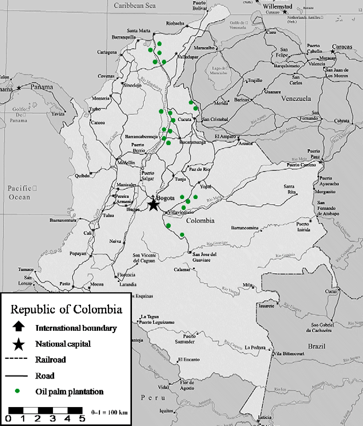
The selection of oil palm crops damaged by Pestalotiopsis was done in municipalities in Colombia, assisted by the Ministry of Agriculture, the Colombian Agriculture Institute, the Inter-American Agriculture Institute, the National Federation in Oil Palm and farmers with the knowledge of plantations affected by defoliator insects. The municipalities were Aracataca (10°35′31″N, 74°11′23″W), Barranca de Upía (04°34′11″N, 72°58′00″W), Bosconia (09°58′20″N, 73°53′19″W), Cantagallo (07°22′34″N, 73°55′11″W), Castilla (03°49′42″N, 73°40′48″W), Ciénaga (11°00′25″N, 74°14′52″W), Cumaral (04°19′09″N, 72°27′43″W), El Copey (10°09′01″N, 73°57′41″W), El Zulia (07°55′57″N, 72°36′04″W), Fundación (10°31′14″N, 74°11′06″W), Granada (03°32′19″N, 73°42′02″W), Paratebueno (04°22′33″N, 73°12′56″W), Puerto Wilches (07°20′54″N, 73°53′46″W), Sabana de Torres (07°23′29″N, 73°29′45″W), San Alberto (07°45′40″N, 73°23′32″W), San Martín (08°00′05″N, 73°30′41″W), San Pablo (07°28′39″N, 73°55′21″W), San Vicente (06°52′52″N, 73°24′35″W), Tibu (08°38′20″N, 72°44′09″W) and Villanueva (04°21′37″N, 72°33′06″W). Sampling was done in dry and rainy seasons where insect populations and Pestalotiopsis fluctuated with the following characteristics: altitude (0–500 m), temperature (27–35 °C), relative humidity (75–85%), sunshine (135–220 h/year) and annual precipitation (1800–2500 m) (Fig. 1). The variety and age of the palms were not taken into account as factors for the selection of areas and insect collections.
(colour online) Map of the study area in Colombia: sampling in oil palm plantations affected by lepidopteran defoliators and Pestalotiopsis.

Species identification
Insect sampling was done four times a year on the leaves of E. guineensis between 2010 and 2011. Lepidoptera larvae collections were done manually. The specimens collected were kept under laboratory conditions with temperature (26 ± 2 °C), humidity (75 ± 5%) and light (12 h:12 h light:dark) conditions, and were fed E. guineensis leaves until adult emergence. Adults were killed and transferred to the Entomology Laboratory of the National University, Colombia. Insects were identified to species level with recent taxonomic keys (Epstein, Reference Epstein1996; Deml and Dettner, Reference Deml and Dettner2002; Freitas and Brown, Reference Freitas and Brown2004; Kaila, Reference Kaila2004) set out by taxonomic specialists, and comparisons found in the entomological collections were made in the Entomological Museum of Agriculture and the Natural Science Museum, National University of Colombia.
Insect–disease interaction
Thirty larvae of Lepidoptera species were isolated from the leaves of E. guineensis with tissue traps (10 × 50 × 90 cm) under field conditions. Each trap contained five leaflets with a larva that remained until the end stage of development. The control was represented by cutting punch leaves, using a paper cutter. The development of Pestalotiopsis was evaluated around the damage site caused by the insects after 60 days. Images of the affected area on each leaf by Pestalotiopsis were taken using a digital camera (D40, 18–55 mm; Nikon Corporation, Japan), fixed to 15 cm with macro focus daylight and fluorescent light (SB-700; Nikon Corporation, Japan). Collection of the images was archived in a JPEG format with a colour resolution of 300 dpi and analysed using digital analysis with Image Tool software (University of Texas, USA). The area (cm2) affected by Pestalotiopsis was measured in pixels based on the RGB histogram (red 700 nm, green 546.1 nm and blue 435.8 nm). The severity and duration of the symptoms of Pestalotiopsis were calculated by the following formula:
 $$\begin{eqnarray} \end{eqnarray}$$
where t 1 is the final evaluation time; t 0 is the initial evaluation time; X 1 is the final severity; and X 0 is the initial severity (Stover, Reference Stover1971). Data were estimated by ANOVA; a Tukey's honestly significant difference test (HSD) was also used for comparisons of severity means with significance level P< 0.05 (Tukey, Reference Tukey1949). All statistical parameters were analysed with the GLMMIX procedure using SAS for Windows (SAS, 2002).
Results
Lepidoptera species
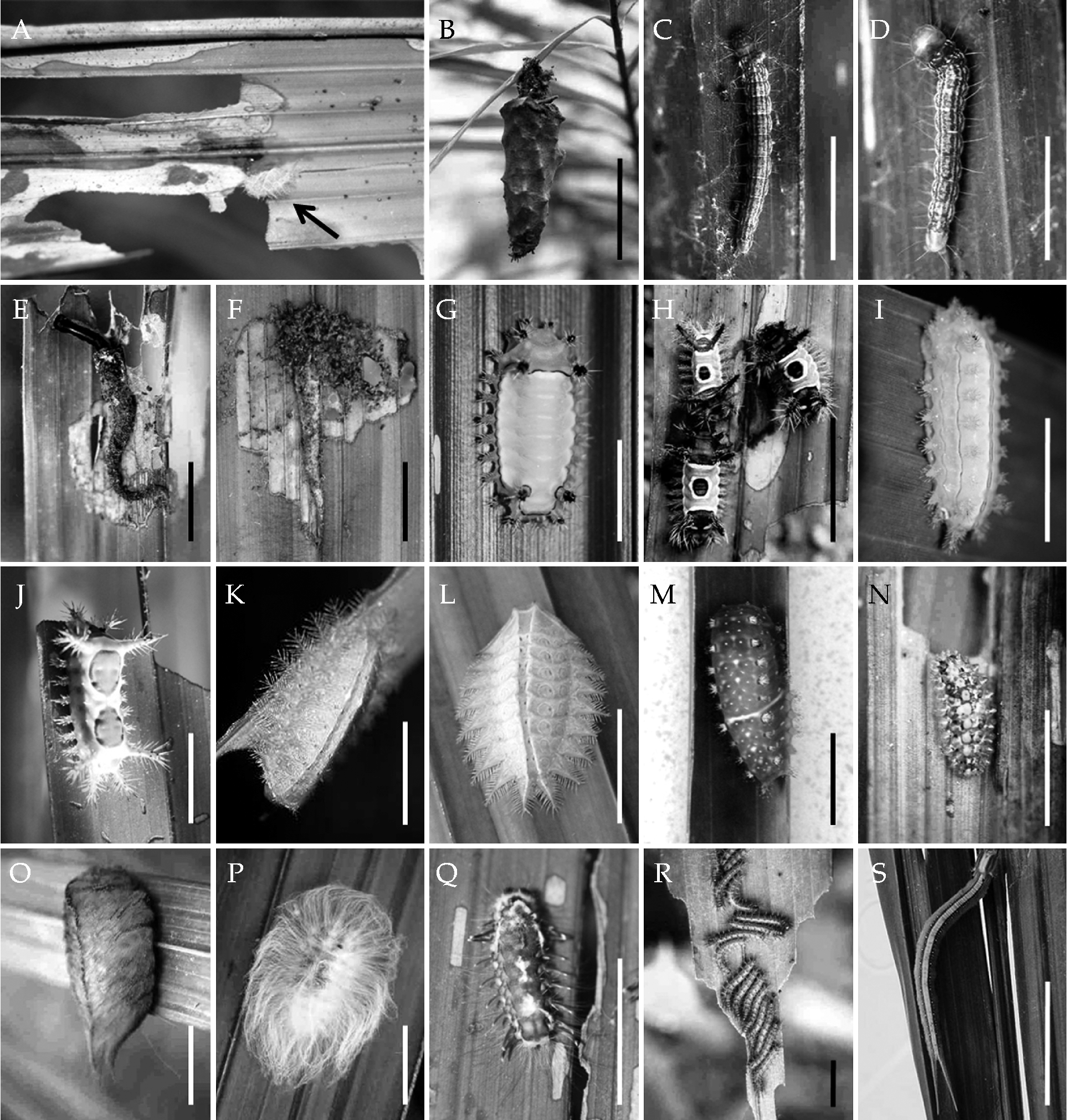
A total of 32 species of Lepidoptera were collected. Eighteen species of the families Dalceridae (one), Elachistidae (four), Limacodidae (eight), Megalopygidae (two), Nymphalidae (one), Psychidae (one) and Saturniidae (one) were identified as vectors of the disease after causing lesions on the leaves of E. guineensis (Fig. 2A).
Lepidoptera species associated with Pestalotiopsis disease in Elaeis guineensis: A, disease damage; B, Oiketicus kirbyi Guilding; C, Antaeotricha phaeoneura Meyrick; D, Durrantia arcanella Busck; E, Loxotoma elegans Zeller; F, Stenoma impressella Busck; G, Acharia fusca Stoll; H, Acharia hyperoche Dognin; I, Acharia pallescens Dognin; J, Euclea diversa Druce; K, Euprosterna elaeasa Dyar; L, Natada michorta Dyar; M, Natada pucara Dognin; N, Acraga ochracea Walker; O, Megalopyge albicollis Walker; P, Mesocia pusilla Stoll; Q, Norape argyrrhorea Hubner; R, Dirphia gragatus Bouvier; S, Opsiphanes cassina Felder. Scale bar: 10 mm (C, D, F, J, K, L, M, N, O, Q); 20 mm (B, E, G, P); 30 mm (I, S); 40 mm (H); 50 mm (R).
Psychidae
Oiketicus kirbyi Guilding, cylindrical body with developed thoracic legs, four pairs of abdominal prolegs, a pair of extensions anal, cuticle without secondary setae, spiracles oval, grey with black spots, length 60 mm (Fig. 2B).
Elachistidae
Dorso-ventrally flattened body with deep intersegmental constriction, primary setae, spiracles circular and undifferentiated pinnacles; Antaeotricha phaeoneura Meyrick, orange with six longitudinal bands purple, 18 mm (Fig. 2C); Durrantia arcanella Busck, orange with four longitudinal bands brown, 20 mm (Fig. 2D); Loxotoma elegans Zeller, yellow with squares spot brown, 35–40 mm (Fig. 2E); Stenoma impressella Busck, yellow with purple longitudinal bands, 20–25 mm (Fig. 2F).
Limacodidae
Larvae with legs and prolegs modified without crochets, setae and scoli variables, stinging; Acharia fusca Stoll, thoracic segments green, abdominal segments yellow, 27–35 mm (Fig. 2G); Acharia hyperoche Dognin, body brown at the edges, green back with a big spot brown; scoli largest 35–40 mm (Fig. 2H); Acharia pallescens Dognin, light green with yellow dorsal bands, 30–34 mm (Fig. 2I); Euclea diversa Druce, yellow with two brown dorsal spots, 22–24 mm (Fig. 2J); Euprosterna elaeasa Dyar, green, flattened body with protuberances, scoli dorsal and lateral, yellow spot on the third abdominal segment, 17–18 mm (Fig. 2K); Natada michorta Dyar, green, flattened body with protuberances, scoli dorsal and lateral, 20 mm (Fig. 2L); Natada pucara Dognin, green with yellow spots, red protuberances, 25 mm (Fig. 2M).
Dalceridae
Acraga ochracea Walker, larva covered with colourless gelatinous tubercles, spiracles circular, yellow, 12–14 mm (Fig. 2N).
Megalopygidae
Verricules and setae long, stinging; Megalopyge albicollis Walker, brown, 45 mm (Fig. 2O); Mesocia pusilla Stoll, white, 30 mm (Fig. 2P); Norape argyrrhorea Hubner, brown with white bands, 12 mm (Fig. 2Q).
Saturniidae
Dirphia gragatus Bouvier, brown with chalazae, scoli branched, stinging, elliptical spiracles, 50 mm (Fig. 2R).
Nymphalidae
Opsiphanes cassina Felder, larva with horns cephalic and caudal appendages, cuticle smooth, elliptical spiracles, green with yellow bands, 70–90 mm (Fig. 2S).
Insect–disease interaction
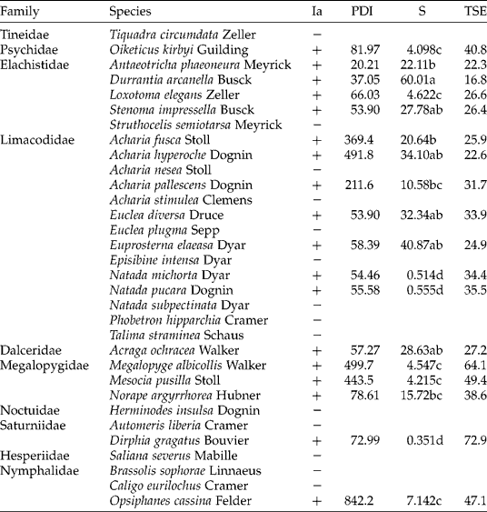
The severity and progression of the disease showed different rates and duration of the symptoms around the lesions made by each species on the leaves of E. guineensis (Table 1). A higher severity of Pestalotiopsis was caused by D. arcanella (60.01), E. elaeasa (40.87), A. hyperoche (34.10), E. diversa (32.34), A. ochracea (28.63) and S. impressella (27.78) (F 2,28= 49.78, P< 0.05). Other species such as N. michorta, N. pucara and D. gragatus showed low percentages of severity. There was a variation between 16.8 and 72.9 days in the development of Pestalotiopsis on the leaves of E. guineensis. There was a lower duration of the symptoms of Pestalotiopsis caused by D. arcanella (16.8 days), A. phaeoneura (22.3 days), A. hyperoche (22.6 days), E. elaeasa (24.9 days) and A. fusca (25.9 days).
Severity and duration of Pestalotiopsis disease symptoms caused by Lepidoptera in Elaeis guineensis
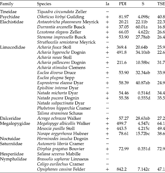
Ia, positive and negative vectors; PDI, percentage damage by insects; S, severity; TSE, time of symptom evolution. The means followed by different letters (a, b, c, and d) are significantly different (P< 0.005).
Discussion
Lepidoptera larvae were found in commercial plantations affected by Pestalotiopsis. Lepidoptera species of the families Dalceridae, Elachistidae, Limacodidae, Megalopygidae, Nymphalidae, Saturniidae and Psychidae can attend and spread the Pestalotiopsis fungus when they gnaw or consume the parenchyma, leaf nervature or petiole. Food preferences of these insects can be classified as monophagous, with the phytochemical traits being common among the species of Arecaceae (Bjorholm et al., Reference Bjorholm, Svenning, Skov and Balslev2005; Asmussen et al., Reference Asmussen, Dransfield, Deickmann, Barfod, Pintaud and Baker2006). The pathogenicity of Pestalotiopsis may result from the interaction of multiple factors such as environmental conditions and susceptibility of the plant and pathogen virulence (Gehlot et al., Reference Gehlot, Bohra and Purohit2008). The presence of Pestalotiopsis in adult palms offers optimal conditions for the development of the disease due to the number of leaves. The density of the leaves and plants may be the main cause for the presence of herbivores and disease onset. Pestalotiopsis species cause diseases on various species of Arecaceae, when weather conditions are associated with the high density of plants grown (Hyde and Fröhlich, Reference Hyde and Fröhlich1995; Hyde, Reference Hyde1996; Jeewon et al., Reference Jeewon, Liew and Hyde2004). High plant density in the plantations of Stryphnodendron microstachyum (Fabales: Fabaceae) has been reported to show increased damage caused by Euclystis spp. (Lepidoptera: Noctuidae) larvae and the occurrence of Pestalotiopsis on leaves (Folgarait et al., Reference Folgarait, Marquis, Ingvarsson, Braker and Arguedas1995).
Simultaneously, the progression of the disease was observed around the damage area caused by the insects. The area affected by the disease on the leaves of E. guineensis was higher in the damage caused by D. arcanella, E. elaeasa, A. hyperoche, E. diversa, A. ochracea and S. impressella (F 2,28= 49.78, P< 0.05). Pestalotiopsis produces abundant spores that are dispersed by the movement of wind and water (Sajeewa et al., Reference Maharachchikumbura, Guo, Chukeatirote, Bahkali and Hyde2011); however, the damage caused by these insects on palms allows the entry of these spores and disease development. Commercial plantations of oil palm in the world are affected by Pestalotiopsis and insect vectors have been recognized (Gitau et al., Reference Gitau, Gurr, Dewhurst, Fletcher and Mitchell2009). Severity and duration of the symptoms was observed between 16.8 and 72.9 days after the damage caused by insects on oil palm. A lower duration of symptoms was caused by D. arcanella, A. phaeoneura, A. hyperoche, E. elaeasa and A. fusca. A direct relationship between the leaf consumption of each insect species and the severity and duration of the symptoms of Pestalotiopsis was not found. Palm trees serve as centres of interactions between micro-organisms and insect vectors, establishing a tripartite relationship with the plant. Direct and indirect interactions between herbivorous insects and pathogenic micro-organisms can occur when the infection or infestation by the first attacker causes damage to the plant, the second attacker increases the damage and both have a spatial or temporal relationship (Bostock, Reference Bostock1999; Stout et al., Reference Stout, Thaler and Thomma2006). Field studies of plant–insect–disease relationships have shown that the attack by pathogens and insects can alter the physiology of the plant, severity of the pathogen and insect dynamics (Hunter, Reference Hunter2000; Rostás and Hilker, Reference Rostás and Hilker2002; Hatcher et al., Reference Hatcher, Moore, Taylor, Tinney and Paul2004).
The results of this study contribute to the identification of Lepidoptera larvae that attend and spread the Pestalotiopsis fungus in oil palm plantations in Colombia. Additionally, we demonstrated a significant association between the severity of Pestalotiopsis and several species of insects, which may arise from a relationship vector or the ease of entry of the pathogen by damage caused by insects on the leaves of E. guineensis. This may contribute to the knowledge of the disease and the association with insects, and incorporate future control strategies for management of oil palm plantations.
Acknowledgements
The authors thank the following oil palm plantations for the contributions made to this study: Agropalma, Indupalma, La Cabaña, Oleagionosas Las Brisas, Palmeras del Casanare, Palmeras del Oriente, Palmeras Puerto Wilches, Palmeras del Cesar, Promociones Monterrey and National University of Colombia. The authors also thank O Conselho Nacional de Desenvolvimento Científico e Tecnológico CNPq (Brazil), Coordenação de Aperfeiçoamento de Pessoal de Nível Superior CAPES (Brazil) and Fundação de Amparo a Pesquisa do Estado de Minas Gerais FAPEMIG (Brazil).