Summations
• Autism is a neurobehavioural disease that is difficult to study in human subjects. Animal model experimentation can provide excellent support in autism research.
• Autistic model animals can be generated by certain drug treatments and mutagenesis.
• This is a review article that discusses the success, limitation and future direction of animal model experimentation in autism research.
Considerations
• Difficult to generate the autistic model of animals.
• Psychologically and physiologically, the animal brain differs from the human brain. Therefore, the animal model cannot give the exact study model.
• Clinical implication of autism research is very limited.
Autism: a neurogenetic disorder
Autism is a highly heritable complex disorder of neural development characterised by impaired social interaction and communication, and by restricted and repetitive behaviour (Reference Klauck1). The hallmark feature of autism is impaired social interaction (Reference Myers and Johnson2). Although autism varies significantly in characteristics and severity, it occurs in all ethnic and socio-economic groups and affects every age group (Reference Newschaffer, Croen and Daniels3). The number of reported cases of autism has markedly increased since the 1990s and early 2000s, and the prevalence of autism is 10–20 per 10 000 (Reference Wing and Potter4,Reference Fombonne5). At present, there is no cure for autism (Reference Myers and Johnson2). Therapies and behavioural interventions are designed to remedy specific symptoms that can cause substantial improvement (Reference Howlin6). However, the treatment is expensive, and publicly supported programmes are often inadequate or inappropriate for a given child (Reference Montes and Halterman7).
Although the exact aetiology of the condition is not known, family and twin studies strongly indicated genetic factors behind autism. Genome-wide scans suggest several susceptibility loci that may contain one or more predisposing genes. Moreover, it does not seem to be a single gene disorder (Reference Shastry8). The large number of autistic individuals with unaffected family members may result from copy number variations, spontaneous deletions or duplications in genetic material during meiosis (Reference Nomura and Takumi9,Reference Cook and Scherer10). Environmental factors such as exposure to certain chemicals including thalidomide, misoprostol and valproic acid (VPA) in early pregnancy are also believed to contribute to autism (Reference Landrigan11). Autism may result from the impairment of foetal brain development caused by the defects in genes that control brain growth and regulate brain cell communications, possibly because of the influence of environmental factors on gene function (Reference Courchesne, Pierce and Schumann12,Reference Chomiak and Hu13).
The brain mechanisms responsible for moderating social behaviour, the specific neuronal circuitry and the chemicals involved in these processes are relatively mysterious. This is mostly because of the difficulty in studying the fundamental neurophysiological processes directly in the human brain. Finding an animal model system with similar behavioural characteristics as human is thus vital for understanding the brain mechanisms in autism.
Why animal model of autism study is necessary
Recent autism research has changed focus from behavioural observations of autism to translating findings from animal models and genomic manipulation studies to pharmacological agents, with the aim to reverse the interior symptoms of autism (Reference Smile and Anagnostou14). The brain mechanisms responsible for autism are not fully understood and the animal models, which parallel related emotional and cognitive impairments, may be helpful in sorting out these mechanisms (Reference Meyza, Defensor and Jensen15). Although it is difficult to reflect human pathophysiology in animal models, some aspects of molecular pathways, brain anatomy, cognitive and behavioural phenotypes can be addressed. Some research groups have created animal models of autism, which are based on the human central nervous system. These mouse models display some brain anatomical and behavioural abnormalities, providing insight into human neuropsychiatric disorders that will contribute to the screening of novel drugs.
Mouse models are advantageous in many respects, primarily because they are genetically developed and appropriate for invasive studies. Knockout mouse variants, in which existing genes have been inactivated via genetic engineering, offer invaluable opportunities to test the functional and behavioural repercussions of manipulating a particular aspect of the nervous system (Reference Watson and Platt16). An ideal animal model of autism would exhibit face validity, in which the behaviour of the model is compromised in a manner that is consistent with autism; construct validity, similar to the underlying biological mechanisms of the disorder; and predictive validity, in which interventions effective in treating autism induce the expected response in the model (Reference Crawley17). Similarly, the underlying causes of the disorder and predictive validity are nearly impossible to address in rodent models, as the underlying causes of autism are unknown and it is difficult to establish face validity (Reference Nestler and Hyman18).
Non-human primates provide an attractive, complementary model for autism owing to the complexity and dynamics of social structures, reliance on vision for social signalling and striking homology in the anatomy of neural circuits mediating social behaviour (Reference Watson and Platt16). The non-human primates have a large behavioural repertoire with a high degree of isomorphism to human behaviour (Reference Dantzer and Kelley19). Neural manipulations in non-human primates that affect behaviour in a manner reminiscent of autism can thus be used as a tool for the study of the aetiology of autism, or as a model for testing therapeutic agents that ameliorate autism symptoms. However, translational research is needed to discover pharmacological targets and treatments of autism. Animal models with phenotypic relevance to diagnostic criteria offer clear experimental strategies to test the efficacy and safety of novel treatments (Reference Silverman, Babineau, Oliver, Karras and Crawley20).
Generation of animal models for autism research
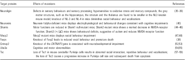
Autism is characterised by impairments in reciprocal social communication and stereotyped verbal and non-verbal behaviours (Reference Ey, Leblond and Bourgeron21). A variety of features of autism can be simulated in rodents, including repetitive behaviours, deficits in social interaction and communication. Neuropathological and some characteristic neurochemical changes, which are frequently seen in autism, and alterations in the immune status in the brain and periphery are found in some of the models by drug treatment or mutagenesis (Tables 1 and 2).
Strategies in generating autism model by drug treatment

ICV, intracerebroventricular; PPA, propionic acid; VPA, valproic acid.
Strategies in generating autism model by mutagenesis

NL, neuroligin; NMDA, N-methyl-d-aspartate.
Drug-induced autism model
VPA
Experiments on rodents have indicated that maternal VPA exposure has permanent adverse effects on neurological and behavioural development. The animal model induced by prenatal exposure of VPA has been proposed to study autism, as prenatal VPA exposure produces many neuroanatomical and behavioural deficits associated with autism (Reference Roullet, Wollaston, Decatanzaro and Foster22,Reference Lukose, Schmidt, Wolski, Murawski and Kulesza23). Several studies compared the effects of different time intervals of prenatal VPA exposure for inducing the altered social behaviours relevant to autism. Anatomically, there was a reduction in brain weight and cortical thickness along with decreased dendritic branching in the orbitofrontal cortex (OFC) and medial prefrontal cortex (mPFC) and decreased spine density in the mPFC, OFC and cerebellum (Reference Bambini-Junior, Rodrigues, Behr, Moreira, Riesgo and Gottfried24–Reference Mychasiuk, Richards, Nakahashi, Kolb and Gibb26). These studies reported that VPA-exposed rats showed significantly fewer neurons and significant alterations in neuronal morphology, and thus prenatal VPA exposure in rats appears to produce similar dysmorphology as reported in the autistic human brain. Kim et al. (Reference Kim, Kim and Go27) reported that prenatal exposure of VPA in rat model shows social behaviour impairment, crooks tail phenotype, reduces expression of GABAergic neuronal marker GAD and increases the expression of glutamatergic neuronal marker vGluT1. Favre et al. (Reference Favre, Barkat, Lamendola, Khazen, Markram and Markram28) reported that rat offspring exposed to VPA leads to the neocortex and amygdala hyperreactive and hyperplastic because of hyperconnectivity between the neurons. They treated Wistar dams with a single intra-peritoneal injection of 500 mg/kg VPA on embryonic day E12.5 and found that offspring exposed to VPA show smaller brain mass on postnatal day 1 and 14, as well as abnormal nest-seeking behaviour at postnatal day 10 in the olfactory discrimination test and physical malformation. Go et al. (Reference Go, Kim and Choi29) reported that prenatal exposure of VPA increases the neural progenitor cell pool and induces macrocephaly in rat brain via a mechanism involving the GSK-3β/β-catenin pathway. Zhang et al. (Reference Zhang, Sun, Wang, Wang, Peng and Li30) suggested that the upregulation of the Wnt/β-catenin signalling pathway induced by VPA administration during early pregnancy produces an imbalance in oxidative homeostasis that facilitates susceptibility to autism.
Propionic acid (PPA)
PPA is a dietary short-chain fatty acid produced in the body during normal cellular metabolism and a metabolic end-product of enteric bacteria. Shultz et al. (Reference Shultz, Macfabe and Martin31) reported that intracerebroventricular (ICV) injection of PPA results in anatomical and behavioural abnormalities in rats similar to the humans suffering from autism. They administered ICV injection of PPA to Long-Evans hooded rats and showed marked impairment in the water maze task and beam task. MacFabe et al. (Reference MacFabe, Cain, Boon, Ossenkopp and Cain32) administered PPA to adolescent male rats and immunohistochemical analysis of the brain tissue from PPA rats revealed reactive astrogliosis and activated microglia, indicating an innate neuroinflammatory response. El-Ansary et al. (Reference El-Ansary, Ben Bacha and Kotb33) administered neurotoxic dose of PPA (250 mg/kg body weight/day) to Western albino male rats and they reported an impairment of social behaviour, typical cognitive disability, repetitive behaviour and object-directed behaviour coupled with the increasing oxidative stress markers, a decrease in glutathione level, decreased activity of glutathione peroxidase and catalase. Thus, the neurotoxicity induced by PPA might be an environmental factor that plays a central role in the aetiology of autistic biochemical features.
Mutagenesis in animal model
A number of mouse models based on the genetic variants have been developed that are associated with autism or autistic features. The genetic heterogeneity of autism poses a major challenge for identifying mechanism-based treatments. A number of rare mutations are associated with autism and it is unclear whether these result in common neuronal alterations (Reference Baudouin, Gaudias and Gerharz34). Mouse models have been generated that carry mutations similar to those identified in patients. These models offer the opportunity to investigate in vivo the physiological and behavioural consequences of the mutations in the genes coding for the proteins associated with autism, including neuroligin (NL), neurexin, shank, Mecp2, Foxp, Cntnap2, Ube3a and Tsc1/Tsc2.
NL
NL is a member of the class of cell adhesion proteins that mediate synapse development and have been implicated in autism (Reference Chadman, Gong and Scattoni35). NL mutants are defective in a subset of sensory behaviours and sensory processing, and are hypersensitive to oxidative stress and mercury compounds; the behavioural deficits are strikingly similar to traits frequently associated with autism (Reference Hunter, Mullen, McManus, Heatherly, Duke and Rand36). In one study, Ellegood et al. (Reference Ellegood, Lerch and Henkelman37) reported that the NL-3 knockin mouse model has marked volume differences in many different structures in the brain: grey matter structures, such as the hippocampus, the striatum and the thalamus, were all found to be smaller in the NL-3 knockin. In addition, knockout of NL-3 and NL-4 in mice diminishes social behaviour and vocalisations (Reference Fischer and Hammerschmidt38). NL-3 and NL-4 knockout mice represent a useful animal model for understanding pathophysiological events in autism and for developing novel treatment strategies in this devastating disorder (Reference Radyushkin, Hammerschmidt and Boretius39).
Neurexins
Neurexins are presynaptic membrane proteins that bind to postsynaptic cell adhesion proteins, NLs (Reference Hunter, Mullen, McManus, Heatherly, Duke and Rand36). Etherton et al. (Reference Etherton, Blaiss, Powell and Südhof40) reported that neurexin-1alpha-deficient mice display a decrease in prepulse inhibition, an increase in grooming behaviours, an impairment in nest-building activity and an improvement in motor learning. Their report indicated that deletion of neurexin-1-alpha is correlated to the electrophysiological and behavioural changes in mice, which is consistent with cognitive impairments observed in human patients (Reference Etherton, Blaiss, Powell and Südhof40).
Shanks
SHANK genes encode a family of synaptic scaffolding proteins located postsynaptically on excitatory synapses and mutations in SHANK genes have been detected in several autistic individuals. Silverman et al. (Reference Silverman, Turner and Barkan41) reported that adult Shank1-null mice are on standardised measures of gen health, neurological reflexes and sensory skills; however, their motor functions are reduced, suggesting that Shank1 has a role in motor functions, although Shank1-null mice do not show autism-relevant social interaction problem directly (Reference Silverman, Turner and Barkan41). Won et al. (Reference Won, Lee and Gee42) showed that Shank2-mutant mice, carrying a mutation identical to the autism-associated micro-deletion in the human SHANK2 gene, exhibit autism-like behaviours including reduced social interaction and reduced social communication. These mice showed a marked decrease in N-methyl-d-aspartate (NMDA) glutamate receptor (NMDAR) function (Reference Won, Lee and Gee42). The protein complex ProSAPs/Shanks at the excitatory synapses organises the complex protein machinery of the postsynaptic density. Genetic deletion of ProSAP1/Shank2 upregulates ionotropic glutamate receptors at the synapse in a brain region-specific manner (Reference Schmeisser, Ey and Wegener43). These mutants were found to display autistic-like behavioural alterations such as repetitive grooming as well as abnormalities in vocal and social behaviours. Bangash et al. (Reference Bangash, Park and Melnikova44) created a mouse genetic model associated with autism that mimics a human mutation of Shank3, which deletes the C terminus. Shank3 (+/ΔC) mice showed behavioural deficits suggestive of autism and reduced NMDA receptor function. Direct stimulation of NMDARs with d-cycloserine (a partial agonist of NMDARs) or treatment with a positive allosteric modulator of metabotropic glutamate receptor 5 normalises NMDAR function and improves social interaction in Shank-mutant mice (Reference Won, Lee and Gee42,Reference Deutsch, Pepe, Burket, Winebarger, Herndon and Benson45). Peca et al. (Reference Peca, Feliciano and Ting46) generated Shank3-mutant mice displaying different levels of severity in synaptic defects and phenotypes in Shank3-related autism. They showed that mice with SHANK3 gene deletions could lead to varying degrees of functional defects such as self-injurious repetitive grooming and deficits in social interaction, which may contribute to the genesis of autistic-like behaviours in mice.
Mecp2
Mouse models of autism with targeted mutations in the MECP2 gene (encodes a methyl-CpG-binding protein) show a high degree of phenotypic consistency with the clinical syndrome. A variety of studies have demonstrated that Mecp2-mutant mice display impaired social behaviour (Reference Johnson, Lam and Punzo47). Derecki et al. (Reference Derecki, Cronk and Lu48) transplanted wild-type bone marrow into irradiation-conditioned Mecp2-null murine model and reported that wild-type Mecp2-expressing microglia within the context of a Mecp2-null mouse arrested disease development and improved locomotor activity. In this study, they suggested bone marrow transplantation as a possible therapeutic approach for autism (Reference Derecki, Cronk and Lu48). However, Pearson et al. (Reference Pearson, Defensor and Pobbe49) performed a series of classic and refined social behaviour tasks in hypomorphic Mecp2 (308/Y) mutant mice, and they suggested that mutations in the Mecp2 are not associated with autism-relevant social impairment in mice (Reference Pearson, Defensor and Pobbe49).
Foxps and Cntnap2
There is emerging evidence that the FOXP family of genes is strongly linked to autism. The FOXP family of genes includes three genes expressed in the central nervous system: FOXP1, FOXP2 and FOXP4 (Reference Bowers and Konopka50). According to the report of Fischer and Hammerschmidt (Reference Fischer and Hammerschmidt38), knockout of FoxP2 leads to reduced vocal behaviour and eventually premature death. They suggested that the introduction of human variant of FoxP2 protein in mice might result in shifts in the frequency and modulation of pup ultrasonic vocalisations. Fujita et al. (Reference Fujita, Tanabe, Momoi and Momoi51) showed a genetic connection between FOXP2 and CNTNAP2 and reported that Cntnap2 mRNA level is significantly increased in the cerebellum of Foxp2 (R552H) knockin pups. Cntnap2 and CtBP (co-repressor of Foxp2) are ubiquitously expressed, whereas Foxp2 co-localised with CtBP only in Purkinje cells (Reference Fujita, Tanabe, Momoi and Momoi51). Peñagarikano et al. (Reference Peñagarikano, Abrahams and Herman52) characterised a mouse knockout of the CNTNAP2 gene, which is strongly associated with autism and related neurodevelopmental disorders. There are some other strong neurobiological, genetic and imaging evidence that the CNTNAP2 gene is a risk factor for autism spectrum disease, which has been reviewed in detail by Peñagarikano and Geschwind (Reference Peñagarikano and Geschwind53).
Ube3a
The most widely used model for autism is the UBE3A-maternal-deficient mouse that recapitulates most of the essential features of autism, including cognitive and motor abnormalities (Reference Jana54). Jiang et al. (Reference Jiang, Pan and Zhu55) generated mutant mice with a 1.6-Mb chromosomal deletion from UBE3A and GABRB3 genes, and suggested that mutant mice with this maternal deletion provide an autistic mouse model that is more similar to the contiguous gene deletion form of autism in humans in molecular biological aspect (Reference Jiang, Pan and Zhu55).
Tuberous sclerosis complex (TSC)
Mutation in TSC is an intriguing model to investigate the cerebellar contribution to the underlying pathogenesis of autism (Reference Reith, Way, McKenna, Haines and Gambello56). Tsai et al. (Reference Tsai, Hull and Chu57) showed that both heterozygous and homozygous loss of Tsc1 in mouse cerebellar Purkinje cells resulted in autistic-like behaviours, including abnormal social interaction, repetitive behaviour and vocalisations, in addition to decreased Purkinje cell excitability (Reference Jiang, Pan and Zhu55). Zeng et al. (Reference Zeng, Rensing, Zhang, Gutmann, Gambello and Wong58) characterised a novel mouse model involving conditional inactivation of the TSC2 gene in glial-fibrillary acidic protein (GFAP)-positive cells [Tsc2(GFAP1)C knockout mice]. Carson et al. (Reference Carson, Van Nielen, Winzenburger and Ess59) reported that Tsc1 conditional knockout animals die by 25 days of life; their brains have increased size and contain prominent large cells within the cerebral cortex that have considerably increased mTORC1 signalling and decreased mTORC2 signalling (Reference Carson, Van Nielen, Winzenburger and Ess59). These findings define a molecular basis for a cerebellar contribution to autism.
Future research
The aetiology of autism is not fully known, and there are very few drugs for ameliorating the basal symptoms of this disease. The introduction of efficient neuroprotective drugs, which might ameliorate secondary neuronal cell damage in various brain regions to the therapy, is the main objective of numerous experimental studies (Reference Budziszewska, Lasoń, Steczkowska and Gergont60). Genetic evidence of the involvement of multiple susceptibility genes in autism will enable future studies to move from single-gene models to models with multiple modified loci, with the promise of better representing the complexity of autism (Reference Flint and Shifman61). Transplantations of transfected cells capable of producing neurotrophic factors or stem cells to the brain might be an efficient method for improving brain function in the future (Reference Flint and Shifman61). If the validity of this animal model is confirmed by future research, translational studies focusing on the interaction between early experience and opioid neurotransmission may provide useful insights for identifying endophenotypes of autism (Reference Cinque, Pondiki and Oddi62). In future, these animal models will be invaluable preclinical tools for the development of highly specific and efficacious treatments for children affected with autism (Reference Piedimonte, Wailes and Weiner63).
Conclusions
Autism is a common socially devastating disorder. The brain mechanisms responsible for autism are not understood properly and animal models paralleling related emotional and cognitive impairments may prove helpful in unravelling them. Better animal models may play a critical role in understanding the aetiology of autism and develop more effective therapies. Through careful behavioural analyses, most of the core characteristics of autism, that is, impairments in social interaction, difficulty with language, and repetitive and stereotyped motor behaviours can be modelled in mice. Integrating clinical studies on humans, genetic manipulations in mice and neural system studies on non-human primates will offer the most promising approach for understanding the biology and allowing the development and testing of therapeutic agents. Finally, the use of intelligent animals in any research programme demands close scrutiny and ethical concerns, and associated financial and social supports.
Acknowledgements
This work has been supported by The National Basic Research Program of China (2012CB517902) and National Natural Science Foundation of China (81070081). Authors are thankful to all the teachers and students of State Key lab of Medical Genetics, Central South University.
Conflict of Interest
The authors report no conflicts of interest.