Kawasaki disease is a systemic vasculitis occurring predominantly in medium-sized muscular arteries, particularly the coronary arteries. Approximately 5% of Kawasaki disease patients develop coronary artery lesions later on,Reference Kato, Sugimura and Akagi 1 which carry an increased risk of myocardial infarction and other adverse eventsReference Gordon, Kahn and Burns 2 in the long-term follow-up. Sudden death due to fatal arrhythmias might occur in such patients, but has most often been reported in patients with reduced left ventricular function.Reference Tsuda, Hirata and Matsuo 3 In this study, we report the cases of two adult patients with largely unpredictable sudden cardiac arrest caused by presumed Kawasaki disease, as well as consider the underlying mechanisms with reference to the literature.
Case report
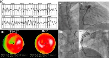
Case 1 involved a 29-year-old man who was transferred to a local emergency centre after experiencing ventricular fibrillation during a music concert. He received by-stander cardiopulmonary resuscitation and was revived using an automated external defibrillator (Fig 1a). The patient had a history of Kawasaki disease when he was 10-months old. Coronary angiography at 8 years of age showed segmental stenosis of the right coronary artery and severe stenosis of the left anterior descending artery with calcification of a giant aneurysm. Myocardial perfusion imaging showed ischaemic changes, and treatment had included coronary artery bypass grafting at 9 years of age – left and right internal thoracic artery to the left anterior descending artery and to the right coronary artery, respectively. Percutaneous transluminal rotational atherectomy for bypass graft had been performed at 17 years of age. He had been asymptomatic and was taking 15 mg of nicorandil, 200 mg of ticlopidine, and 100 mg of aspirin daily. Myocardial perfusion imaging had shown ischaemic changes in the anterior septal area at 24 years of age. In the past 4 years, he had not been regularly consulting the hospital. His height was 178 cm and body weight was 83 kg; his blood pressure was 130/80 mmHg and total cholesterol was 164 mg/dl. The patient had no family history to note, and did not drink alcohol or smoke.
( a ) Ventricular fibrillation recorded in an automated external defibrillator. ( b ) Myocardial perfusion imaging showed diffuse ischaemic changes of the anterior septum area. ( c – f ) Coronary angiography showed total occlusion of the left ( c ) and right ( d ) internal thoracic arteries, severe stenosis of the left anterior descending artery ( e ), and development of collateral circulation of the right coronary artery ( f ).

On admission, electrocardiography and laboratory findings did not reveal acute myocardial infarction. Echocardiography revealed normal left ventricular ejection fraction. A decision was made not to proceed with emergency cardiac catheterisation and thrombolysis. Myocardial perfusion imaging showed no marked changes from earlier reports (Fig 1b), coronary angiography showed total occlusion of the left and right internal thoracic arteries (Figs 1c–f), and programmed ventricular stimulation could not induce ventricular fibrillation. We then decided to proceed to a second coronary artery bypass graft – radial artery to the left anterior descending artery and right gastroepiploic artery to the right coronary artery – and suspended implantation of an implantable cardiac defibrillator. The patient has remained asymptomatic as of the last follow-up.
Case 2 involved a 26-year-old man who suddenly collapsed due to ventricular fibrillation while playing futsal and was hospitalised after revival using an automated external defibrillator. He had a history of Kawasaki disease when he was 1 year old, complicated with bilateral giant coronary aneurysms. Coronary angiography at 13 years of age had shown segmental stenosis of the left anterior descending artery with calcification. He had undergone catheter intervention comprising rotational atherectomy to the left anterior descending artery at 14 years of age. Myocardial perfusion imaging showed ischaemic changes in the anterior septum area at 25 years of age. He had been treated with 50 mg of clopidogrel and 100 mg of aspirin daily and had remained asymptomatic. His height and body weight were 167 cm and 58 kg, respectively, blood pressure was 114/91 mmHg, and total cholesterol was 149 mg/dl. His mother had hypertension and the patient drank a small amounts of alcohol, but did not smoke.
On admission, no evidence of acute myocardial infarction was observed on electrocardiography or in laboratory findings. Echocardiography showed normal left ventricular ejection fraction. He underwent coronary angiography, which showed severe stenosis of the left anterior descending artery and segmental stenosis of the proximal site of the right coronary artery (Fig 2a). Myocardial perfusion imaging showed the same findings as before (Fig 2b), and thus a decision was made to proceed with coronary artery bypass grafting – left internal thoracic artery to the left anterior descending artery. The patient has remained asymptomatic as of the last follow-up.
( a ) Coronary angiography showed severe stenosis of the LAD artery (arrow). ( b ) Myocardial perfusion imaging showed diffuse ischaemic changes of the anterior septum area. ( c and d ) Coronary CT angiography showed aneurysm of the LAD artery (arrow head) ( c ) and severe stenosis with calcification in it ( d ). LAD=left anterior descending; LCX=left circumflex.

Discussion
Fatal arrhythmias are one of the major causes of sudden death in patients with Kawasaki disease complicated by coronary artery lesions. The frequency of ventricular tachycardia appearing after myocardial infarction is reportedly around 75% in 25 years,Reference Tsuda, Hirata and Matsuo 3 and non-sustained ventricular tachycardia and low left ventricular ejection fraction (<45%) have been mentioned as risk factors for fatal arrythmia;Reference Tsuda, Arakaki and Shimizu 4 however, our cases did not show left ventricular dysfunction or symptomatic arrhythmia, and thus predicting the possibility of fatal arrhythmia was difficult. The mechanisms underlying life-threatening arrhythmias in our cases remain unclear. Animal studies have shown that myocardial ischaemia causes differences in action potentials between epicardial and endocardial muscles, resulting in phase 2 re-entry,Reference Yan, Joshi and Guo 5 which subsequently progresses to ventricular fibrillation. In our cases, transient myocardial ischaemia might have been induced by exercise or vasospasm of the coronary arteries with excitement of the sympathetic nervous system. In addition, no significant relationship was identified between coronary stenosis or obstruction and electrophysiological study parameters,Reference Sumitomo, Karasawa and Taniguchi 6 and we should evaluate individual potential risk factors for fatal arrhythmia in each case. We would like to emphasise the importance of periodic arrhythmia evaluations including ambulatory electrocardiogram, electrocardiogram under exercise stress, and signal average electrocardiogram, even if no serious left ventricular dysfunction is detected. If some risk factors are identified, precise evaluations using electrophysiological studies and indications for anti-arrhythmia drugs or an implantable cardioverter defibrillator should be considered. Appropriate intervention for coronary artery lesions is essential, because fatal arrhythmia might be induced by ischaemia.
To this end, young asymptomatic adults tend to be lost to follow-up, and we therefore encourage visits to hospitals and continued education on medication. Our experiences may suggest the need for a paradigm shift in the management of Kawasaki disease patients with complicating coronary artery lesions in the course of long-term follow-up.
Acknowledgement
The authors express their deepest gratitude to Associate Prof. Hiroshi Kamiyama, who advised them about managing these cases, and Prof. Shori Takahashi, who provided constant encouragement.
Financial Support
This research received no specific grants from any funding agency, commercial, or not-for-profit sectors.
Conflicts of Interest
None.
Ethical Standards
The authors assert that all procedures contributing to this work comply with the ethical standards of the Helsinki Declaration of 1975, as revised in 2008.