Implications
Surgical castration is a painful practice performed on millions of pigs every year and finding novel easily applicable pain assessment tools is necessary for the assessment of pain reduction protocols. Results of this study showed that in castrated piglets behavioural and physiological alterations occur mainly in the first 3 h from treatment. Latency to move, alterations in posture and walking, and IR thermography for the detection of eye temperature are promising tools for pain assessment in piglets. However, IR thermography needs additional handling of the animals during the process for temperature acquiring.
Introduction
Surgical castration in piglets is the most commonly performed management practice in order to avoid boar taint in swine meat. It is estimated that 100 million male pigs are castrated every year in the 25 EU member states (European Food Safety Authority, 2004). This practice is widely executed without pain relief, even though it appears to cause distress regardless of piglet age (Carrol et al., Reference Carroll, Berg, Strauch, Roberts and Kattesh2006; von Borell et al., Reference von Borell, Baumgartner, Giersing, Jäggin, Prunier, Tuyttens and Edwards2009). Moreover, it represents an animal welfare issue with an increasing negative public perception owing to animal suffering (Guatteo et al. Reference Guatteo, Levionnois, Fournier, Guémené, Latouche, Leterrier, Mormède, Prunier, Servière, Terlouw and Le Neindre2012; Sutherland et al., Reference Sutherland, Davis, Brooks and Coetzee2012); hence, the European Declaration on alternatives to surgical castration of pigs (EU Commission, 2010) exhorts to abandon the practice by 2018. However, the same document recognises the unavoidability of castration in case of pig meat produced for traditional products, in order to meet their current quality standards. For this reason pigs reared under protocols for ‘traditional guaranteed specialties’ or for ‘geographical indications’ (Protected Geographical Indication or Protected Designation of Origin (PDO)) should be therefore submitted to surgical castration using analgesia and/or anaesthesia. The waiver include the Italian heavy pig production for the typical cured ham in which castration is unavoidable considering that pigs are slaughtered at around 160 kg and 9 months of age, when the sexual maturity has been reached and might be responsible for pronounced boar taint, but an appropriate protocol for pain reduction during castration is required.
Pain is a complex and individual sensation, which means that it is difficult to measure and compare among animals (Sneddon and Gentle, Reference Sneddon and Gentle2000) and handling could also interfere with the behavioural and physiological responses of young pigs (Prunier et al., Reference Prunier, Mounier and Hay2005). During the last years, methods to reduce pain with anaesthesia and/or analgesia were investigated (Sutherland et al., Reference Sutherland, Davis, Brooks and McGlone2010; Hansson et al., Reference Hansson, Lundeheim, Nyman and Johansson2011; Kluivers-Poodt et al., Reference Kluivers-Poodt, Houx, Robben, Koop, Lambooij and Hellebrekers2012). In order to monitor suffering after mutilation, several pain indicators are commonly considered such as physiological (including a clinical and a neuroendocrine approach) and behavioural parameters (Weary et al., Reference Weary, Niel, Flower and Fraser2006; Sutherland et al., Reference Sutherland, Davis, Brooks and Coetzee2012). Within physiological parameters, clinical indicators such as rectal temperature, heart rate, respiratory rate and blood pressure are feasible to measure in restrained awake piglets (White et al., Reference White, DeShazer, Tressler, Borcher, Davey, Waninge, Parkhurst, Milanuk and Clemens1995), while capillary perfusion and pupillary dilatation are usually assessed during general anaesthesia (Hodgson, Reference Hodgson2007) or sedation (Axiak et al., Reference Axiak, Jaggin, Wenger, Doherr and Schatzmann2007). Regarding neuroendocrine indicators, surgical castration has shown to cause the activation of the hypothalamus–pituitary–adrenal and the sympathetic axes (Prunier et al., Reference Prunier, Mounier and Hay2005). This leads to several consequences such as the increase of ACTH and plasma cortisol up to 60 min after castration (Marchant-Forde et al., Reference Marchant-Forde, Lay, McMunn, Cheng, Pajor and Marchant-Forde2009; Keita et al., Reference Keita, Pagot, Prunier and Guidarini2010; Sutherland et al., Reference Sutherland, Davis, Brooks and Coetzee2012) with a possible second peak 3 h later (Moya et al., Reference Moya, Boyle, Lynch and Arkins2008), the increase of lactate owing to glycogen mobilisation (Prunier et al., Reference Prunier, Mounier and Hay2005), and the rapid and transient increase of epinephrine followed by a longer lasting increase in plasma noradrenaline (Prunier et al., Reference Prunier, Hay and Servière2002; Mühlbauer et al., Reference Mühlbauer, Zöls, Otten, Palzer, Ritzmann and Heinritzi2010). Moreover, variations of sympathetic and parasympathetic systems seem to alter eye temperature in animal subjected to painful practice, as recently described in cattle by Stewart et al. (Reference Stewart, Verkerk, Stafford, Schaefer and Webster2010a and Reference Stewart, Webster, Stafford, Schaefer and Verkerk2010b). Surgical castration also causes behavioural changes (Taylor et al., Reference Taylor, Weary, Lessard and Braithwaite2001; Hay et al., Reference Hay, Vulin, Genin, Sales and Prunier2003). Changes in behaviour are considered relevant parameters to assess the overall pain and discomfort following this practice and are studied with methodologies such as scan or focal sampling (Hay et al., Reference Hay, Vulin, Genin, Sales and Prunier2003; Moya et al., Reference Moya, Boyle, Lynch and Arkins2008; Sutherland et al., Reference Sutherland, Davis, Brooks and Coetzee2012). Alteration of latency to move is considered to indicate distress in pigs (Chaloupková et al., Reference Chaloupková, Illmann, Neuhauserová, Tománek and Valis2007). Increased restlessness in animals suffering from pain could be interpreted as an adaptation attempt to stop other animals from inflicting more pain (Hay et al., Reference Hay, Vulin, Genin, Sales and Prunier2003). Moreover, stiffness can be considered as a protective mechanism, allowing the animals to avoid or reduce the stimulation of the painful tissues after castration (Molony and Kent, Reference Molony and Kent1997; Mellor et al., Reference Mellor, Cook and Stafford2000). Moreover, Keita et al. (Reference Keita, Pagot, Prunier and Guidarini2010) observed that an animal in pain assumes a different standing position and changes walking behaviour in order to relive pain after castration.
The present study aimed to test if some innovative parameters such as latency to move, differences in postures and walking, and eye temperature are useful for detection of pain in piglets in response to surgical castration. These parameters have been compared with those generally used as indicators of pain. Considering the pressure of the European Declaration (EU Commission, 2010), which is requiring research on alternatives to surgical castration in piglets used for production of traditional high quality raw ham, the study was conducted in the specific rearing context of the heavy pig, with the perspective to subsequently test analgesic and anaesthetic protocols.
Material and methods
Animal, housing and surgical procedure
All procedures carried out on animals were approved by the Ethical Committee for Animal Experiments of the University of Padova with the permission number 56 BIS-2012.
The study was conducted in a 2500 sow commercial pig farm located in the north-east of Italy. Data were collected over a 16-week period. All piglets in the study belonged to a commercial hybrid (75% Large White and 25% Belgian Landrace) selected for the production of the PDO cured ham.
Farrowing accommodations consisted of eight identical rooms. Each room held 12 (1.50×2.00 m) farrowing crates, with two rows of six crates separated by a corridor. Crates had fully slatted floors made of wire mesh covered with a plastic carpet. Ventilation and temperature were automatically controlled by fans and air heating, and an electric radiant lamp was used to heat the creep area in each pen. Immediately after farrowing, fostering was performed in litters for size and BW of piglets. According to the regular managerial practices of the hosting farm, at day 4 after birth, all piglets were subjected to intramuscular injection of iron dextran and males were castrated immediately after. Piglets were not submitted to tail docking nor tooth resection. All male piglets within litter were randomly allocated either to handling (H) or to surgical castration (C) experimental treatments excluding underweight or clinically sick piglets. The veterinarian entered the farrowing crate, took each piglet from under the sow or from the nest and individually identified piglets with a different coloured number on their back depending on treatment group. Surgical castration of the C group was performed following the common castration technique without using anaesthesia and analgesia, as allowed by the European Directive in force (EU Council Directive 2008/120/EC, 2008) if castration is carried out within the 7th day of life. The piglets were restrained between the handler’s legs in a head down position, then an incision on each side of the scrotum was carried out with a scalpel and the testes removed by cutting the spermatic cord. Finally, a chlorhexidine-based antiseptic was applied on both the open wounds. For handled piglets (H) castration was simulated only by manipulation without making an incision, whereas iron injection and antiseptic application were performed as for the C group. After the procedure, piglets were returned to the farrowing crate.
To avoid confounding effects and excessive stress owing to manipulations during data collection, different sets of piglets were used for different experimental measurements, except for rectal and eye temperature. For each parameter considered, H and C piglets were randomised within each litter, choosing to have a higher number of castrated piglets (one or two H v. two or three C piglets). Within litter, H and C piglets were numbered consecutively and they were submitted to experimental measurements at different time points from treatment with the same order.
Experimental measurements
Plasma cortisol, lactate and glucose levels were measured on samples collected from 32 male piglets (12 H and 20 C) belonging to eight randomly chosen litters. Blood samples were collected from the anterior vena cava using 2.5 ml syringe and then stored in vacuum tubes without anticoagulant (Vacutest Kima srl, Arzergrande, PD, Italy). Blood was sampled 1 h before treatment (−1 h) to obtain individual basal levels and then repeated within 20 min after castration (20 min). To decrease the stress owing to repeated blood sampling, half the piglets were sampled 3 h after treatment (3 h), whereas the other half were sampled 5 h after treatment (5 h); 24 h after treatment all the piglets were sampled again. Glucose levels were measured immediately after sampling at the farm using a human self-monitoring system for assessing glycemic status (Accu-Chek®), never used in swine but already validated in cattle as a convenient field monitor (Rumsey et al., Reference Rumsey, Kahl and Elsasser1999). The system allowed the measurement of glucose levels instantaneously using a drop of blood that remained in the syringe after the collection. For cortisol and lactate, blood samples were stored at 4°C and transferred to the laboratory within 8 h where they were centrifuged at 2500×g for 10 min at 20°C. Serum cortisol concentration was determined with chemiluminescent assay (LKCO1; Medical System, Genova, Italy) performed with automated analyzer Immulite One (Siemens Healthcare Diagnostics Products Ltd, Gwynedd, UK). The assay sensitivity was 5.5 nmol/l. The intra- and inter-assay CVs were 5.8% and 6.3%, respectively. The method was linear for up to 1380 nmol/l. Lactate was determined with a commercial colorimetric assay kit (L-Lactate; Randox Laboratories Ltd, Crumlin, UK) performed with an automated analyzer Cobas 501 (Roche Diagnostics, Mannheim, Germany). The assay sensitivity was 0.165 mmol/l. The intra- and inter-assay CVs were 0.9% and 5.7%, respectively. The method was linear for 0.165 to 19.7 mmol/l.
Rectal temperature was measured using a digital paediatric thermometer that was disinfected after each measurement and lubricated with gel for rectal examination in order to decrease the stress to the animals. In the first part of the experiment, rectal temperature was measured on 10 H and 14 C piglets belonging to six litters. The measurement was performed 1 h before treatment in order to obtain the basal temperature (−1 h), then it was repeated within 20 min, 4 and 24 h after treatment. On the basis of the results of the first set of piglets, showing an increment of temperature around 4 h after treatment, a second set of piglets (19 H and 43 C piglets belonging to 16 litters) was submitted to temperature detection 1 h before and 1, 3, 5 and 24 h after treatment.
Eye temperature was measured on 12 H and on 16 C piglets belonging to seven litters. The measurements were carried out 1 h before and repeated within 20 min, 2 and 4 h after treatment. The same piglets were submitted also to measurement of rectal temperature in order to correlate eye to rectal temperature. Piglets eye images were scanned using a portable IR camera (ThermaCam P25; Flir Systems, Boston, MA, USA), which was calibrated to room temperature and absorptive conditions on each sampling day. While piglets were restrained by an operator, a second operator took two pictures of piglets’ left half of the face. To reduce the effects of environmental factors on thermographic readings, all images were scanned within the barn at a right angle and at the same distance from the subject for all the piglets and all the time of measurement. The settings of the camera were as follows: range of temperature 7 to 34°C; emissivity of skin 0.93; reflected air temperature 20°C; distance between camera and skin surface 0.8 m; and field of view 23°.
The detector consisted of a focal plane array uncoiled microbolometer with 320×240 pixels resolution, thermal sensitivity of 0.08°C at 30°C, spatial resolution (instantaneous field of view) of 1.3 mrad, spectral range between 7.5 and 13 μm, accuracy ±2°C. Automatic corrections based on user input were conducted for reflected ambient temperature, distance, relative humidity and atmospheric transmission. Maximum and mean eye temperatures (°C) of the medial posterior palpebral margin of the lower eyelid and the lacrimal caruncle (Stewart et al., Reference Stewart, Stafford, Dowling, Schaefer and Webstera2008) were then extracted from images. Image analysis was performed using the ThermaCam Researcher Basic software (Flir Systems) only on images depicting completely open eyes.
Latency to move was measured on 287 piglets (130 H and 157 C) from 48 litters. Latency to move was defined as the time required by piglets to start walking when placed back in the farrowing crate after H or C treatment. This measurement was performed once by two veterinarians. The first one took all the male piglets of the litter away from each sow (one at a time), applied the treatments (H or C) and right after the treatment placed back each piglet in its farrowing crate. The other veterinarian (always the same one) started the stop-watch at the moment of the placement of the piglet in the farrowing crate. Every piglet was placed in the same point on the back of the farrowing crate at a constant distance from the sow which was standing and from the heated nest.
Occurrence of alterations in posture and gait such as weaker and protracted forward hind limbs (hind limbs positioned more forward than normal under the belly), hind limb non-weight bearing (walking with one hind limb off the ground), hind tip-toe walking pattern (contact with the floor only by the distal part of the hoof) and kyphosis (hunchback) were observed on a total of 97 (20 H and 77 C) male piglets belonging to 25 litters. Direct observations were carried out always by the same veterinarian 1 h before, within 20 min and 1, 2, 3, 5 and 24 h after treatment. As for latency, all piglets of the litter were taken from the sow and placed back one at a time with the same order by one veterinarian while the other carried out the observations. Each piglet was observed for a maximum of 1 min. When a piglet showed at least one alteration it was classified as ‘altered’, otherwise as ‘normal’.
Statistical analysis
All statistical analyses were performed using SAS (SAS 9.3 Institute Inc., Cary, NC, USA). Data on cortisol, lactate, glucose, rectal temperature and eye temperature were analysed using an unequally spaced repeated measures model (PROC MIXED), assuming as covariance structure among repeated measures a spatial power law (Littell et al., Reference Littell, Henry and Ammerman1998 and Reference Littell, Milliken, Stroup and Wolfinger2006) at different times that included the fixed effects of treatment (H v. C), time and their interaction, the litter was the random effect and the animal the repeated factor. For statistical differences (P<0.05) showed by time and time×treatment effects, contrast were also calculated. The variable latency to move was analysed using a mixed model procedure considering the treatment as fixed effect and the litter as random. Normality of residuals was evaluated by Shapiro–Wilk test (PROC UNIVARIATE) and values ⩾0.9 were considered as normal, whereas not normally distributed variables (glucose and latency to move) were log transformed before analysis. Independence of residuals was graphically checked.
Pearson correlations within treatment were also calculated among blood parameters (cortisol, lactate and glucose) and between rectal and eye temperature. Percentage of animals with locomotory alterations were analysed by a repeated GLM (PROC GENMOD) with the Poison distribution and the log link function. The model considered the effects of treatment (H v. C), time and their interaction and piglet within litter was the repeated effect.
Results
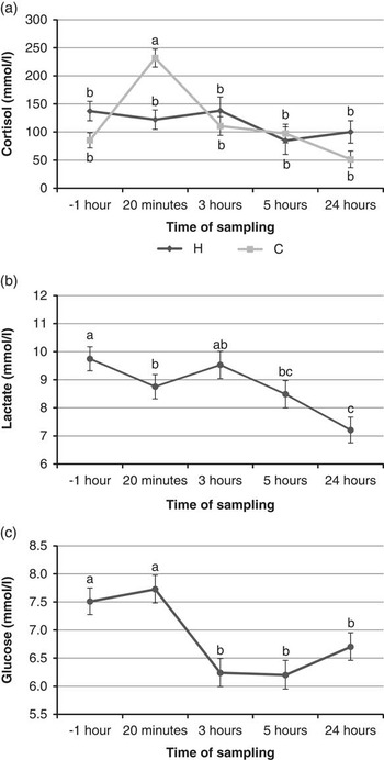
Cortisol levels were significantly affected by the interaction between time and treatment (P<0.001) and its concentration showed the highest value within 20 min after surgery in C piglets (Figure 1a). Time of sampling affected significantly lactate concentrations (P<0.001) and the lowest values were observed 24 h after treatments (Figure 1b). Glucose was also affected only by time (P<0.001; Figure 1c). In particular, the concentration was higher before and within 20 min compared with 3, 5 and 24 h after treatment. In C piglets glucose was positively correlated to lactate (r p =0.30; P<0.05) and cortisol concentrations (r p =0.38; P<0.01).
Effect of interaction treatment×time on (a) cortisol (LS means±s.e.m.) in handled (H) and castrated (C) piglets at different time of sampling, and effect of time of sampling on (b) lactate (LS means±s.e.m.) and (c) glucose (back transformed LS means±lower and upper 95% confidence interval). LS means with different letters differ significantly per P<0.05. LS=least square.

In this first set of piglets, rectal temperature was significantly affected by time (P<0.05), but not by treatment or interaction between time and treatment (Figure 2a). In particular, rectal temperature at 4 h after treatment was higher compared with the baseline and with those at 20 min and 24 h after treatment. In the second set of piglets, rectal temperature was affected by interaction between time and treatment (Figure 2b), in particular at 3 h after treatment it was higher in castrated compared with handled piglets (P<0.01).
Effect of time of measurement on (a) rectal temperature in piglets of the first trial (LS means±s.e.m.), and effect of the interaction treatment×time on (b) rectal temperature in handled (H) and castrated (C) piglets of the second trial (LS means±s.e.m.). Effect of the interaction treatment×time on (c) mean eye temperature in handled (H) and castrated (C) piglets (LS means±s.e.m.), and effect of time of measurement on (b) maximum (max) eye temperature in piglets (LS means±s.e.m.). LS means with different letters differ significantly per P<0.05. LS=least square.

Mean eye temperature was affected by time of measurement (P<0.001) and by the interaction between time of measurement and treatment (P<0.05). Baseline mean eye temperature was lower and similar between H and C piglets, the highest value was observed for castrated piglets 4 h after castration (Figure 2c). Maximum eye temperatures were significantly affected by treatment (P<0.05) and by time of measurement (P<0.001) but not by their interaction. Maximum eye temperature was higher in castrated piglets compared with handled (35.6±0.08 v. 35.4±0.09; P<0.05) and it was higher 4 h after treatments compared with the previous measurements (Figure 2d). Rectal temperature was correlated with maximum eye temperature in both H (r p =0.45; P<0.01) and C piglets (r p =0.31; P<0.01) and with mean eye temperature in both H (r p =0.38; P<0.05) and C piglets (r p =0.28; P<0.05).
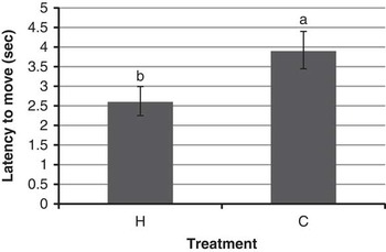
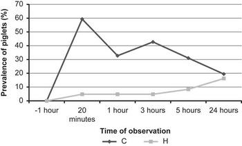
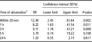
Latency to move was significantly affected by treatment (P<0.01), and H piglets hesitated less before walking after being handled, whereas C piglets showed a latency of almost 4 s (Figure 3). Prevalence of alterations in locomotory behaviours (postures and gait) were the highest within 20 min from castration (Figure 4). Moreover, surgically castrated piglets had a higher risk to show alterations than handled piglets within 20 min, 1 and 3 h after treatment (Table 1). At the other two observations carried out 5- and 24-h post-treatment the risk was not statistically different between handled and castrated piglets.
Latency to move in piglets (LS means±s.e.m.) immediately after handling (H) or surgical castration (C). LS means with different letters differ significantly (P<0.01). LS=least square.

Prevalence (%) of locomotory behavior alterations in handled (H) and castrated (C) piglets at different time of observation.

Relative risk (RR) of locomotory behavior alterations in surgically castrated piglets (C) compared with handled piglets (H) (P=0.037) at different time of observation

1 Time of observation: right after treatment (within 20 min), 1, 3, 5 and 24 h after treatment.
Discussion
The present study aimed to test if some innovative parameters such as latency to move, differences in postures and walking, and eye temperature are useful for detection of pain in piglets in response to surgical castration, besides the physiological indicators commonly used for pain assessment. Regarding blood parameters, the highest cortisol concentration observed for castrated piglets at the first sampling after surgical castration (within 20 min) is in line with previous studies that reported increased cortisol levels for up to 120 min after castration (Prunier et al., Reference Prunier, Mounier and Hay2005; Waldmann et al., Reference Waldmann, Rittershaus, Kietzmann and Puppe2010; Sutherland et al., Reference Sutherland, Davis, Brooks and Coetzee2012). This is indicative of acute stress and could be related to an increased adrenal output owing to pain and tissue damage (Prunier et al., Reference Prunier, Mounier and Hay2005). After a painful event, epinephrine stimulates the mobilisation of muscular glycogen and its consequent hepatic metabolism, resulting in an increment of plasma lactate and glucose concentrations (Mayes, Reference Mayes1995). Lactate and glucose concentrations were therefore expected to vary between treatments and to be higher in castrated piglets, likely experiencing greater visceral pain. The lack of a significant treatment effect on glucose could be because of insufficient hepatic glycogen stores in 4-day-old piglets as hypothised by Prunier et al. (Reference Prunier, Mounier and Hay2005). The high lactate levels in piglets of both treatments in the current study, that were in the range of peak values observed 5 min after castration (Prunier et al., Reference Prunier, Mounier and Hay2005) or other acute stressors (Merlot et al., Reference Merlot, Mounier and Prunier2011), could be related to a high level of stress owing to manipulation.
In the present study, castrated piglets had higher rectal temperature than handled ones only 3 h after treatment. Body temperature was reported to increase in response to anxiogenic or stress-inducing stimuli and injury (surgery and trauma) for the activation of the endogenous mechanisms related to inflammation (Takakazu et al., Reference Takakazu, Kae and Tetsuro2001; Olivier et al., Reference Olivier, Zethof, Pattija, van Boogaerta, van Oorschota, Leahyc, Oostinga, Bouwknechta, Veeningf, van der Gugtena and Groeninka2003; Roth et al., Reference Roth, Rummel, Barth, Gerstberger and Hubschle2009). Inflammatory mediators such as TNF-α and IL-1β are considered the main endogenous pyrogens (Roth et al., Reference Roth, Rummel, Barth, Gerstberger and Hubschle2009) and they were observed to increase in piglets 3 h after castration (Moya et al., Reference Moya, Boyle, Lynch and Arkins2008). This could explain the tardive hyperthermia observed in the present study in castrated piglets, even if other external factors interfering with body temperature (exposure to heat lamps or time from milk intake) cannot be completely excluded. Moreover, the stress due to extra manipulation for temperature measurement could be associated to the late (5 and 24-h post-treatment) increase in rectal temperatures in both, handled and castrated piglets.
Maximum eye temperature was also higher in castrated compared with handled piglets and it increased in both treatments over time. Stewart et al. (Reference Stewart, Verkerk, Stafford, Schaefer and Webster2010a and Reference Stewart, Webster, Stafford, Schaefer and Verkerk2010b) and Dockweiler et al. (Reference Dockweiler, Coetzee, Edwards-Callaway, Bello, Glynn, Allen, Theurer, Jones, Miller and Bergamasco2013) described an increase of eye temperature in calves right after castration (up to 10 min) due to an increase in parasympathetic nervous system activity associated with deep visceral pain. Results from these studies suggest that it would be beneficial to investigate eye temperature in a closer time interval to castration increasing the number of samplings but at the same time avoiding repeated handling that would not allow discrimination between pain and handling stress. Considering eye thermography, presence of different amounts of tears might have also influenced eye temperature as reported by Kamao et al. (Reference Kamao, Yamaguchi, Kawasaki, Mizoue, Shiraishi and Ohashi2011) studying the dry eye syndrome in humans. In this case, it could be speculated that the difference between castrated and handled piglets detected in our study would be even underestimated.
Among behavioural parameters, latency to move differed between treatments. Castrated piglets needed more time to start walking and this gives evidence that castration causes pain in the region of the hind legs. Similar hypothesis were formulated by McGlone et al. (Reference McGlone, Nicholson, Hellman and Herzog1993) and Keita et al. (Reference Keita, Pagot, Prunier and Guidarini2010) who reported that walking is easily involved in alterations probably due to pain at the hind region subsequent to surgical castration of the piglet. Further support to this assumption comes from results of this study pointing out that none of the piglets showed locomotory alterations before treatments and that castrated piglets had a higher risk of occurrence of weaker and protracted forward hind limbs, hind limb non-weight bearing, hind tip-toe walking or kyphosis within 20 min, 1 and 3 h after treatment. Similar results were found by Lonardi et al. (Reference Lonardi, Brscic, Scollo and Gottardo2011) and Kluivers-Poodt et al. (Reference Kluivers-Poodt, Zonderland, Verbraak, Lambooij and Hellebrekers2013) although they are in contrast with those of Hay et al. (Reference Hay, Vulin, Genin, Sales and Prunier2003) who reported abnormal behaviour for a few days after treatment.
Results of the study pointed out that detectable alterations of behaviour and of physiological parameters occur in the first 3 h from castration. Latency to move, alterations in posture and walking, and eye temperature acquired by IR thermography appear to be novel applicable pain assessment measures. The first two measurements are particularly promising since they require minimal handling of the piglets although they should be validated with inter- and intra-observer reliability tests. On the other hand, acquisition of data on eye temperature requires animals to be constrained in standardised conditions and needs further refinement of the methodology on pigs.
Acknowledgement
The authors would like to thank Animal Ethics Pty Ltd (Australia) for funding this project. The authors are grateful to Dr Meredith Sheil for advice and comments.