INTRODUCTION
Strongyloides stercoralis (Bavay, 1876) is an intestinal nematode, which causes strongyloidiasis, helminthiasis usually responsible for chronic asymptomatic infections of the gastrointestinal tract in immunocompetent human hosts and may remain undetected for decades. In immunocompromised patients due to immunosuppressive therapy (particularly steroids) (Genta, Reference Genta1992; Keiser and Nutman, Reference Keiser and Nutman2004; Lam et al. Reference Lam, Tong, Chan and Siu2006), infection with human immunodeficiency virus (HIV) (Ferreira, Reference Ferreira2003; Keiser and Nutman, Reference Keiser and Nutman2004; Silva et al. Reference Silva, Ferreira, Borges and Costa-Cruz2005), infection with human T-lymphotropic virus 1 (HTLV- 1) (Porto et al. Reference Porto, Muniz, Oliveira Júnior and Carvalho2002; Keiser and Nutman, Reference Keiser and Nutman2004), solid organ transplantation (Keiser and Nutman, Reference Keiser and Nutman2004; Concha et al. Reference Concha, Harrington and Rogers2005), haematological malignancies (Graeff-Teixeira et al. Reference Graeff-Teixeira, Leite, Sperhacke, Fassina, Petry, Mucenic, Pandolfi, Barcelos and Job1997; Keiser and Nutman, Reference Keiser and Nutman2004), solid-organ malignancies (Safdar et al. Reference Safdar, Malathum, Rodriguez, Husni and Rolston2004), hypogammaglobulinaemia (Keiser and Nutman, Reference Keiser and Nutman2004), chronic alcoholism (Oliveira et al. Reference Oliveira, Ribeiro, Mendes, Oliveira and Costa-Cruz2002; Silva et al. Reference Silva, Ferreira, Borges and Costa-Cruz2005; Marques et al. Reference Marques, Zago-Gomes, Gonçalves and Pereira2010) severe malnutrition (Keiser and Nutman, Reference Keiser and Nutman2004) or diabetes mellitus (Mendonça et al. Reference Mendonça, Gonçalves-Pires, Rodrigues, Ferreira and Costa-Cruz2006), this infection may develop into a hyperinfection syndrome or disseminated disease (Keiser and Nutman, Reference Keiser and Nutman2004; Marcos et al. Reference Marcos, Terashima, DuPont and Gotuzzo2008).
This parasite infects humans through skin penetration, and has a complex life cycle involving 2 stages of development: a phase of free-living that occurs in the environment, good conditions of temperature and humidity, and the other stage as a parasite, which occurs within the host. The parthenogenetic female of S. stercoralis lays few eggs per day, which are deposited in the intestinal epithelium, the rhabditiform mature larvae exit in the feces of the patients, and constitute in the diagnostic stage viewed in optical microscopy. By a process of autoinfection, there may be a rapid increase in the number of infective larvae; strongyloidiasis consequently persists for decades (Grove, Reference Grove1996; Costa-Cruz, Reference Costa-Cruz, Neves, Melo, Linardi and Vitor2005). The fact that the free-living forms of the parasite in the soil depend on favourable environmental conditions such as humidity and high temperature, makes conditions in many regions of Brazil ideal for the development and maintenance of intestinal parasites (Carmo et al. Reference Carmo, Perez, Gerolomo, Silva and Alves2005).
Definitive diagnosis of strongyloidiasis is performed mainly by the identification of larvae in fecal specimens, through concentration techniques or culture methods. Nevertheless, these larvae are excreted in low numbers and not daily, making the parasitological methods unreliable in most cases, requiring the analysis of 7 stool samples to increase the positivity (Dreyer et al. Reference Dreyer, Fernandes-Silva, Alves, Rocha, Albuquerque and Addiss1996; Uparanukraw et al. Reference Uparanukraw, Phongsri and Morakote1999). Several techniques are described for the parasitological diagnosis of strongyloidiasis, including the modified Baermann method by Moraes (BM) (Baermann, Reference Baermann1917; Moraes, Reference Moraes1948), the method of Rugai (R) (Rugai et al. Reference Rugai, Mattos and Brisola1954), Harada-Mori (HM) (Harada and Mori, Reference Harada and Mori1955) and culture method on agar plates (AP) (Arakaki et al. Reference Arakaki, Hasegawa, Asato, Ikeshiro, Kinjo, Saito and Iwanaga1988). However, the increased number of stool samples and the inconvenience of fresh stool specimens, make these parasitological diagnostic methods unusable in routine laboratory practice. Larvae can also be found in other body fluids (sputum, broncho-alveolar lavage, among others), through endoscopy, colonoscopy, or intestinal biopsy (Costa-Cruz, Reference Costa-Cruz, Neves, Melo, Linardi and Vitor2005; Minematsu et al. Reference Minematsu, Hokama, Makishi, Arakaki, Kinjo and Fujita2011). The failure to diagnose this parasitosis is reflected in the frequent reports of fatal cases associated with hyperinfections, brought on by corticosteroid therapy. The similarity between corticosteroids and an ecdyses hormone of the parasite, which promotes maturation of infective larvae in the intestine of the patient, is a possible explanation for this association (Hernandez-Chavarria, Reference Hernandez-Chavarria2001). Thus, the serological methods present a good alternative for early diagnosis and for evaluating the effectiveness of treatment (Sato et al. Reference Kobayashi, Hasegawa, Forli, Nishimura, Yamana, Shimabukuro and Sato1995; Paula et al. Reference Paula, Castro, Gonçalves-Pires, Marçal, Campos and Costa-Cruz2000; Siddiqui and Berk, Reference Siddiqui and Berk2003; Costa-Cruz, Reference Costa-Cruz, Neves, Melo, Linardi and Vitor2005; Page et al. Reference Page, Dempsey and McCarthy2006; Sudré et al. Reference Sudré, Macedo, Peralta and Peralta2006; Varatharajalu et al. Reference Varatharajalu, Parandaman, Ndao, Andersen and Neva2011).
Many epidemiological aspects of Strongyloides infection are not well known, such as prevalence rates that can be expected in different sectors of the population, environmental and zoonotic aspects, geographical variations, besides transmission and clinical presentation, host immunity and the risk factors for dissemination. The prevalence of S. stercoralis is underestimated, especially by the low sensitivity of parasitological diagnostic methods employed in routine procedures that do not include specific methods to search for larvae, and also the shortage of parasitological surveys. Thus, S. stercoralis infections in many areas remain without information (Hernandez-Chavarria, Reference Hernandez-Chavarria2001; Olsen et al. Reference Olsen, van Lieshout, Marti, Polderman, Polman, Steinmann, Stothard, Thybo, Verweij and Magnussen2009). Strongyloidiasis has a heterogeneous worldwide distribution, and prevalence has been divided into 3 categories: sporadic (<1%), endemic (1–5%) and hyperendemic (>5%) (Pires and Dreyer, Reference Pires and Dreyer1993). It is estimated that strongyloidiasis affects 30–100 million individuals around the world. All hyperendemic areas are located in the tropics, the endemic areas in the subtropics, but the parasite is also present, sporadically, in the United States, Europe and Asia (Olsen et al. Reference Olsen, van Lieshout, Marti, Polderman, Polman, Steinmann, Stothard, Thybo, Verweij and Magnussen2009).
The World Health Organization (2005) recommended that the Strongyloides infection could be included in the ‘Parasite Control Program’ in areas where the infection is endemic. However, now the infection is only a possible target and presumably, not effectively integrated in helminth control programmes. Recognizing that intestinal parasites are still prevailing in the country, the Brazilian government created the National Surveillance and Control of Enteroparasitoses in 2005 (Carmo et al. Reference Carmo, Perez, Gerolomo, Silva and Alves2005) and in 2007, the Growth Acceleration Program (PAC) (Ministério da Fazenda, 2007) providing for actions in sanitation, targeting the reduction of infectious and parasitic diseases, including intestinal parasitosis.
Despite scientific knowledge and technological advances available for its treatment and prevention, strongyloidiasis is still a parasitic disease of great importance to public health in Brazil, but information on strongyloidiasis is based on studies that show local epidemiological data. The objective of this review is to outline an epidemiological profile of this intestinal helminth in Brazil by parasitological and serological diagnosis in inhabitants of rural and urban areas, and associate this profile with different immunosuppression situations.
METHODOLOGY
We conducted a review of the literature on epidemiological aspects of strongyloidiasis in Brazil, using the keywords ‘Strongyloides stercoralis’, ‘strongyloidiasis’, ‘intestinal parasites’ and ‘Brazil’ in different combinations. We searched in the Scientific Eletronic Library Online (SciELO) and the U.S. National Library of Medicine (PubMed) over 20 years (1990–2009). There have been many studies regarding intestinal parasites from which the specific results for S. stercoralis were extracted. We used as inclusion criteria any study that gave values of the occurrence of S. stercoralis in Brazil in accordance with the region, location by state, rural and urban areas, considering the population by age, immunity, the method of diagnosis, or parasitological and serological association with other diseases (alcoholism, diabetes, cancer, infection with AIDS/HIV and HTLV-1, among others). As exclusion criteria: any study published before 1990 and after 2009, and papers that did not show the occurrence of S. stercoralis, when we used the keyword ‘intestinal parasite’. To analyse the information the value of the mean (X) frequencies reported was chosen.
RESULTS AND DISCUSSION
Epidemiological profile of S. stercoralis by parasitological methods
In the period from 1990 to 2009 it was found that the occurrence of infection by S. stercoralis in Brazil, using parasitological methods of diagnosis was 5·5%, characterizing the country as a hyperendemic area for strongyloidiasis. There was a variation of 3·9% to 7·9% among the 5 regions of Brazil, but there was no variation between the rural (4·8%) and urban (5·0%) areas. It is known that the marked socio-economic inequalities observed in Brazil, combined with the disorderly urbanization process, cause portions of the population to live in precarious living conditions. The occupation of the peripheral areas of cities where sanitary conditions make the quality of life style not different from rural areas, and often in worse conditions that favour the transmission of intestinal parasites (De Carli et al. Reference De Carli, Mentz, Rott, Silva, Wendorff, Tasca, Castilhos, De Carli, Montenegro and Mylius1997).
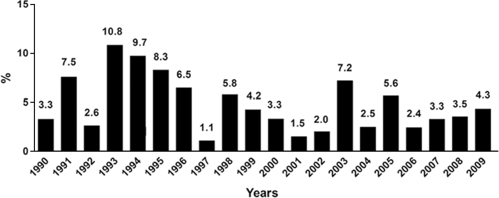
Changes in the prevalence of Strongyloides stercoralis in Brazil in the period 1990–2009 as assessed by parasitological methods of diagnosis are shown in Fig. 1. The variation was from 1·1 to 10·8% during the period studied. The occurrence of S. stercoralis infection in humans, using parasitological methods, distributed by region, state of the federation and the population studied is shown in Table 1. The diversity of parasitological diagnostic methods used in parasitological survey, make epidemiological knowledge of this parasite irregular. Of the 64 studies analysed and shown in Table 1, the diversity of techniques used as a diagnostic tool can be observed, in which 39 of the studies (60·9%) used non-specific methods for the detection of S. stercoralis larvae. We included as non-specific parasitological methods the parasitological techniques used in the routine of clinical laboratories, for example: the Direct (Amato Neto and Correa, Reference Amato Neto and Correa1991), Faust (Faust et al. Reference Faust, D'Antoni, Odom, Miller, Peres, Sawitz, Thomen, Tobie and Walker1938), Lutz (Lutz, Reference Lutz1919), or Ritchie/Blagg/MIFC (Ritchie, Reference Ritchie1948) methods that are not suitable or specific for the detection of nematode larvae. Only 25 (39·1%) of the studies present results based on at least 1 specific method (BM, R, HM or AP) for the detection of these helminth larvae.
Changes in the prevalence of Strongyloides stercoralis in Brazil in the period 1990–2009 as assessed by parasitological methods of diagnosis.

Occurrence of Strongyloides stercoralis infection in humans, considering the parasitological diagnosis, region, state and population in Brazil from 1990 to 2009

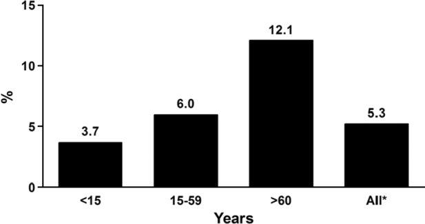
The concept that children are a group largely involved in parasite infections (Moura et al. Reference Moura, Bragazza, Coelho and Aun1997; Carvalho et al. Reference Carvalho, Carvalho and Mascarini2006; Silva et al. Reference Silva, Silva, Almeida, Sousa and Freitas2009) was not observed for this parasite in this period. When we analysed the articles that were available to characterize the age group studied, it was noted that the occurrence of S. stercoralis increased with age (Fig. 2). The mean occurrence of 12·1% for S. stercoralis, for age 60 and over (Hurtado-Guerrero et al. Reference Hurtado-Guerrero, Alencar and Hurtado-Guerrero2005; Aguiar et al. Reference Aguiar, Gonçalves, Sodré, Pereira, Bóia, Lemos and Daher2007; Miné and Rosa, Reference Miné and Rosa2008), suggests epidemiological conditions of concern for the elderly population in Brazil since, in this group, the immune system is weakened, particularly for those with poor health. In a parasitological study in the Amazon region, Hurtado-Guerrero et al. (Reference Hurtado-Guerrero, Alencar and Hurtado-Guerrero2005) drew attention to the effect that intestinal parasites have on the nutritional status and health of the elderly population, and, moreover, mentioned an occurrence of 9·1% for S. stercoralis in this age group. The greater occurrence of S. stercoralis in the elderly was obtained from the study of Miné and Rosa (Reference Miné and Rosa2008) in São Paulo state, which detected 20·4% infection in the elderly, considering both sexes, but 40% in elderly males of >80 years of age.
Changes in the prevalence of Strongyloides stercoralis, by the age of the Brazilian population, in the period 1990–2009 as assessed by parasitological methods of diagnosis. * The population was not weighted by the age distribution of the sampled members.

Epidemiological profile of S. stercoralis by serological methods
Owing to the limited sensitivity of parasitological methods for diagnosis of infection by S. stercoralis, serological methods have been resorted to, which have high sensitivity and specificity. Immunological tests such as enzyme-linked immunosorbent assay (ELISA), immunofluorescence antibody test (IFAT) and Western-blotting, have been described for the diagnosis of strongyloidiasis (Costa-Cruz et al. Reference Costa-Cruz, Bullamah, Gonçalves-Pires, Campos and Vieira1997; Sudré et al. Reference Sudré, Siqueira, Barreto, Peralta, Macedo and Peralta2007; Machado et al. Reference Machado, Faccioli, Costa-Cruz, Lourenço, Roque-Barreira, Gonçalves-Pires and Ueta2008c; Feliciano et al. Reference Feliciano, Gonzaga, Gonçalves-Pires, Gonçalves, Rodrigues, Ueta and Costa-Cruz2010). Among the various techniques used in immunodiagnosis of strongyloidiasis, the ELISA test has been shown to be superior with respect to practicality, safety and the availability of reagents (Liu and Weller, Reference Liu and Weller1993), in addition to high sensitivity and specificity (Schaffel et al. Reference Schaffel, Nucci, Carvalho, Braga, Almeida, Portugal and Pulcheri2001). The ELISA was the test of choice for most of the articles analysed as a single technique for serological diagnosis (Rossi et al. Reference Rossi, Partel, Teixeira, Takahashi, De Barros-Mazon and Da Silva1993; Costa-Cruz et al. Reference Costa-Cruz, Madalena, Silva, Sopelete, Campos and Taketomi2003; Machado et al. Reference Machado, Ueta, Gonçalves-Pires, Oliveira, Faccioli and Costa-Cruz2003; Rodrigues et al. Reference Rodrigues, Oliveira, Sopelete, Silva, Campos, Taketomi and Costa-Cruz2007), or in association with the IFAT and/or Western blot (Silva et al. Reference Silva, Barcelos, Passos-Lima, Espindola, Campos and Costa-Cruz2003; Rodrigues et al. Reference Rodrigues, Sopelete, Silva, Cunha-Junior, Taketomi and Costa-Cruz2004; Mota-Ferreira et al. Reference Mota-Ferreira, Gonçalves-Pires, Ferreira, Sopelete, Adballah and Costa-Cruz2009). The difficulty in obtaining S. stercoralis larvae and thus obtaining a sufficient amount of antigenic material has been a limiting factor for the immunodiagnosis of strongyloidiasis (Rossi et al. Reference Rossi, Partel, Teixeira, Takahashi, De Barros-Mazon and Da Silva1993). This is why many researchers have been using heterologous antigens, Strongyloides ratti or Strongyloides venezuelensis, in the diagnosis of strongyloidiasis, since they are easy to obtain and are a safe source without risk to their handlers, besides having high sensitivity and specificity (Costa-Cruz et al. Reference Costa-Cruz, Bullamah, Gonçalves-Pires, Campos and Vieira1997; Machado et al. Reference Machado, Ueta, Gonçalves-Pires, Oliveira, Faccioli and Costa-Cruz2001, Reference Machado, Ueta, Gonçalves-Pires, Oliveira, Faccioli and Costa-Cruz2003; Silva et al. Reference Silva, Barcelos, Passos-Lima, Espindola, Campos and Costa-Cruz2003; Rodrigues et al. Reference Rodrigues, Sopelete, Silva, Cunha-Junior, Taketomi and Costa-Cruz2004, Reference Rodrigues, Oliveira, Sopelete, Silva, Campos, Taketomi and Costa-Cruz2007; Feliciano et al. Reference Feliciano, Gonzaga, Gonçalves-Pires, Gonçalves, Rodrigues, Ueta and Costa-Cruz2010).
Of the 3 seroepidemiological studies conducted in the general population in Brazil, the mean positivity in serum samples of S. stercoralis infection was 21·7% and 29·2%, using serological testing by IFAT-IgG and ELISA-IgG, respectively, studies were concentrated in the Southeast region (Costa-Cruz et al. Reference Costa-Cruz, Machado and Campos1998; Machado et al. Reference Machado, Teixeira, Paula, Gonçalves-Pires, Ueta and Costa-Cruz2007; Mota-Ferreira et al. Reference Mota-Ferreira, Gonçalves-Pires, Ferreira, Sopelete, Adballah and Costa-Cruz2009).
Occurrence of S. stercoralis infection associated with different types of immunosuppression
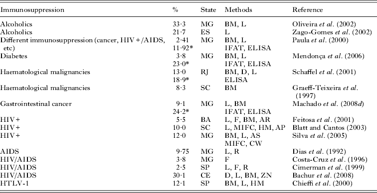
The occurrence of S. stercoralis infection associated with different types of immunosuppression compared to parasitological diagnosis and/or serology, is shown in Table 2. The variety of techniques may be noted, but most studies have used techniques suitable for the detection of larval helminths since the suspicion of infection by S. stercoralis could be mentioned in the population studied. The average occurrence was 11·8% using parasitological diagnostic methods. In 4 studies of immunosuppression using parasitological and immunological diagnosis it was found that the mean occurrence increased from 7·1% to 19·5% respectively (Paula et al. Reference Paula, Castro, Gonçalves-Pires, Marçal, Campos and Costa-Cruz2000; Schaffel et al. Reference Schaffel, Nucci, Carvalho, Braga, Almeida, Portugal and Pulcheri2001; Mendonça et al. Reference Mendonça, Gonçalves-Pires, Rodrigues, Ferreira and Costa-Cruz2006; Machado et al. Reference Machado, Teixeira, Gonçalves-Pires, Loureiro, Araújo and Costa-Cruz2008d), emphasizing the usefulness of serological methods as a tool for diagnosis of strongyloidiasis, especially when the question involved is immunosuppression.
Occurrence of Strongyloides stercoralis infection in humans using parasitological methods and/or serological diagnosis, distributed according to the type of immunosuppression (1990–2009)

* Positive by serological methods (ELISA – enzyme-linked immunosorbent assay; IFAT, indirect fluorescent antibody test); parasitological methods (BM, Baermann-Moraes; D, direct; F, Faust/flotation; HM, Harada-Mori; L, Lutz; MIFC, Ritchie/Blagg; AP, culture agar plate, R, Rugai) and staining (AR, acid fast; ZN, Ziehl-Neelsen; AS, Blue metilen Safranin; CW, Chromotropic Weber's).
Although the association of strongyloidiasis and immunosuppression are important, case reports are scarce. In Brazil, the occurrence of strongyloidiasis in transplant patients (Dulley et al. Reference Dulley, Costa, Cosentino, Gamba and Saboya2009; Villela et al. Reference Vilela, Clemente, Mira, Torres, Veloso, Fonseca, Fonseca, França and Lima2009), the use of corticosteroids and/or with neoplasias (Godoy et al. Reference Godoy, Campos, Costa and Castro1998; Rodrigues et al. Reference Rodrigues, Fróes, Anefalos and Kobayasi2001; Lambertucci et al. Reference Lambertucci, Leão and Barbosa2003; Porto et al. Reference Porto, Alcântara, Leal, Castro and Carvalho2005; Salluh et al. Reference Salluh, Bozza, Pinto, Toscano, Wellers and Soares2005; Giavina-Bianchi et al. Reference Giavina-Bianchi, Silva, Toledo-Barros, Birolini, Kalil and Rizzo2006; Benincasa et al. Reference Benincasa, Azevedo, Canabarro, Valentim, Silva, Superti and Dias2007; Ben-Horin et al. Reference Ben-Horin, Barshack, Chowers and Mouallem2008; Veloso et al. Reference Veloso, Porto and Moraes2008), and haemolytic anaemia (Ferreira et al. Reference Ferreira, Barbosa, Ferreira, Souza, Neves, Carvalho and Barbosa2008) have been emphasized. Many of these reports show that the late diagnosis of infection may be associated with the severity of infection, causing patient death (Rodrigues et al. Reference Rodrigues, Fróes, Anefalos and Kobayasi2001; Salluh et al. Reference Salluh, Bozza, Pinto, Toscano, Wellers and Soares2005; Benincasa et al. Reference Benincasa, Azevedo, Canabarro, Valentim, Silva, Superti and Dias2007; Ferreira et al. Reference Ferreira, Barbosa, Ferreira, Souza, Neves, Carvalho and Barbosa2008; Veloso et al. Reference Veloso, Porto and Moraes2008; Villela et al. Reference Vilela, Clemente, Mira, Torres, Veloso, Fonseca, Fonseca, França and Lima2009). In a study of prevalence of symptomatic parasitosis in adult renal transplant recipients, in Porto Alegre, Brazil, from 2001 to 2005, Valar et al. (Reference Valar, Keitel, Dal Prá, Gnatta, Santos, Bianco, Sukienni, Pegas, Bittar, Oliveira and Garcia2007) showed that out of 657 transplants, 11 (1·67%) had symptoms and confirmed S. stercoralis diagnosis by the finding of larvae in feces (8 cases), by broncho-alveolar lavage fluid (in 2 cases), duodenal biopsy and sputum (in 1 case), and from these, 3 cases developed hyperinfection, and 1 died. The diagnosis of strongyloidiasis in these reported cases occurred through the finding of larvae in feces (Dulley et al. Reference Dulley, Costa, Cosentino, Gamba and Saboya2009), in other fluids (Porto et al. Reference Porto, Alcântara, Leal, Castro and Carvalho2005; Salluh et al. Reference Salluh, Bozza, Pinto, Toscano, Wellers and Soares2005; Ferreira et al. Reference Ferreira, Barbosa, Ferreira, Souza, Neves, Carvalho and Barbosa2008; Villela et al. Reference Vilela, Clemente, Mira, Torres, Veloso, Fonseca, Fonseca, França and Lima2009), at biopsy (Rodrigues et al. Reference Rodrigues, Fróes, Anefalos and Kobayasi2001; Lambertucci et al. Reference Lambertucci, Leão and Barbosa2003; Salluh et al. Reference Salluh, Bozza, Pinto, Toscano, Wellers and Soares2005; Giavina-Bianchi et al. Reference Giavina-Bianchi, Silva, Toledo-Barros, Birolini, Kalil and Rizzo2006; Ben-Horin et al. Reference Ben-Horin, Barshack, Chowers and Mouallem2008) and at autopsy (Godoy et al. Reference Godoy, Campos, Costa and Castro1998; Benincasa et al. Reference Benincasa, Azevedo, Canabarro, Valentim, Silva, Superti and Dias2007; Veloso et al. Reference Veloso, Porto and Moraes2008).
Co-infection by S. stercoralis and HTLV-1 has been reported to decrease in response to anti-helmintic treatment and increased severity of viral infection in these patients (Porto et al. Reference Porto, Oliveira Filho, Neva, Orge, Alcântara, Gam and Carvalho2001, Reference Porto, Muniz, Oliveira Júnior and Carvalho2002). In a coproparasitological survey, Chieffi et al. (Reference Chieffi, Chiattone, Feltrim, Alves and Paschoalotti2000) called attention to the higher prevalence of S. stercoralis among HTLV-1 positive blood donors in relation to the negative HTLV-1.
HIV/AIDS infection associated with S. stercoralis has been reported in some studies (Dias et al. Reference Dias, Mangini, Torres, Vellosa, Silva, Silva, Corrêa and Coletti1992; Feitosa et al. Reference Feitosa, Bandeira, Sampaio, Badaró and Brites2001; Blatt and Cantos, Reference Blatt and Cantos2003; Silva et al. Reference Silva, Ferreira, Borges and Costa-Cruz2005) using parasitological methods and/or serological diagnosis. However, only 3 reports demonstrated the seriousness of helminthiasis when associated with HIV/AIDS, previously considered an opportunistic parasite (Takayanagui et al. Reference Takayanagui, Lofrano, Araújo and Chimelli1995; Ferreira et al. Reference Ferreira, Nishioka, Borges, Costa-Cruz, Rossin, Rocha, Silvestre and Nunes-Araújo1999; Bachur et al. Reference Bachur, Vale, Coêlho, Queiroz and Chaves2008).
The association of chronic alcoholics with strongyloidiasis is an interesting aspect, since this group of patients may be suffering from severe forms of strongyloidiasis, probably due to dulled mental function, breakdown of local protective barriers, aspiration, exposure to pathogens and malnutrition caused by alcoholism (Oliveira et al. Reference Oliveira, Ribeiro, Mendes, Oliveira and Costa-Cruz2002; Zago-Gomes et al. Reference Zago-Gomes, Aikawa, Perazzio, Gonçalves and Pereira2002). The corticosteroid metabolites in alcoholics may increase the fecundity of the females, and thus the number of rhabditiform larvae coming out in the feces (Zago-Gomes et al. Reference Zago-Gomes, Aikawa, Perazzio, Gonçalves and Pereira2002). However, Genta (Reference Genta1992) suggested that the ecdysoid-like action of the steroids would increase the developmental rate and moulting of the larvae in the intestine causing autoinfection. The increase in autoinfection would lead to an increased number of adult female worms, leading to an increase in the number of larvae (L1) in the feces.
Complementary aspects
Domestic animals such as dogs infected with Strongyloides, particularly in areas where sanitary conditions are unsatisfactory, can become reservoirs for this helminth and sources of human infection (Ferreira-Junior et al. Reference Ferreira-Junior, Gonçalves-Pires, Silva, Gonçalves and Costa-Cruz2006; Gonçalves et al. Reference Gonçalves, Machado, Gonçalves-Pires, Ferreira, Silva and Costa-Cruz2007; Campos-Filho et al. Reference Campos-Filho, Barros, Campos, Braga, Cazorla, Albuquerque and Carvalho2008). Thus, the presence of feces from infected humans or animals contaminating the soil are epidemiological factors that may influence the onset, maintenance and propagation of human strongyloidiasis as well as favourable conditions of temperature and humidity, and inadequate hygienic habits (Costa-Cruz, Reference Costa-Cruz, Neves, Melo, Linardi and Vitor2005).
We would like to emphasize that one drawback of immunological assays for antibody demonstration is that it reflects the history of infection and not necessarily the current on-going infection. It is difficult in many cases to know whether or not low-level autoinfection is continuing. This occurs mainly because antibody levels remain detectable for years after antihelmintic treatment. (Liu and Weller, Reference Liu and Weller1993; Grove, Reference Grove1996).
Currently, the detection of coproantigen by ELISA, standardized in experimental models (Nageswaran et al. Reference Nageswaran, Craig and Devaney1994; Gonçalves et al. Reference Gonçalves, Silva, Ueta and Costa-Cruz2010), and for human strongyloidiasis (El-Badry, Reference El-Badry2009) allows the identification of cases of active infection. The development of Polymerase Chain Reaction (PCR) in faecal samples of animals with low experimental infection by S. venezuelensis (Marra et al. Reference Marra, Chiuso-Minicucci, Machado, Zorzella-Pezavento, França, Ishikawa, Amarante, Sartori and Amarante2010) and also in human faecal samples (Verweij et al. Reference Verweij, Canales, Polman, Ziem, Brienen, Polderman and van Lishout2009) are promising as an alternative for the diagnosis of strongyloidiasis.
CONCLUSIONS
The non-application of techniques for the accurate diagnosis of this helminthiasis, either parasitological or immunological and molecular biology in the large centres, contributes to the underestimation of the prevalence of strongyloidiasis. Thus, many epidemiological aspects of infection by S. stercoralis are unknown or only poorly understood, such as national endemic areas, in which prevalence rates and intensities can typically be expected in different settings and populations, the zoonotic aspects, and geographical variations involved in infection. Therefore, we recommend what we consider to be the way forward and the priorities to reducing the prevalence of strongyloidiasis. (1) Health education campaigns on the disease. (2) Parasitological diagnosis including specific methods for analysing the nematode larvae. (3) Need to screen patients from endemic areas of strongyloidiasis prior to receiving corticosteroid therapy or organ transplantation. (4) Follow-up of patients with immunosuppression. (5) Development of novel diagnostic assays, and finally (6) the notification of positive cases.
Considering that Brazil is a tropical country and the character of chronicity and auto-infection of this parasite which can result in severe forms of hyper-infection or dissemination makes strongyloidiasis an important medical and social problem that unfortunately is neglected by health authorities.