Introduction
Isolated pulmonary valve stenosis accounts for approximately 10% of all CHDs. Reference van der Linde, Konings and Slager1 Since 1982, balloon pulmonary valvuloplasty has been considered the treatment of choice. Reference Rao2 Despite advances, pulmonary regurgitation remains the main outcome related to the procedure, Reference Feltes3–Reference Harrild, Powell and Tran8 and it seems to impact right ventricle function in long-term follow-up. Reference Mercer-Rosa, Ingall and Zhang9,Reference Massoud, Botros and Yehia10 In the last decade, few studies have objectively investigated this relationship. Reference Merino-Ingelmo, Santos-de Soto, Coserria-Sánchez, Descalzo-Señoran and Valverde-Pérez5–Reference Massoud, Botros and Yehia10 This may be in part due to the difficulties of right ventricle imaging. Recently, strain analysis by 2D speckle-tracking echocardiography has emerged as a promising technique for the evaluation of right ventricle systolic function in several clinical settings. Reference Colquitt and Pignatelli11,Reference Mondillo12
We sought to evaluate the right ventricle function by conventional echocardiographic methods and strain analysis in the long term after balloon pulmonary valvuloplasty. In addition, we investigated the relationship between pulmonary regurgitation, demographic data at the time of the procedure, and right ventricle dysfunction in late follow-up.
Methods
Patients
This is a single-centre observational study with patients with isolated pulmonary stenosis who underwent balloon pulmonary valvuloplasty between January 2001 and December 2015. Initially, a retrospective analysis of the hospital records of the patients was carried out, and it included demographic, clinical, haemodynamic, and echocardiographic data before, at the time of the procedure, and during follow-up. Patients were excluded from this first analysis if any CHD other than small atrial or ventricular septal defects or persistent ductus arteriosus without haemodynamic significance was found, and if they had less than 5 years of follow-up.
The second part of the study was a cross-sectional analysis, named derived cohort, which comprised an updated clinical and echocardiographic evaluation of the patients included in the retrospective analysis. The patients with a residual gradient across the pulmonary valve > 35 mmHg after balloon pulmonary valvuloplasty were excluded. Those who could be contacted by either telephone or letter and met inclusion criteria were included. Analysis included anthropometrics, oxygen saturation, functional class,Reference Russell, Saval and Robbins 13 medications, physical examination, and echocardiography. Informed consent was obtained from all participants and their families.
Echocardiographic evaluation
In the derived cohort, the echocardiographic exams were performed according to international guidelines Reference Lopez, Colan and Frommelt14 using a commercially available system (GE Vivid 7; Horten, Norway) with a 3 MHz transducer.
The cardiac dimensions and the systolic function parameters were obtained as recommended. Reference Lopez, Colan and Frommelt14 When available, z score values from the paediatric population were used as references. Reference Pettersen, Du, Skeens and Humes15–Reference Koestenberger, Ravekes and Everett17 In the absence, reference values were obtained from data for adult subjects. Reference Lang18 The size of the right atrium was determined by the systolic area (cm2) and volume (ml), both indexed by the body surface area (m2). The right ventricle was considered enlarged if both basal and medium-cavity diastolic diameters had a z score > 2. Reference Pettersen, Du, Skeens and Humes15–Reference Cantinotti, Scalese and Murzi16 Conventional parameters of right ventricle systolic function included fractional area change, tricuspid annular plane systolic excursion, and tissue Doppler-derived tricuspid lateral annular systolic velocity.
The systolic longitudinal strain of the right ventricle was measured by the speckle-tracking technique adjusting the frame rate of two-dimensional images to 60 – 80 frames/s. Reference Badano19 The images were acquired under continuous electrocardiographic monitoring. A right ventricular–focused four-chamber view was obtained. The strain analysis was performed using the echocardiographic system software (GE EchoPAC – GE Healthcare version 12.1). The software provided semiautomatic myocardial delineation with manual adjustment if necessary. The analysis was accepted only when the software showed adequate tracking for all segments. The right ventricle was divided into six segments: three in the septal wall (basal, middle, and apical) and three in the free wall (basal, middle, and apical). The right ventricular global systolic longitudinal strain was obtained by the arithmetic mean of the values of the six segments. The systolic strain analysis of the left ventricle was performed as previously described. The normal reference value considered was −25 for the right ventricular global systolic longitudinal strain, −27 for the right ventricle free wall strain, Reference Levy, Sanchez Mejia, Machefsky, Fowler, Holland and Singh20 and −20 for the left ventricle strain. Reference Levy, Machefsky and Sanchez21
Restenosis was considered if the maximum gradient across the pulmonary valve, after a successful balloon pulmonary valvuloplasty, returned to values > 35 mmHg. The pulmonary regurgitation was assessed by a single observer using the combined use of colour Doppler flow mapping and spectral Doppler (pulsed and continuous). It was considered mild (grade 1) when the colour flow mapping detected a small holodiastolic regurgitant jet close to the pulmonary valve and the continuous wave Doppler flow curve had a slow deceleration throughout the diastole; moderate (grade 2) if the colour flow mapping detected diastolic flow in the pulmonary trunk and by the continuous wave Doppler the deceleration of the flow velocity of the regurgitant jet was faster but occurred throughout the diastole. Pulmonary regurgitation was considered severe (grade 3) if the regurgitant jet was wide and filled the entire right ventricular outflow tract, there was reverse diastolic flow in the pulmonary trunk and branches, and the continuous wave Doppler velocity curve showed rapid deceleration (deceleration time less than 260 ms and pressure half-time less than 100 ms). Reference Van Berendoncks, Van Grootel and McGhie22
Statistical analysis
Continuous variables are presented as mean ± SD for symmetric distributions or as median and range for asymmetric distributions; categorical variables are presented as percentages. Normality was assessed using Lilliefors and Kolmogorov–Smirnov tests. Comparisons employed t-tests or Mann–Whitney/Wilcoxon tests, with Pearson’s or Spearman’s correlation as appropriate. ANOVA was used for multiple means, the binomial test for two categorical groups, and Williams’ G test for three or more. Statistical significance was set at p < 0.05.
Results
Retrospective analysis
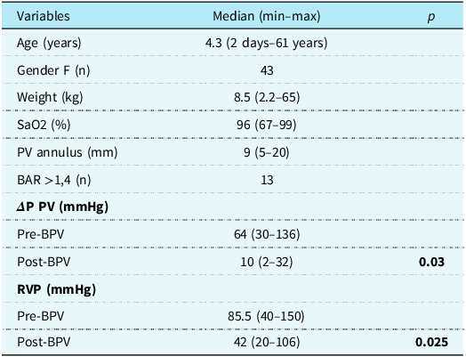
A total of 73 patients were included; the demographic and haemodynamic data are summarised in Table 1.
Demographic and haemodynamic data of the patients included in the retrospective analysis at the time of balloon pulmonary valvuloplasty

N = number of patients; F = female; PV = pulmonary valve; BAR = balloon/annulus ratio; ΔP = pressure gradient; PV = pulmonary valve; RVP = right ventricular systolic pressure; SaO2 = arterial oxygen saturation; BPV = pulmonary balloon valvuloplasty; statistical analysis = t test.
Immediate success was achieved in 69/73 patients (94.5%). Dysplastic pulmonary valve was seen in three of the four unsuccessful procedures. The balloon/pulmonary annulus ratio > 1.4 was observed in 13 patients; no patients were treated with a balloon/pulmonary annulus ration < 1.2 (Table 1). The mortality was 2.7% (2/73); both patients were female with critical pulmonary stenosis and weight < 3.5 kg. Other major complications occurred in eight patients; seven were newborns (p = 0.008) and included sinus of Valsalva dissection, dissection of the inferior vena cava, spontaneous reversed atrioventricular block, atrial fibrillation, atrial flutter, and ventricle perforation with cardiac tamponade. There were no cases of restenosis. Pulmonary regurgitation was observed in all echocardiographic records available in the follow-up. No patient required replacement of the pulmonary valve due to severe pulmonary regurgitation.
Derived cohort
Of the 73 initial patients, 6 were excluded (2 deaths and 4 with residual gradients > 35 mmHg). Among the 67 patients eligible for the derived cohort analysis, 21 returned to their referral hospital and no data were available, 26 were unreachable, and 2 declined participation.
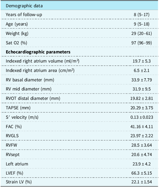
The derived cohort was composed of 18 patients (Table 2). Three patients were classified as NYHA II, while the others were in class I; no patients were taking medication.
Demographic and echocardiographic data of the 18 patients of the derived cohort undergoing an updated analysis

RV = right ventricle; RVOT = RV outflow tract; EDA = end-diastolic area; TAPSE = tricuspid annular plane systolic excursion; S′ = tissue Doppler-derived tricuspid lateral annular systolic velocity; FAC = fractional area change; RVGLS = right ventricle global systolic strain; RVFW = right ventricle free wall strain; LVE = left ventricle ejection fraction; LV = left ventricle; data are expressed as the median (min–max) or mean and standard deviation.
At the time of pulmonary valvuloplasty, these patients had a median age of 4 months (7 days – 3.8 years), weight of 6.4 kg (2.3 – 15 kg), body surface area of 0.32 m2 (0.17 – 0.63), and oxygen saturation of 90% (67 – 99%). Compared to the original cohort at the time of the procedure, there were no statistically significant differences in age (p = 0.32), weight (p = 0.28), pulmonary valve annulus diameter (p = 0.39), pulmonary valve pressure gradient before (p = 0.34), and after balloon pulmonary valvuloplasty (p = 0.07).
Echocardiographic measurements
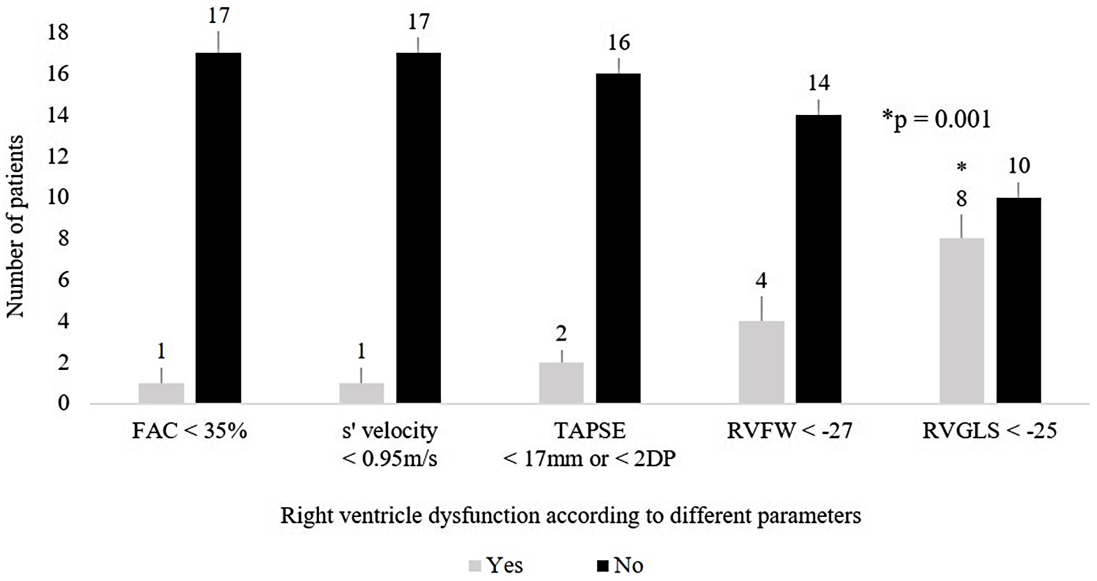
The cardiac dimensions and the function parameters are described in Table 2. The right ventricle was the only chamber dilated. The proportion of patients with abnormal right ventricle global strain was significantly higher when compared to the other methods (p < 0.01; Figure 1).
Right ventricle function according to conventional echocardiographic parameters and strain imaging.
* Statistical significance by binomial test. FAC: fractional area change; TAPSE: tricuspid annular plane systolic excursion; S’ velocity: tissue Doppler–derived tricuspid lateral annular systolic velocity; RVFW: right ventricle free wall strain; RVGLS: right ventricular global systolic longitudinal strain.

The mean right ventricular global systolic strain was significantly reduced in patients who underwent balloon pulmonary valvuloplasty < 1 year of age (23.8% ± 1.9) compared with those treated at > 1 year of age (27% ± 3.1; p = 0.03). The duration of follow-up did not differ significantly between patients with abnormal (6.8 ± 2.0 years) and normal strain values (9.3 ± 3.6 years; p = 0.07).
The three patients with cardiac symptoms had abnormal values of both right ventricle global and free wall strain, in addition to right ventricular dilation and severe pulmonary regurgitation.
Pulmonary regurgitation was observed in all 18 patients of the derived cohort, with a significant long-term increase in moderate or severe grade when compared to their previous echocardiographic records (p < 0.001). Immediately post-valvuloplasty, pulmonary regurgitation was absent in 12 and mild in 6; at 1 – 5 years of follow-up, it was mild in 12 and moderate in 6; and at the final analysis, it was mild in 1, moderate in 11, and severe in 6.
At the time of balloon pulmonary valvuloplasty, age < 1 year (p = 0.006), weight < 3 kg (p = 0.03), and body surface area < 0.3 m2 (p = 0.04) were associated with severe pulmonary regurgitation in late follow-up, while pulmonary annulus hypoplasia (p = 0.064), sex (p = 0.17), and balloon/pulmonary annulus ratio (p = 0.71) were not. Of the six patients with severe pulmonary regurgitation, a balloon/pulmonary annulus ratio > 1.4 was used in two patients.
There was a significant correlation between pulmonary regurgitation severity and enlargement of the right ventricle mid-diameter and an inverse correlation with both global and free wall strain abnormal values (Figure 2). There was no correlation between pulmonary regurgitation severity and abnormal conventional methods of the right ventricle function (p = 0.52).
Correlation between the pulmonary regurgitation grade and RV dimensions (A and B) and strain (C and D).
RV: right ventricle; A and B: Spearman’s test; C and D: Pearson’s test.

Discussion
Despite the small derived cohort, the demographics and haemodynamic data did not statistically differ from the initial patients, which allowed us to infer that the results found in the cross-sectional analysis could represent that general sample.
As observed in other studies, pulmonary regurgitation was the main residual lesion after balloon pulmonary valvuloplasty. Reference Stanger, Cassidy, Girod, Kan, Lababidi and Shapiro4–Reference Loureiro, Cardoso, Gomes, Martins and Pinto7,Reference Bouhlel, Ajmi, Slim, Gribaa and Naffati23,Reference Devanagondi, Peck and Sagi24 There is controversy in the literature about whether pulmonary regurgitation worsens over time. Reference Merino-Ingelmo, Santos-de Soto, Coserria-Sánchez, Descalzo-Señoran and Valverde-Pérez5,Reference Saad, Roushdy and Elsayed6,Reference Fawzy, Hassan and Fadel25 The present results showed a higher proportion of severe pulmonary regurgitation in long-term follow-up compared to their immediate post-pulmonary valvuloplasty. Nonetheless, the absence of standardised grading criteria for pulmonary regurgitation in both the derived cohort and initial group limits the reliability of the true progression of pulmonary regurgitation. It cannot be ruled out that the greater severity observed in the derived cohort is due to differences in the inter- and intra-observer variability or evolving diagnostic practices over time, rather than a progression of the regurgitation itself.
A balloon/pulmonary annulus ratio > 1.4 was not associated with the degree of pulmonary regurgitation in the late follow-up, consistent with conflicting results in the literature. Reference Merino-Ingelmo, Santos-de Soto, Coserria-Sánchez, Descalzo-Señoran and Valverde-Pérez5,Reference Fawzy, Hassan and Fadel25–Reference Yin, Wu and Xiang27 In contrast, the procedure performed in children < 1 year, <3 kg, or < 0.3 m2 was associated with severe pulmonary regurgitation, as reported in some previous studies. Reference Harrild, Powell and Tran8,Reference Mercer-Rosa, Ingall and Zhang9,Reference Devanagondi, Peck and Sagi24,Reference Fawzy, Hassan and Fadel25
The severity of pulmonary regurgitation has also been related to impairment of the right ventricle performance. Reference Harrild, Powell and Tran8,Reference Mercer-Rosa, Ingall and Zhang9,Reference Eyskens, Brown and Claus35 Possibly, overload leads to geometric changes and rearrangement of the ventricular contractility, which turns to a worse function. Reference Eyskens, Brown and Claus35 In the derived cohort, abnormal right ventricular strain values were correlated with severe pulmonary regurgitation, whereas this association was not observed with conventional parameters. This may reflect the small sample size of the cohort, as well as the higher sensitivity of strain imaging compared with conventional functional measures.
Only 2 of the 18 patients presented right ventricle dysfunction as measured by conventional echocardiographic parameters. Despite being a practical and validated tool, a criticism of tricuspid annular plane systolic excursion and tissue Doppler-derived tricuspid lateral annular systolic velocity is that the evaluation of the right ventricle function is based on the displacement of the lateral extremity of tricuspid valve annulus and extrapolated as a parameter for a global analysis. Thus, segmental dysfunction can be missed. Reference Haddad, Hunt, Rosenthal and Murphy28,Reference Muraru, Haugaa and Donal29
In contrast, right ventricle dysfunction was detected in four patients by free wall strain and in eight patients by global strain. As seen in other clinical settings, strain seems to have a greater ability to detect dysfunction, even with normal conventional parameters. Reference Cameli, Righini, Lisi and Mondillo30–Reference Meris, Faletra and Conca32 Unlike the conventional parameters, strain allows automatic and objective global and regional assessment of the right ventricle contractility, and it seems to be less dependent on loading. Reference Stassen, van der Bijl and Galloo33,Reference Morris, Krisper and Nakatani34 In the setting of pulmonary regurgitation, the right ventricle preload is increased and the conventional tools may remain within normal ranges despite underlying myocardial dysfunction. Also, Sirico et al. reported a persistent impairment of right ventricle function 24 hours after valvuloplasty when assessed by strain, while the global parameters recovered soon, again suggesting the better sensitivity of this method to detect dysfunction.Reference Sirico, Spigariol and Mahmoud 36 Thus, these phenomena could explain the observed disparity between strain and conventional parameters in the current findings, emphasising the sensitivity of deformation imaging in detecting dysfunction.
As in any cross-sectional study, correlation results should be interpreted with caution, as no prior right ventricular strain data were available. The patients were evaluated at least 5 years post-valvuloplasty, suggesting that late dysfunction observed is more likely due to pulmonary regurgitation than initial pressure overload from stenosis.
The size and function of the left ventricle were normal. Harrild et al. concluded that in the absence of severe right ventricle dilation, there is no significant interventricular interaction that affects the left ventricle. Reference Harrild, Powell and Tran8
The present study may have some clinical implications. A difference between the number of symptomatic patients and right ventricle dysfunction was observed when strain analysis was applied: it was observed in eight patients with right ventricle dysfunction, while three patients reported cardiac symptoms. According to guidelines, follow-up is based on the presence of residual stenosis; if it is mild, the recommendation for clinical evaluation is to be done every 5 years. Reference Feltes3, Reference Baumgartner 37 Since all patients had a mild residual gradient, the use of only this parameter could delay the detection of the right ventricle dysfunction. It is reasonable that there could be some benefit to a more frequent evaluation of patients with severe pulmonary regurgitation, despite the clinical symptoms reported.
Although incipient, these results suggest the benefit of using right ventricle strain analysis in the follow-up of patients undergoing balloon pulmonary valvuloplasty. Long-term studies with large numbers of patients will be able to better define the prognostic value of this right ventricle parameter in this clinical scenario.
Study limitations
The small number of patients enrolled in the revised cohort may have caused a bias in accurately estimating the general prevalence of pulmonary regurgitation and right ventricle dysfunction in our population.
Conclusion
The right ventricle dysfunction late after balloon pulmonary valvuloplasty was more frequently detected by strain analysis even in asymptomatic patients, and it was related to the severity of pulmonary regurgitation. Severe pulmonary regurgitation was related to balloon pulmonary valvuloplasty performed in children < 1 year of age, < 3 kg, and < 0.3 m2.
Acknowledgement
The authors are grateful to Prof. Francinelson de Lima – Federal University of Rondônia for the statistical assistance.
Financial support
This research received no specific grant from any funding agency, commercial, or not-for-profit sectors.
Competing interests
None.
Ethical standard
This study was approved by Sao Paulo Federal University Ethics Committee (Approval Number: 97895218.10000.5505).