Hemodynamic changes caused by physical exercise alter the loading conditions of the heart, and there is certain cardiac adaptation which serves to normalize this loading stress.1, 2 Left ventricular adaptation to long-term intensive training includes increases in cavity diameter, wall thickness, and left ventricular mass. The degree of these changes is consistent with the type of sporting activity and exercise programme.3 The number of prepubertal boys and girls that are active in sports increases each year. This imposes on paediatric cardiologists the need to explore, and become familiar, with the physiological changes in the hearts of children caused by conditioning. Despite the increasing involvement of child athletes in intensive training programmes, little is known about the influence of such training on cardiac structure and function compared to adults. Values derived from adult athletes cannot be extrapolated to younger athletes, who are less physically mature, and are exposed to shorter periods of intense training.4 We undertook the present study, therefore, to evaluate the physiologic changes in left ventricular dimensions and function, as well as the features of mural motion, in prepubertal male swimmers.
Materials and methods
We studied 22 male prepubertal swimmers during the peak of their competitive season. The swimmers have been training for at least 3 years, and at least 8 hours per week. Specifically, the swimmers were trained once a day, over 5 or 6 days each week. The typical training started with 30 to 45 minutes of isometric exercises, such as gymnastics or stretching, and continued with 75 to 90 minutes of swimming. The total training programme consisted of 10% of warm-up exercises, 15% of technical training, and 75% of three-staged aerobic exercises. In the first stage of aerobic training, accounting for three-fifths of all the aerobic training, 65 to 70% of the maximal heart rate, equivalent to 130 to 145 beats per minute, was achieved. In the second and third stages, making up 25% and 15% of all the aerobic training, respectively, 70 to 80% of the maximal heart rate, or 145 to 165 beats per minute, and 80 to 85% of the heart rate or 165 to 180 beats per minute, were achieved.
The control group consisted of 21 boys at similar age, height and weight as the swimmers, but who were not participating in regular sporting activities, their weekly physical activity being less than 2 hours. All the parents gave informed consent for their offspring to participate in this study, and they filled out a questionnaire confirming that there was no family history of cardiac disorders, which impose increased risk for sudden cardiac death,5 or symptoms like chest pain, dizziness or syncope present in any of the participants. Any cardiac or systemic pathology was ruled out by detailed physical examination. Echocardiographic examinations were undertaken by two paediatric cardiologists using a Philips EnVisor C Version B.0 machine, interfaced with a 3.5 Megahertz transducer.
Left ventricular systolic function and dimensions were examined with M-mode echocardiography. The measurements were indexed to body surface area. Body surface area was calculated by using the formula of Mosteller.6 Left ventricular mass was calculated according to the Penn convention, and indexed for height as height to the power of 2.7, the so-called Cornell correction.7 Stroke volume was obtained by the left ventricular Doppler outflow method.8 Fractional shortening was calculated from left ventricular internal dimensions by using the formula of Teicholz.
The velocities and durations of flow across the mitral valve were assessed with pulsed Doppler. Parameters measured in the apical four-chamber view included the peak early (E) and peak late (A) velocities and their ratio. The deceleration and isovolumic relaxation times were also measured for the left ventricular inflow.
The regional systolic, early diastolic, and late diastolic velocities of the lateral mitral annulus in four-chamber position, the left ventricular posterolateral wall, and the midseptum in long-axis position, were examined with pulsed wave tissue Doppler echocardiography. The isovolumic relaxation and contraction times were also noted. The method used for tissue Doppler assessments was similar to our previous study made in healthy children.9
For all echocardiographic measurements, we used three consecutive cycles for each echocardiographic parameter, and their average was used for statistical analysis. Correction for heart rate was done for all the time related measurements by dividing the measured time by the square root of the simultaneous R-R, although the resting heart rates of the swimmers and their controls were similar.
The data is given as mean and standard deviation, and is compared between the groups of swimmers and controls using the Mann–Whitney U-test. The correlation of the measured data with variables like age, height, body surface area, total and weekly training time are analyzed with Spearman correlation constant. p values less than 0.05 are considered significant.
Results
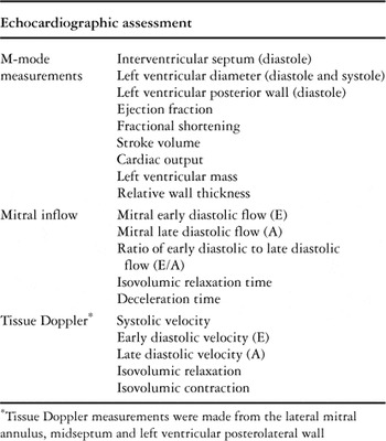
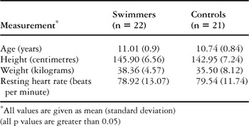
Table 1 summarizes all our echocardiographic measurements. The characteristics of the swimmers versus their controls are presented in Table 2. Mean age, mean height, and mean weight were similar in both groups, as well as the resting heart rates (p is more than 0.05). The mean total training time was 3.91 years, with a standard deviation of 1.10 years, and a range from 3 to 6 years. The mean weekly training time was 10.0 hours, with standard deviation of 1.70 hours, and a range from 8 to 12 hours.
Table 1. Measured parameters.

Table 2. The characteristics of the swimmers versus the controls.

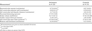
Among the echocardiographic measurements, the thickness of the ventricular septum, the left ventricular posterior wall, along with the left ventricular mass and the relative mural thickness, were all increased in swimmers (p is less than 0.05) (Table 3). The left ventricular dimensions, left ventricular mass, stroke volume, cardiac output, and early diastolic velocity of the septum were all positively correlated to height and weight (p is less than 0.05). After correcting these measurements for body surface area, except for the relative wall thickness, they were still increased in the swimmers (p for ventricular septum is 0.049, p for posterior wall is 0.015, p for left ventricular mass is less than 0.001).
Table 3. M-mode echocardiographic measurements in swimmers and controls.

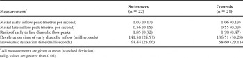
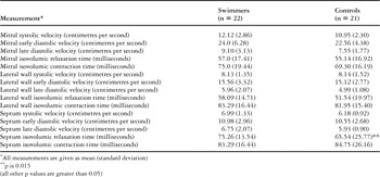
The velocities and durations of flow across the mitral valve were similar in both groups (Table 4). Except for the isovolumic relaxation time and corrected isovolumic relaxation time of the septum, all the tissue Doppler measurements were also similar. Although statistically insignificant, there was a trend toward increased myocardial velocities among swimmers (Table 5). The M-mode or the Doppler echocardiographic parameters were not significantly correlated to the total or weekly training times (p is more than 0.05).
Table 4. Transmitral Doppler echocardiographic measurements in swimmers and controls.

Table 5. Left ventricular tissue Doppler echocardiographic measurements in swimmers and controls.

Discussion
Our study reveals that prepubertal boys, who are trained regularly for swimming, have significantly thicker ventricular septums and left ventricular posterior walls compared to their non-trained peers. These measured values, nonetheless, are within the normal limits determined for healthy children.
Individual and team sports have been classified as having low, moderate and high dynamic and static components for competition. According to this classification, swimming is a highly dynamic and moderately static sport.5, 10 The echocardiographic description of the changes seen in so-called athlete's heart was primarily shown in skeletally mature male athletes. Less change was noted in younger athletes and female athletes, suggesting that the cardiac adaptation of prepubertal athletes to exercise may be different than adult athletes.4, 11–13
Volume load caused by dynamic exercise leads to enlargement of the internal diameter of the left ventricle and a proportional increase in mural thickness, an arrangement known as eccentric hypertrophy.1, 2 Fractional shortening, however, considered the index of systolic function, is usually not different between athletes and non-athletes. Evidence is accumulating that left ventricular diastolic function is enhanced in the exercising endurance-trained athletes compared to their controls, favouring adequate filling of the ventricle when the diastolic period gets shorter at higher heart rate.1
Doppler tissue imaging, a relatively new tool that provides quantitative assessment of systolic and diastolic mural motion,3 appears suitable for documentation of the influence of sporting activity on left ventricular myocardial function in different left ventricular segments. Tissue Doppler measurements in endurance-trained adult athletes have revealed that mitral annular early diastolic velocity (E) is increased, and/or late diastolic velocity (A) is decreased, with a resultant increase in their ratio.3, 14–16
The results of adult studies certainly cannot be extrapolated to young prepubertal athletes. Although Mitchell et al.,10 and Rowland et al.,17 determined similar responses to exercise challenges in young athletes, Sharma et al.4 observed that adolescent and adult athletes clearly differed with respect to range of measurements left ventricular mural thickness.
There is little data concerning prepubertal athletes, and the results are conflicting. In some of the previous studies, no difference was found between the left ventricular dimensions of sportsmen and those of controls.18, 19 The study groups, however, were not homogenous in terms of the type of the training exercises. In other studies, greater left ventricular diastolic diameter was observed in prepubertal swimmers, while the thickness of the ventricular septum and left ventricular posterior wall were similar.12, 20, 21 We are aware of one study where swimmers of school age had augmented thicknesses of the ventricular septum and left ventricular posterior wall, as well as the left ventricular diastolic diameter.22 These latter findings are similar to those observed in adult athletes, but children of various ages and pubertal stages are included in that study. Our own findings, in contrast, conflict with these previously published data. We observed an increased thickness of the ventricular septum and left ventricular posterior wall, greater left ventricular mass, and increased relative mural thickness in swimmers, but the left ventricular diastolic dimensions were similar to controls.
Pelliccia et al.13 have observed that one of the major determinants of the left ventricular cavity dimension among different athletes was greater body surface area. Due to regular conditioning, the body composition of the athletes may be modified, and height, weight and body surface areas of the athletes may frequently be more than the age-matched controls, suggesting that the left ventricular diastolic diameter of athletes may be greater than those of non-athletes purely because of unmatched body proportions. Correction of these dimensions for body surface area, therefore, is more favorable in terms of comparison. In our study, the height, weight and the body surface areas were similar in swimmers and controls. Furthermore, the dimensions of the left ventricular wall remained augmented in swimmers, even after correcting for body surface area.
The increase in mural thickness, rather than the left ventricular internal diameter, was more pronounced than expected in the swimmers examined in our study. This finding may partly be explained by the fact that swimming and related training regimens are not exclusively dynamic. Therefore the load on the heart is not purely of the volume type.1 Another possible explanation is that the left ventricular adaptive response in prepubertal athletes may be different than that of adolescents or adults. In an effort to investigate the age at which specific traits of the athlete's heart first appear, Manolas et al.23 have studied athletes between the ages of 9 and 20 years. In their study, the left ventricular mass was larger in athletes of 11 or 12 years age, and was maintained with advancing age. Most of this increase was attributed to the increase in wall thickness that became manifest first in the athletes aged 13 and 14 years. A significantly larger left ventricular cavity was only found in the age group of 15 to 16 years.23 We hypothesize that our swimmers were younger than the group in which significant left ventricular diastolic enlargement was expected, since they were between 9 and 12 years of age.
Among the tissue Doppler parameters, we observed prolonged isovolumic relaxation of the middle part of the septum in swimmers. We believe this is mostly due to changes in preload of the heart, and the different septal behaviour as a result of additive wall stress derived from the right ventricle.3 The prolonged isovolumic relaxation time may be a reflection of reduced preload, which seems to disagree with the general knowledge that exercise increases preload in athletes. This may be true for adult athletes, who have increased left ventricular stroke volume both at rest and during exercise compared to non-athletes.24 The cardiovascular system of children responds to exercise differently than does that of an adult. Children have a lower cardiac output at a given rate of work or uptake of oxygen. This is attributed to a lower stroke volume, which is compensated for by a higher heart rate and increased extraction of oxygen by the muscles.24–26 The lower stroke volume compared to adults results in an attenuated venous return, or preload, thereby decreasing the pressure in the atriums, which in turn delays the opening of the mitral valve and lengthens the time available for isovolumic relaxation. Whether a similar physiological change is true when a child athlete is compared to a non-athlete peer, as suggested in this study, is a subject of further research.
We failed to observe any correlations between the measured echocardiographic parameters and the total or weekly training times. This is probably because the small number of swimmers who were similarly trained comprised a relatively homogeneous group in our study. When larger groups of athletes with different load and frequencies of training are studied, significant correlations might be observed. The echocardiographic parameters derived from a large enough sample may provide reference values in prepubertal male swimmers. Furthermore, the trend toward increased myocardial velocities observed among the swimmers in our study may translate into significant changes in a larger group undergoing a longer period of training.
As a conclusion, our finding of more concentric rather than eccentric hypertrophy in prepubertal swimmers is contrary to the previous observations on adult swimmers. Whether such structural changes reflect the unique cardiac adaptation of the heart to exercise during childhood will only be disclosed by longitudinal studies of prepubertal athletes.