I. INTRODUCTION
Zirconium carbide (ZrCx) is one of the refractory transition metal carbides with the rocksalt structure. Similar to the other transition metal carbides such as TiCx and VCx, ZrCx is a highly nonstoichiometric compound with a wide homogeneity composition region and high concentration of vacancies on the octahedral sites of the metalloid sublattice.Reference Pierson1 The presence of carbon vacancies has a significant influence on properties, such as hardness,Reference Lipatnikov, Rempel and Gusev2–Reference Valeeva, Davydov, Rempel and Rempel6 yield stressReference Miracle and Lipsitt7, Reference Tsurekawa, Kurishita and Yoshinaga8 electrokineticReference Obata and Nakazawa9 and magnetic susceptibilityReference Gusev and Rempel10 of transition metal carbides. For the nonstoichiometric carbides, the ordered carbon vacancies are considered to be the thermodynamic equilibrium at room temperature.Reference Gusev11 However, using the conventional sintering routes such as hot-pressing and hot isostatic pressing (HIP), the synthesis of nonstoichiometric carbides usually leads to the disordered carbon vacancies due to the high melting point of carbides and the low self-diffusion of carbon atoms.Reference Bulychev, Andrievskii and Nezhevenko12, Reference Gusev and Rempel13 The disordered carbon vacancies are considered to be metastable at room temperature. In order to achieve the ordered carbon vacancies, long-duration post annealing or other special kind of thermal treatment becomes necessary. For the ordered carbon vacancies in ZrCx, three stable phases of Zr2C, Zr3C2, and Zr6C5 have been predicted, depending on the carbon or vacancy concentrations.Reference Gusev and Rempel13 By the measurements of neutron and electron diffractions, the existence of Zr2C with the space group of Fd3m has been claimed in ZrC0.51 after post-annealing time duration.Reference Goretzki14–Reference Moisy-Maurice, Lorenzelli, De Novion and Convert16 However, the long-time annealing or thermal treatment induces fast growth in grain size, which is detrimental to the mechanical properties.
The spark plasma sintering (SPS) is one of the novel sintering techniques developed in recent years. In comparison to those conventional sintering routes such as hot pressing (HP) and HIP, the SPS has characteristics of the rapid heating by the pulsed DC current and the simultaneous application of pressure, leading to dense sintering of the compacted powders at relatively low temperatures and with short dwell times.Reference Fu, Gu, Shearwood, Luo, Flewitt and Milne17–Reference Zhao, Jia, Duan, Yang and Zhou24 Hence, the SPS at low temperature is possible to limit the grain growth during the densification process.Reference Sreenivasulu, Gopalan, Chandrasekaran, Markandeyulu, Suresh and Murty25–Reference Kumar, Prakash, Cheang and Khor29 In the present work, the nonstoichiometric zirconium carbide of ZrC0.61 was synthesized by the SPS sintering of the mechanochemically synthesized ZrCx nanopowders. Investigations were undertaken on the ordered carbon vacancies in the sintered ZrC0.61.
II. EXPERIMENTAL PROCEDURE
The starting nanopowders of ZrCx were prepared by ball milling of Zr powders (−325mesh, purity 99.5%) in toluene using a planetary ball miller (P4, Fritsch, Idar-Oberstein, Germany).Reference Xiang, Liu, Hu, Zhang, Chen, Wang, He, Yu, Xu, Lu, Tian and Liu30 The collected products after a 6-h milling were dried in a vacuum chamber (∼10-3Pa) at 80 °C for 20 h to remove any residue of toluene. The dried nanopowders were loaded in a graphite die (20 mm in diameter) and sintered in a DR. SINTER type SPS 3.20 MK-IV (Sumitomo Coal Mining Co. Limited, Tokyo, Japan). The sintering process started by the first application of a 50 MPa compressive pressure and then the rapid heating of 50 °C/min to 700 °C. After soaking at 700 °C for 30 min, the heating of 50 °C/min was continued to 1600 °C. After keeping at 1600 °C for 5 min, the applied current was reduced and the pressure was released with the specimen cooling down in the chamber. After being polished to remove any surface contamination, the finally obtained sample is 10 mm in thickness and 20 mm in diameter.
The carbon content x of the sintered ZrCx was determined to be 0.61 via the complete oxidation to ZrO2 in air at 1000 °C for 24 h in a home-made tubular furnace (Pansen Technology Co. Ltd., Xian, China). Morphologies of the fracture surfaces and the etched surfaces after mechanically polishing were checked using a field-emission scan electron microscopy (SEM; Model S4800, HITACHI, Tokyo, Japan). The etching was performed in a twin jet electro-polisher (Tenupol-5, Struers, Ballerup, Denmark) using solution of HClO4 and CHOOCH3 in a ratio of 2:1. The crystal structures of as-milled nanopowders and SPS-sintered ZrC0.61 (ground into powders) were characterized by x-ray diffraction (XRD; Rigaku D/MAX-2500PC, Tokyo, Japan). Cu Kα radiation (λ = 1.5418 Å) at 200 mA and 40 kV) and high-resolution transmission electron microscopy (HRTEM; JEM-2010, JEOL, Tokyo, Japan, operating at 200 kV).
III. RESULTS AND DISCUSSION
Figure 1(a) shows the XRD patterns of the as-supplied Zr powders and the as-milled ZrCx nanopowders after of Zr powders with toluene. The average size of as-milled nanopowders is determined to be about 7 nm. Small amounts of Zr are still detectable in XRD pattern. Figure 1(b) presents the XRD pattern for the SPS-sintered ZrC0.61. In addition to the strong reflection peaks corresponding to the rocksalt-structured ZrC (JCPDS #73-0477), some extra weak peaks (marked with green triangles) are clearly observed in the XRD pattern. These extra weak peaks are identified to originate from the cubic superstructure of ordered carbon vacancies, which will be discussed in detail later. The average grain size is determined to be 480 nm from the Rietveld refinement of powder XRD spectrum by using the Material Analysis Using Diffraction (MAUD) software.
(a) The XRD spectra for the as-received Zr powders and the 6 h ball-milled ZrCx nanopowders. The vertical bars on the bottom represent the reflection positions of the hexagonally close packed (hcp)_ Zr (black) and the fcc ZrC (red). (b) The XRD spectrum (+) together with the Rietveld refinement result (Solid line). The vertical bars on the bottom show the positions of Bragg reflections from the rocksalt-structured ZrC0.61 with the superstructure of the ordered carbon vacancies, and solid curve above the bars represent the error of the Rietveld refinement.

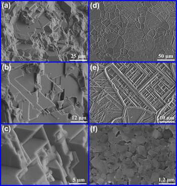
Figure 2 shows the SEM images of the fracture surface and the etched surfaces for the SPS-sintered ZrC0.61. The SEM images of the fracture surface [Figs. 2(a)–2(c)] demonstrate the sintering densification and an interesting laminated microstructure. The SEM images of the etched surfaces in Figs. 2(c)–2(e) reveal more interesting microstructural features. On the image of Figs. 2(d) and 2(e), big domains separated by thick walls are observed, and the domains are found to be constructed by the interlaced laminar sheets. By properly etching the fracture surface, the SEM image in Fig. 2(f) reveals that the laminar sheets are actually constructed by many ultrafine grains of ∼500 nm in average size. This observed grain size is consistent with the estimated one from the XRD pattern.
(a), (b), (c) The SEM images of fracture surface of the sintered ZrC0.61. (d), (e), (f) The SEM images of the sintered ZrC0.61 after polishing and etching.

The interesting microstructure of the sintered ZrC0.61 should be related to the novel SPS technique and the agglomeration of as-milled ZrCx nanopowders. Generally, the nanopowders have strong tendency to agglomerate. The nanopowders inside the agglomerates are in close contact with each other, but big gaps might exist between the agglomerates. Thus, discharge may occur mainly between the agglomerates during the SPS process, leading to high temperature locally around the agglomerates. The local high temperature induced by discharge could be so high that the agglomerates are melted outside, forming a liquid shell to enclose the nanopowders inside. Owing to the simultaneous application of uniaxial pressure during the sintering, the agglomerates covered with a liquid shell are easy to be pressed into the laminated sheets, while the nanopowders inside the liquid shell grow into the ultrafine grains. The slow growth rate should benefit strongly from the applied uniaxial pressure, the rapid heating, and the very short soaking time. This special sintering process has been investigated in our previous studies on the SPS-sintered TiCx from the mechanochemically synthesized TiCx nanopowders.Reference Xiang, Hu, Liu, Chen, Zhang, Wang, Wang, Wen, Xu, Yu, He, Tian and Liu31 In the sintered TiCx by SPS, big domains have been observed to be composed of nanopowders covered with a shell, and the shell has been considered to be formed by the melted outside surface of the agglomerate of nanopowders.
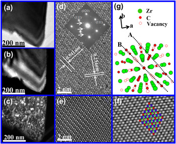
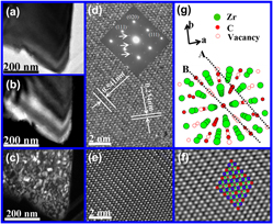
For the nonstoichiometric transition metal carbides with the rocksalt structure (for example, TiCx,Reference Tsurekawa, Kurishita and Yoshinaga8 ZrCx,Reference De Novion and Moisy-Maurice15 VCx,Reference Lipatnikov and Gusev32 etc.) sintered by the conventional sintering routes such as HP or HIP, the carbon vacancies are usually disordered. The realization of the ordering of carbon vacancies involves complicated post-annealing at high temperature for a long time up to several days.Reference De Novion and Moisy-Maurice15, Reference De Novion and Landesman33 However, we find that the ordered carbon vacancies are achieved directly in the SPS-sintered ZrC0.61, and no post-annealing treatment for long time is necessary after the sintering. The cubic superstructure formed by the ordered carbon vacancies in the sintered ZrC0.61 is confirmed by the measurements of XRD, HRTEM images, and selected area electron diffraction (SAEDs). As shown in the XRD pattern of the sintered ZrC0.61 [Fig. 1(b)], extra weak peaks are observed to appear in addition to the strong reflection peaks of the rocksalt-structured ZrC. The positions of these extra weak peaks are consistent with those predicted ones by Gusev et al.,Reference Gusev and Rempel13 that were calculated by taking into consideration of the ordered carbon vacancies in the rocksalt-structured ZrCx. On the basis of the consideration of Gusev et al.,Reference Gusev and Rempel13 a crystal structure model for the ordered cubic phase of Zr2C is reconstructed and shown in Fig. 3(g). The atoms of C and Zr are located at positions of 16d (5/8 5/8 5/8) and 32e (3/8 3/8 3/8), respectively, while the carbon vacancies are considered to be at position of 16c (1/8 1/8 1/8). On the (111)C planes of C sublattice, the atomic positions are occupied in two different ways. One is that 2/3 positions are occupied by C atoms and the rest are the vacancies [named as A in Fig. 3(f)], and the other is that only 1/3 positions are occupied by C atoms with the rest being the vacancies [named as B in Fig. 3(f)]. On the (111)C plane of type A, some of the C atoms are located consecutively along the direction of [ ], forming the C columns, and the rest are located alternatively with the vacancies along the direction of [
 ], forming the atoms-vacancies (C-V) strings. On the (111)C plane of type B, the arrangement of C atoms and vacancies is in a different way. Some of the vacancies lie consecutively along the direction of [
 ], forming the vacancy channels, while the rest vacancies and the C atoms form the C-V strings by lying alternatively along the direction of [
 ] axes. Along the [111] direction, the (111)C planes of type A and B are stacked alternatively in sequence of ABAB…AB or BABA…BA. This ordering arrangement of the carbon vacancies gives a superstructure with the space group of Fd3m.. Based on the reconstructed structure model, the Rietveld refinement is performed, giving a good agreement with the experimental XRD spectrum as shown in Fig. 1(b), and the cell parameter of Zr2C-type cubic phase is determined to be 0.9323 nm. Thus, the observed extra weak peaks result from the cubic superstructure of Zr2C, which is formed by ordered carbon vacancies in the parent rocksalt structure of ZrC0.61. Moreover, the refinement results indicate that the C atoms and vacancies are located at the ideal position of 16d and 16c, respectively, but the Zr atoms have a small displacement of 0.0034 nm away from the vacancies.
(a) The BF TEM image of a broken piece from the sintered ZrC0.61. (b) The DF TEM image of the broken piece taken via one of the relatively intense reflections in SAED pattern [Inset in (d)]. (c) The DF TEM image of the broken piece taken via one of the superstructure reflections in SAED pattern [Inset in (d)], showing the particle-like nanodomains in contrast. (d) The HRTEM image and the SAED pattern (Inset) taken along the direction of  zone axis. In the SAED, in addition to the fundamental reflections, the superstructure reflections (marked with white arrows) appear due to the ordering carbon vacancies. In the HRTEM image, the C-V strings and the vacancy channels along the
 direction are demonstrated with different contrasts than the Zr atoms columns and C atoms columns, giving rise to the visible superstructure modulation features. The superstructure modulation features can be observed more clearly in the HRTEM image obtained after Fourier filtering process (e) and the enlarged HRTEM image after the deconvolution process (f). Inset in (f) is a schematic showing the superstructure modulation features, in which red circle, blue circle with a red point in the center, red point, and green point represent the vacancy channel, the C-V string, the C atoms column, and the Zr atoms column, respectively. (g) The constructed structure model by Gusev et al.,Reference Gusev and Rempel13 showing the ordering arrangement of the carbon vacancies viewed along the direction of
 zone axis.

For further confirmation, the TEM investigations were also carried out on the sintered ZrC0.61 with ordered carbon vacancies. The superstructure modulation, which induced by the ordering of the carbon vacancies, can be confirmed by the appearance of additional spots in addition to the fundamental reflections in the ED patterns. Because of the specific arrangement of the vacancies along the  zone axis, the carbon atoms columns, the C-V strings, and the vacancy channels are demonstrated in the HRTEM image with different contrasts,Reference Valeeva, Tang, Gusev and Rempel34 and they can be observed only along some special zone axes. Figure 3(a) shows the bright-field (BF) image of a broken piece of the sintered ZrC0.61. The SAED pattern [inset in Fig. 3(d)], which was taken along the zone axis of
 , reveals the good crystallinity of the sintered ZrC0.61, and demonstrates the presence of clear superstructure modulation. In addition to the relatively intense reflections of the rocksalt-structured ZrC, the weak superstructure reflections (marked with white arrows) are observed. All of these reflections can be indexed on the cubic superstructure unit cell with lattice parameter of a = b = c = 0.9323 nm. By using one of relatively intense reflections, the dark field image is obtained, as shown in Fig. 3(b). Besides the broad bright-dark fringes induced by the strain and thickness, the DF image in contrast dose not show any visible grain boundaries. Figure 3(c) shows a DF TEM image taken using one of superstructure reflections. Nanodomains induced by the ordered carbon vacancies are observed to be separated by thin walls of dark contrast. Their average size is determined to be ∼30 nm. Figure 3(d) shows the HRTEM image taken with a defocus value of about −7 nm, demonstrating the superstructure modulation along the directions of {111}. The superstructure modulation becomes more obvious in the HRTEM images after the Fourier filtering process [Fig. 3(e)] and the deconvolution process [Fig. 3(f)]. According to the Rietveld refinement results, though the distances of C–C and C-V are the same, the distances of Zr-Zr around the C atom and the vacancy are different owing to a small displacement (0.0034 nm) of the Zr atoms away from the vacancies. This feature is revealed in the arrangement of those bright points in the HRTEM image [Fig. 3(f)]. Thus, those bright points in the HRTEM images can be identified to be the atomic positions of Zr atoms, and then the positions of carbon atoms and vacancies can be recognized according to the different distance of Zr-Zr. For clarity, they are shown schematically in Inset of Fig. 3(f). The distance between the superstructural ordering planes of 1/2(1 1 1) is determined to be 0.541 nm, being precisely two times of the distance between the parent face centered cubic (fcc) (1 1 1) planes. The structure model viewed along the direction of the
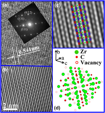
 zone axis is shown in Fig. 3(g). It illustrates the superstructure modulation along the directions of {111} induced by the ordered carbon vacancies. Figure 4(a) and Inset show another HRTEM image and SAED pattern taken along the different zone axis of
 . The superstructure modulation along the direction of
 is also clearly observed. Figures 4(b)–4(d) display the HRTEM images after the Fourier filtering and the deconvolution process, and the structure model viewed along the direction of
 zone axis, respectively. The locations of the Zr columns, C-V strings, and vacancies channels are also marked in Fig. 4(c). Figure 5(a) and Inset show the HRTEM image and SAED pattern taken along the direction of
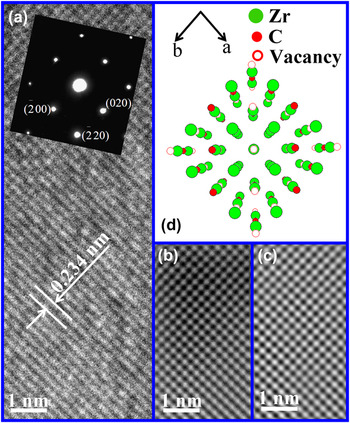
 zone axis. Figures 5(b)–5(d) show the HRTEM images obtained after the Fourier filtering and deconvolution process, and the structure model viewed along the direction of
 zone axis, respectively. The superstructure reflections are absent in the SAED, and no superstructure features can be observed in the HRTEM image. With the help of TEM investigations along the three different directions of
 ,
 , and
 zone axes, further confident evidence is provided for cubic Z2C-type superstructure in our sintered ZrC0.61. The space group of the superstructure is determined to be Fd3m , which is consistent with the reconstructed structure model and the results of the Rietveld refinement.
(a) The HRTEM image and the SAED pattern (Inset) taken along the direction of  zone axis. In the SAED, in addition to the fundamental reflections, superstructure reflections appear due to the ordering carbon vacancies. In the HRTEM image, the C-V strings and the vacancy channels are demonstrated with different contrasts than the Zr atoms column and C atoms column, giving rise to the visible superstructure modulation features. The superstructure modulation features can be observed more clearly in the HRTEM image obtained after Fourier filtering process (b) and the enlarged HRTEM image after the deconvolution process (c). Inset in (c) is a schematic showing the superstructure modulation features, in which red circle, blue circle with a red point in the center, red point, and green point represent the vacancy channel, the C-V string, the C atoms column, and the Zr atom column, respectively. (d) The same structure model as the one in Fig. 3(g) but viewed along a different direction of
 zone axis.

(a) The HRTEM image and the SAED pattern (Inset) taken from the direction of [001] zone-axis. In the SAED, only the fundamental reflections corresponding to ZrC are observed. Correspondingly, no visible superstructure modulation feature is shown not only in the HRTEM image but also in the HRTEM image after Fourier filtering process (b) and the enlarged HRTEM image after the deconvolution process (c). (d) The same structure model as the one in Fig. 3(g) but viewed along the direction of [001] zone axis.

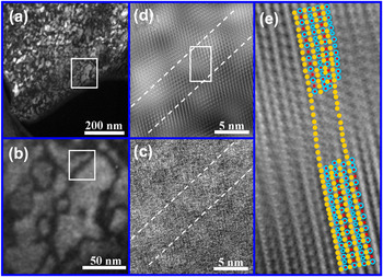
The detailed TEM investigations focused on the thin domain walls were also carried out to reveal the structure features. Figure 6(a) shows the DF image obtained by using one of superstructure reflections shown in the SAED [inset in Fig. 4(a)]. The thin walls separating the superstructural nanodomains display the dark contrast in image, as shown in the Fig. 6(b), which indicates that the walls have no contributions to superstructure reflections. It suggests that the carbon vacancies inside the walls are not able to be in the long-range ordering state. Figures 6(c) and 6(d) show the HRTEM and Fourier filtering HRTEM images obtained from the area marked with white rectangle. The enlarged image of area marked with white rectangle in Fig. 6(d) is shown in Fig. 6(e). Obviously, the lattices inside the walls are distorted slightly, and the distortions is considered to be induced from the stacking sequence alteration of (111)C planes from ABAB…AB in one superstructural domain to BABA…BA in the neighboring one or vice versa. Considering the arrangement of the carbon atoms and vacancies, the thin walls can also be regarded as the antiphase domain boundaries.Reference Nagakura and Kusunoki35 The distortions of (111)Zr planes are resulting from the displacement of the nearest neighbor Zr atoms of the carbon vacancies.Reference Moisy-Maurice and De Novion36 The nanodomains and the walls were also observed in carbon vacancies ordered sintered TiC0.70 by SPS.Reference Xiang, Hu, Liu, Chen, Zhang, Wang, Wang, Wen, Xu, Yu, He, Tian and Liu31
(a) The DF TEM image of the broken piece of ZrC0.61 (The same piece as the one in Fig. 3(a), but it is obtained via one of the superstructure reflections in the SAED pattern taken along the direction of  zone axis. The nanodomains are clearly displayed. (b) The enlarged DF TEM image. (c) The HRTEM image along the direction of
 zone axis, showing the atomic structure inside the thin walls separating the nanodomains; (d) and (e) are the HRTEM image after Fourier filtering process and the enlarged HRTEM image after the deconvolution process, respectively. Inset in (e) is a schematic showing the superstructure modulation features, in which cyan circle, cyan circle with a red point in the center, red point, and yellow point represent the vacancy channel, the C-V string, the C atoms column, and the Zr atoms column, respectively.

The mechanism for the simultaneous formation of the ordered carbon vacancies in the sintered ZrC0.61 during the sintering is not completely clear to us by now. We believe that it is related to the novel SPS process. Considering the tendency of nanopowders to agglomerate and the discharge in the gaps between the agglomerates during the SPS process, high temperature is induced locally around the agglomerates such that the outside surface is melted to form a liquid shell. During the rapid heating process, the temperature inside the liquid shell might be lower than that outside. The similar phenomenon was observed in carbon vacancies ordered sintered TiC0.70 by SPS.Reference Xiang, Hu, Liu, Chen, Zhang, Wang, Wang, Wen, Xu, Yu, He, Tian and Liu31 Since the ordered carbon vacancies are predicted to be stable at low temperature,Reference Gusev11 the low temperature inside the liquid shell could be favorable for the carbon vacancies to be arranged in an orderly manner during the limited grain growth.
IV. SUMMARY
In summary, by SPS of the mechanochemically synthesized ZrCx nanopowders, the prepared nonstoichiometric zirconium carbide, ZrC0.61, exhibits interesting microstructural features. It has been observed that the sintered ZrC0.61 by SPS is composed of big domains separated by thick walls. The big domains are constructed by the interlaced laminar sheets, and each laminar sheet is formed by many ultrafine grains with ∼500 nm in average size. In contrast to long-time post annealing for realization of the ordered carbon vacancies in the nonstoichiometric transition metal carbides with the rocksalt structure, which are sintered by the conventional sintering routes such as HP and HIP, the ordered carbon vacancies in the sintered ZrC0.61 are achieved simultaneously during the sintering process, forming a Zr2C type ordering phase with cubic superstructure. Moreover, the ordering of carbon vacancies is found to form the nanodomains separated by the antiphase domain boundaries. The average superstructure domain size is determined to be ∼30 nm, being indicative of the short ordering length of the carbon vacancies.
ACKNOWLEDGMENTS
Financial supports from the National Basic Research Program of China (Grant No. 2010CB731605, No. 50872118, and No. 58021001), National Science Fund for Distinguished Young Scholars (Grant No. 51025103) are gratefully acknowledged.