




No CrossRef data available.
Article contents
Mechanisms of mitral valvar insufficiency in children and adolescents with severe rheumatic heart disease: an echocardiographic study with clinical and epidemiological correlations
Published online by Cambridge University Press: 21 January 2005
Abstract
We carried out a detailed clinical, epidemiological, and echocardiographic study in 41 patients ≤14 years of age who were admitted in a public hospital in Salvador, Brazil, with severe rheumatic heart disease.
Mitral insufficiency was severe in 90%, and moderate in 10%, of the patients. A posteriorly directed jet was seen in 93% of the patients. We identified three mechanisms producing the regurgitation: prolapse of the aortic leaflet of the mitral valve in 13 (32%) patients, rupture of tendinous cords in 14 (34%), and a retracted, non-coapting mural leaflet in 14 (34%). The mean ages, with standard deviations, for these three groups were 7.0 (1.6) years, 7.9 (2.2) years, and 10.5 (2.4) years, respectively (p < 0.001). Rheumatic activity was diagnosed in 58.5% of them. Evidence of previous rheumatic fever was present in 54% of patients with prolapse, in all patients with rupture, and in 93% of those with non-coapting leaflets (p = 0.002).
Prolapse of the aortic leaflet, rupture of tendinous cords, and a retracted, non-coapting mural leaflet are the mechanisms responsible for mitral valvar insufficiency in children and adolescents with severe rheumatic heart disease. Prolapse seems to be an early phenomenon in the natural history of rheumatic heart disease, while rupture and non-coaption of the leaflets were associated with older age and signs of chronic rheumatic disease.
Information
- Type
- Original Article
- Information
- Copyright
- © 2004 Cambridge University Press
Rheumatic fever, and rheumatic heart disease, still represent major problem for public health in developing countries, where rheumatic disease remains the leading cause of cardiac illness and premature death among children and young adults.1, 2 In the developing world, severe rheumatic heart disease is often observed at a very low age, even in children <10 years old. In these young children, mitral insufficiency is the single most frequent lesion,3 leading to dilation of the left atrium and cardiac failure. Diverse mechanisms have been described as causing mitral insufficiency in such patients, such as fusion and elongation of the tendinous cords, non-coaption of the leaflets, and dilation of the valvar annulus. Prolapse of the leaflets, and rupture of the tendinous cords, have also been recognized as important mechanisms of mitral insufficiency in patients with acute rheumatic carditis and chronic rheumatic heart disease.3–8 Previous echocardiographic observations from a small study of Brazilian children called attention to the role of prolapse and rupture of the tendinous cords underscoring a very severe variant of rheumatic heart disease.9 The objective of our study was to investigate in detail the functional anatomy and the mechanisms of the mitral insufficiency in children and adolescents with severe rheumatic heart disease, and to assess its epidemiological and clinical correlations.
Materials and methods
Our population consisted of 41 children, aged 14 years or less, who were admitted to a public hospital in Salvador-Bahia, Northeast Brazil (Obras Sociais Irma Dulce) from November 1997 to June 2002 with symptomatic and severe rheumatic heart disease. This sample represents 2.5 per 1000 patients of the same ages admitted in the same period. The criterions for inclusion in the study were presence of a murmur, evidence of haemodynamic overload to the heart, as shown by increased size on the postero-anterior chest radiography, chamber dilation on the cross-sectional echocardiogram or signs of congestive heart failure, echocardiographic findings suggestive of rheumatic heart disease, and absence of other diseases that could explain the valvar involvement. We excluded 3 other patients because they did not have evidence of haemodynamic overload to the heart. The aetiologic diagnosis of rheumatic disease was made on the basis of a history of acute rheumatic fever in the past or during the actual admission, or by the presence of a multivalvar disease not explained by other pathology in this age group, with echocardiographic findings suggestive of rheumatic disease, such as non-myxomatous thickening of the leaflets, predominantly at the edges, and diminished mobility of the mural leaflet of the mitral valve. The protocol of the study was reviewed and approved by the Ethics and Research Committee of the Hospital Portugues (Salvador-Bahia). A systematic clinical examination and epidemiological interview were done by the assistant physician and reviewed by one of the investigators. The diagnosis of rheumatic activity was based on the revised Jones’ criterions.10 Titres of anti-streptolysin O, acute phase reactants such as C-reactive protein, erythrocyte sedimentation rate, and serum mucoproteins in tyrosine were measured at the initial laboratory investigation and usually repeated during the hospital stay.
Transthoracic cross-sectional Doppler echocardiograms with colour-flow mapping, using a Sonos 2500 Hewlett-Packard equipment, were performed in all patients and recorded on super VHS videotape for retrospective analysis. Measurements of the cavities and mural thickness were made during the examination, and repeated retrospectively from the videotapes images, according to the recommendations of the American Society of Echocardiography.11, 12 Mitral and tricuspid annular diameters were measured at end-diastole as the distance between annular ridges in the apical four-chamber view. We always tried to measure the maximal velocity of tricuspid insufficiency to estimate pulmonary arterial systolic pressure, adding 10 mmHg to the gradient from the right ventricle to the right atrium calculated by applying the modified Bernoulli equation. From the spectral display of mitral insufficiency, using continuous Doppler, we obtained the maximum rate of rise of the left ventricular pressure using the formula: derivative of pressure over derivative of time where derivative of pressure is equal to 32 mmHg, the pressure difference between the velocities of 1 and 3 m/s, and the derivative of time is equal to the time in milliseconds required for the velocity to increase from 1 to 3 m/s. All of the measurements were averaged over 3 or more cardiac cycles. Mitral valvar prolapse was defined as a systolic displacement of the level of coaptation of the leaflets of at least 2 mm above the mitral annular plane of one or both leaflets as assessed in the longitudinal parasternal and apical four-chamber views. The diagnosis of rupture of tendinous cords was based on the loss of the point of coaptation, with intense prolapse and inversion of the tip of the leaflet, pointing toward the left atrium in two echocardiographic views, and whenever possible also on the visualization of the ruptured cords. Using the same criterions, so as to assess the interobserver and intraobserver variability, the data were independently analysed by two of the investigators and repeated months later by one of them. In either case, the investigators were blinded to the other results. Any identified discordances were reviewed and resolved by consensus between the echocardiographers. Mitral, aortic, and tricuspid insufficiency was graded as none, mild, moderate or severe by standard criterions using colour Doppler echocardiography. The regurgitant jet was specified as pointing posterior, central, anterior or multidirectional. Chronic rheumatic heart disease was identified by a thickened and immobile mural leaflet, or one with significantly reduced mobility, along with thickened cords, and the characteristic dome-shaped opening of the aortic leaflet. These features are not seen during a first episode of acute rheumatic fever, and they are not seen in patients with mitral insufficiency due to other aetiologies in this age group. The valvar thickening was graded as mild when the leaflets were only slightly thickened at the edges and the mid and basal portions had normal thickness, or moderate to severe when whole leaflet was significantly thickened with increased brightness.
Quantitative variables are reported as mean values with one standard deviation, giving also the range and the median. Comparisons between the three groups were done with the Kruskal-Wallis test. Multiple post hoc pairwise comparisons between groups were then made using Bonferroni test. Differences in proportions between two groups were compared by Fisher’s exact test. To compare the three groups, we used a chi-square test for association, along with an r-by-c table, and statistical significance was established by the Likelihood Ratio. Pearson’s correlation was performed to measure the linear association between mitral annular diameter and end-diastolic dimension of the left ventricle. A two-tailed p value <0.05 was used to denote statistical significance. Interobserver and intraobserver variability were analysed by using kappa statistics.
Results
Demographic and clinical characteristics (Table 1)
There were 22 males (54%) and 19 females (46%), with the mean age of 8.5 (2.5) years, the range being from 4 to 14 years, with the median at 9 years. Of the group, 62% were of black–white mixed race, 36% were black, and there was 1 (2%) white patient. Most of the patients (86%) had signs of congestive heart failure. Rheumatic activity was diagnosed in 58.5% of the patients. All children had mitral insufficiency. This was deemed to be severe in 37 (90%), and moderate in 4 (10%). A posteriorly directed jet of the mitral insufficiency (Fig. 1) was seen in 38 (93%) of the patients, this being exclusively posterior in 32 patients, and associated with some anterior streaming in 6. In 2 patients, the jets were directed anterior, while in the other patient it was central. Aortic valvar insufficiency was present in 27 (66%) of the patients, being mild in 15, moderate in 9, and severe in 3. Tricuspid insufficiency was present in 32 (78%) patients, mild in 17, moderate in 10, and severe in 5. The mean of the pulmonary arterial systolic pressure was 47 ± 13.3 mmHg, and pulmonary hypertension, defined as pulmonary arterial systolic pressure higher or equal to 35 mmHg, was present in 70% of the patients. Other echocardiographic measurements are depicted in Table 1. An ejection fraction inferior to 60% was observed in 17% of the patients.

Figure 1. A characteristic posterior jet of rheumatic mitral insufficiency.
Table 1. Comparisons between groups.

Mechanisms of mitral insufficiency
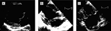
We identified three mechanisms for mitral insufficiency: prolapse of one or both leaflets without signs of cordal rupture; prolapse with signs of rupture of the tendinous cords; and a thickened, retracted, immobile, non-coapting mural leaflet (Fig. 2). Kappa values of 0.70 and 0.82 were obtained for interobserver and intraobserver variance, respectively.

Figure 2. Examples of the mechanisms of rheumatic mitral insufficiency. (a) A patient with prolapse of the aortic leaflet. Note that the leaflets are only mildly thickened. (b) A patient with rupture of tendinous cords and intense prolapse of the aortic leaflet pointing towards the left atrium in systole. (c) An example of a thickened, retracted, non-coapting mural leaflet.
Mitral valvar prolapse
A prolapsed but non-flail leaflet was present in 13 (32%) of the children. The mean age of this group was 7.0 (1.6) years, with a range from 5 to 10 years, and a median of 7 years. All of these patients had prolapse of the aortic leaflet, associated in only one patient with additional prolapse of the mural leaflet. Mitral insufficiency was graded as severe in 11 (85%), and moderate in 2 (15%) patients. The jet was directed posteriorly in 11 (85%) patients, and deemed multidirectional in 2 (15%). Thickening of the leaflets was mild in the majority of the patients of this group, and there was no sign suggestive of chronic rheumatic heart disease in 46% of them.
Rupture of the tendinous cords
Prolapse with a flail leaflet and signs of rupture of tendinous cords was present in 14 (34%) of the children. The mean age of this group was 7.9 (2.2) years, with a range from 4 to 12 years, and a median of 8.5 years. The prevalence of cordal rupture was higher in the group of patients aged 10 years or less (38%) than those aged more than 10 years (14%), with borderline statistical significance (p = 0.058). Rupture involved the cords supporting the aortic leaflet, with the jet directed posteriorly, in 12 (86%) of these patients. The other 2 patients (14%) had rupture of cords supporting the mural leaflet with the jet directed anteriorly. Mitral insufficiency was graded as severe in all of the patients. Thickening of the leaflet in this group was usually moderate or severe, with signs of chronic rheumatic heart disease in all of them.
Retracted and non-coapting mural leaflet
A thickened, retracted, immobile, and non-coapting mural leaflet was seen in 14 (34%) of the patients. Their mean age was 10.5 (2.35) years, with a range from 6 to 14 years, and a median of 10 years. Mitral insufficiency was graded as severe in 12 (86%), and moderate in 2 (14%) of the patients. The jet was directed posteriorly in 9 (64%), anteriorly and posteriorly in 4 (29%), and centrally in 1 (7%) of the patients in this group. Thickening of the leaflets was moderate or severe in all except one patient (93%), suggesting chronic rheumatic heart disease.
Comparisons between groups
Mean ages differed between groups (p < 0.001), being higher in those with a non-coapting mural leaflet and lower in those with prolapse (Fig. 3a). There was no statistically significant difference with regard to age between those having prolapse and ruptured cords, but the median ages of these patients were 7.0 and 8.5 years, respectively. A history of acute rheumatic fever in the past was less frequent in those with prolapse (23%) than in those with ruptured cords (64%), with borderline statistical significance – p = 0.054, odds ratio = 0.36, 95% confidence interval 0.12 to 1.0 – and than in the patients with a non-coapting mural leaflet (50%). The findings did not attain statistical significance for this last group – p = 0.24, odds ratio = 0.3, 95% confidence interval 0.06 to 1.6. Considering the presence of signs of chronic rheumatic heart disease on the echocardiogram, and/or a past history of acute rheumatic fever as criterions for previous rheumatic fever, this finding was less frequent in those with prolapse (54%) compared to those with rupture cords (100%) – p = 0.006, odds ratio = 0.54, 95% confidence interval 0.33 to 0.89, and also less frequent when compared to those with a non-coapting mural leaflet (93%) – p = 0.03, odds ratio = 0.58, 95% confidence interval 0.034 to 0.98 – Table 1 and Figure 3b. At the time of hospitalization, acute rheumatic activity was less frequent in those with ruptured cords (36%) compared to 69% for those with prolapse and 71% for those with a non-coapting mural leaflet, these findings not achieving statistical significance. Left ventricle end-diastolic and end-systolic dimensions, and left atrial dimensions, were greater in the patients with ruptured cords and non-coapting mural leaflets, albeit that children with non-coapting mural leaflets were older and heavier than those making up the other two groups. For this reason, we made the same analysis for children weighing 30 kg or less, and the difference persisted. There was no statistically significant difference between groups with regard to ejection fraction, maximal derivative of pressure over derivative of time, pulmonary arterial systolic pressure, or mitral and tricuspid annular diameters. Mitral annular diameter, however, demonstrated a significant correlation with the left ventricle end-diastolic dimension (r = 0.55, p = 0.002).

Figure 3. (a) Mean age with the 95% confidence interval of the three groups. (b) Proportions of previous rheumatic fever, defined as history of acute rheumatic fever in the past and/or signs of chronic rheumatic heart disease on echocardiogram, in the three groups. *p = 0.006 compared to the group with prolapse; **p = 0.03 compared to the group with prolapse, and p > 0.05 compared to the group with ruptured cords.
Discussion
The results of our study are consistent with findings for Brazilian children and adolescents having the most severe forms of rheumatic heart disease. This may be explained by multiple episodes of rheumatic fever, particularly in populations living at the lowest socioeconomic levels. Most of these small children had echocardiographic signs of chronic, irreversible, severe rheumatic heart disease. This disease is the most common form of acquired cardiac disease, cardiac failure, and premature death in children and young adults in developing countries.1–3 The most frequent valvar lesion in patients of this age is known to be mitral insufficiency, a finding also consistent with our results.
We identified three mechanisms for the mitral insufficiency: prolapse of the aortic leaflet, prolapse with signs of rupture of the tendinous cords, and a non-coapting retracted immobile mural leaflet. In our study, mitral annular dilation was not an independent mechanism for mitral insufficiency. There was no significant difference in mitral annular diameter between the groups. In addition, there was a statistically significant positive linear correlation between mitral annular and left ventricular diastolic diameters. We suppose that mitral annular dilation is a secondary mechanism of mitral insufficiency in rheumatic disease, associated with left ventricular dilation. The prevalences of prolapse of the aortic leaflet, rupture of the tendinous cords, and non-coaption of the retracted and immobile mural leaflet were 32%, 34%, and 34% respectively. This is the highest reported prevalence of rupture of the tendinous cords in patients with rheumatic heart disease. It has been described as existing in one-twentieth to one-quarter of cases analysed in clinical, pathological, and surgical series.3, 5, 7, 8 Mitral valvar prolapse has been reported in from one-tenth to five-sixths of cases of rheumatic mitral insufficiency.3, 6–8 Our rate of mitral valvar prolapse, seen in one-third of patients, is similar to that reported by Kalangos et al.8 It may be partially explained by the inclusion of a flail leaflet with cordal rupture, as seen in a distinct group in our study, and because we have been careful not to include cases of pseudoprolapse, where the mural leaflet is fixed and the mobile aortic leaflet can give the impression of prolapsing, but in reality remaining within the mitral annular plane.8 The reasons for the higher prevalence of cordal rupture in our study are probably related to the characteristics of our sample, and our method of investigation. Thus, first we studied only children and adolescents ≤14 years old, all of them with severe rheumatic heart disease. Second, we obtained good quality cross-sectional Doppler echocardiograms with colour-flow mapping in all of these children, and the mitral valve was analysed with detail by two of the authors. The prevalence of cordal rupture, at 38%, was higher in the group of patients aged 10 years or less than in those aging between 10 and 14 years, when the proportion was 14%. Children of the same social group, admitted to the same hospital with a similar clinical presentation, but who did not have mitral valvar rolapse or rupture of the tendinous cords, were significantly older. This fact probably means that prolapse and rupture of the tendinous cords may be responsible for the earlier symptoms and haemodynamic overload seen in this population.
Inflammation has been recognized as a cause of mitral valvar prolapse, sometimes described as postinflammatory valvar prolapse, due to deformation of the valves and elongation of the tendinous cords.5 Cordal rupture could be a consequence of the increasing tension exerted on elongated cords over time, or as a complication of active rheumatic carditis, which weakens the cords by inflammation.8 It is important to note that all of the patients with cordal rupture had evidence of a previous episode of rheumatic fever, whereas this occurred in only half of the patients with prolapse. Others have reported their experience with mitral valve prolapse in acute rheumatic carditis,7 describing prolapse in 71% of their patients and thickened leaflets in 47%. These data are in accordance with the concept that mitral valvar prolapse is an early phenomenon in the natural history of rheumatic heart disease, with less frequent and less intense signs of chronicity, more often associated with an active carditis, whereas cordal rupture is a consequence of repeated episodes of rheumatic fever, usually associated with echocardiographic signs of chronic rheumatic heart disease. Characteristically, prolapse and rupture of the tendinous cords involved the aortic leaflet, producing a jet that was directed posteriorly. Only one patient had additional prolapse of the mural leaflet, and two patients had cordal rupture involving the mural leaflet.
It is noteworthy that ruptured cords attached to the leaflets may be compounded by vegetations.8 In a condition with a high risk for endocarditis like rheumatic heart disease, this finding can be troublesome to the clinician. The elongated aspect, the similarity in density compared to the other non-ruptured tendinous cords, and the absence of other criterions for endocarditis, can all help in reaching the correct diagnosis.
One limitation of our study is the absence of surgical or pathological confirmation of the echocardiographic findings. Mintz et al.,13 however, reported values for sensitivity, specificity, predictive accuracy and predictive value of 96%, 84%, 89% and 94%, respectively. Similar good agreement between echocardiographic findings and surgical findings have been reported elsewhere.5, 8, 14
In conclusion, we have observed that prolapse of the aortic leaflet of the mitral valve, and rupture of tendinous cords, occur with a high prevalence and are seen just as frequently as non-coaption of the thickened mural leaflet as causes of severe mitral insufficiency in children. Our data suggest that the valvar prolapse is an early phenomenon in the natural history of rheumatic heart disease. It may occur during a first episode of acute rheumatic carditis, while cordal rupture and non-coaption of the retracted and immobile mural leaflet are produced by repeated episodes of rheumatic fever, and are almost always associated with signs of chronic rheumatic heart disease.
Acknowledgments
Dr Gabriel Ferreira Câmara was supported by a grant from Conselho Nacional de Desenvolvimento Científico e Tecnológico. We are grateful to Dr Luiza V. Pires and Dr Diego Miranda for their help during data acquisition. Dr Diego Miranda was supported by a grant from PIBIC.
References
Bhattacharya SK, Jha BN, Somani PN. Carditis in acute rheumatic fever in Varanasi, India. Trop Geogr Med 1974; 26: 271–277.Google Scholar
Snitcowsky R. Rheumatic fever prophylaxis. Revista da Sociedade de Cardiologia do Estado de São Paulo 1993; 3: 17–20.Google Scholar
Marcus RH, Sareli P, Pocock WA, Barlow JB. The spectrum of severe rheumatic mitral valve disease in a developing country. Correlations among clinical presentation, surgical pathologic findings and hemodynamic sequelae. Annals of Internal Medicine 1994; 120: 177–183.Google Scholar
Hwang WS, Lam KL. Rupture of chordae tendineae during acute rheumatic carditis. Brit Heart J 1968; 30: 429–431.Google Scholar
Tomaru T, Uchida Y, Mohri N, Mori W, Furuse A, Asano K. Postinflammatory mitral and aortic valve prolapse: a clinical and pathological study. Circulation 1987; 76: 68–76.Google Scholar
Vasan RS, Shrivastava S, Vijayakumar M, Narang R, Lister BC, Narula J. Echocardiographic evaluation of patients with acute rheumatic fever and rheumatic carditis. Circulation 1996; 94: 73–82.Google Scholar
Zhou L, Lu K. Inflammatory valvular prolapse produced by acute rheumatic carditis: echocardiographic analysis of 66 cases of acute rheumatic carditis. Int J Cardiol 1997; 58: 175–178.Google Scholar
Kalangos A, Beghetti M, Vala D, et al. Anterior mitral leaflet prolapse as a primary cause of pure rheumatic mitral insufficiency. Ann Thorac Surg 2000; 69: 755–761.Google Scholar
Herdy GVH, Pinto CAM, Carrinho M, Olivaes MC, Medeiros CCB, Souza DC. Clinical and echocardiographic study of mitral valve in children with severe rheumatic carditis: aspects of prolapse or rupture. Arquivos Brasileiros de Cardiologia 1996; 66: 125–128.Google Scholar
Special Writing Group on the Committee of Rheumatic Fever E, and Kawasaki Disease of the Council on Cardiovascular Disease in the Young of the American Heart Association. Guidelines for the diagnosis of rheumatic fever: Jones criteria, 1992 update. JAMA 1992; 268: 2069–2073.
Sahn DJ, DeMaria A, Kisslo J, Weyman A. Echocardiography TCoM-MSotASo. Recommendations regarding quantification in M-mode echocardiography: results of a survey of echocardiographic measurements. Circulation 1978; 58: 1072–1083.Google Scholar
Schiller NB, Shah PM, Crawford M, et al. Recommendations for quantitation of the left ventricle by two-dimensional echocardiography. J Am Soc Echocardiogr 1989; 2: 358–367.Google Scholar
Mintz GS, Kotler MN, Parry WR, Segal BL. Statistical comparison of M Mode and Two Dimensional echocardiographic diagnosis of flail mitral leaflets. Am J Cardiol 1980; 45: 253–259.Google Scholar
Enriquez-Sarano M, Freeman WK, Tribouilloy CM, et al. Functional anatomy of mitral regurgitation: accuracy and outcome implications of transesophageal echocardiography. J Am Coll Cardiol 1999; 34: 1129–1136.Google Scholar

A characteristic posterior jet of rheumatic mitral insufficiency.
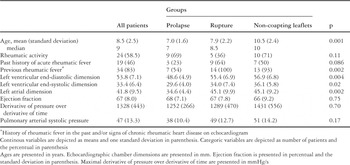
Table 1.
Examples of the mechanisms of rheumatic mitral insufficiency. (a) A patient with prolapse of the aortic leaflet. Note that the leaflets are only mildly thickened. (b) A patient with rupture of tendinous cords and intense prolapse of the aortic leaflet pointing towards the left atrium in systole. (c) An example of a thickened, retracted, non-coapting mural leaflet.

(a) Mean age with the 95% confidence interval of the three groups. (b) Proportions of previous rheumatic fever, defined as history of acute rheumatic fever in the past and/or signs of chronic rheumatic heart disease on echocardiogram, in the three groups. *p = 0.006 compared to the group with prolapse; **p = 0.03 compared to the group with prolapse, and p > 0.05 compared to the group with ruptured cords.
 You have
Access
- 9
- Cited by